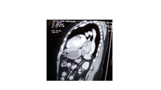
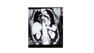
60 years old female H/O experiencing pain about an inch beneath her sternum
and sharp pains in radiating towards her left shoulder. It varies in intensity and is
increased immediately after eating spicy foods. After most meals, she c/o
suffering from mild heartburn. She is on course of Omeprazole, which alleviated
the symptoms, but they returned after a few days if she discontinue the PPI.
Dr Ibrahim Baloch
Resident Thoracic surgeon
CMH Rawalpindi , Pakistan
Anatomy
o The esophageal hiatus is formed by muscle fibers of the right
crus of the diaphragm, with little or no contribution from the
left crus.
o These fibers overlap inferiorly where they attach over and
along the right side of the median arcuate ligament, which is
attached to the lateral aspects of vertebral bodies.
o The phrenicoesophageal ligament is formed by fusion of the
endothoracic and endoabdominal fascia at the
diaphragmatic hiatus. This ligament inserts onto the
esophagus and holds the distal esophagus
Hiatus hernia
• It is defined as “The herniation of abdominal contents into the chest through the esophageal hiatus”.
May occur as a result of a
I. Congenital defect
II. Trauma
III. After antireflux or other hiatal hernia operations
Hiatal Hernia Types
Type I
It is also called a sliding
hiatal hernia, where the
GEJ migrates above the
diaphragm. The stomach
remains in its usual
longitudinal alignment and
the fundus remains below
the GEJ.
Type II
Hernias are pure
paraoesophageal hernias
(PEH); the GEJ remains in
its normal anatomic
position but a portion of
the fundus herniates
through the diaphragmatic
hiatus adjacent to the
oesophagus.
Type III
Hernias are a combination
of Types I and II, with both
the GEJ and the fundus
herniating through the
hiatus. The fundus lies
above the GEJ also called
giant PEH.
Type IV
Hiatal hernias are
characterized by the
presence of a structure
other than stomach, such
as the omentum, colon or
small bowel within the
hernia sac.
Epidemiology
• Sliding hernia greater then 95%
• PEHs account for approximately 5% to 15%.
• 3% to 6% of all patients undergoing surgical repair of hiatal hernias.
• Obesity is also a clear risk factor for developing a hiatal hernia.
• kyphosis or scoliosis may also lead to the development of PEH.
• In children, congenital defects are the most common cause of PEH.
• 30% of patients with PEHs presented with gastric volvulus.
• 29% mortality rate with nonoperative treatment.
Diagnosis
Chest x-ray
Retrocardiac air bubble with or
without an air-fluid level noted on
the lateral view of a standard chest
x-ray
• Barium study of the esophagus helps
establish the diagnosis with greater
accuracy .
• Typical findings include:
o Flattening of the base of the hernia
pocket, above the esophageal hiatus
o Stasis of barium in the herniated
segment above the esophageal hiatus
o Absent peristaltic waves in the
herniated pocket
o Irregularly shaped pouch as compared
to the smooth, pearshaped phrenic
ampula
o Identifiable gastric mucosal folds within
the pouch localization of the EGJ above
the diaphragm
o Upward the esophageal hiatus
o Retrograde filling of a herniated pouch
o A small fundus of stomach
Endoscopy
• Presence or absence of esophagitis.
• Diagnosis of GE reflux associated with PEH.
• Presence of barrett’s esophagus.
• Identify fibrotic stricture, esophageal neoplasm, or epiphrenic diverticulum.
• Identify intragastric ulcers, which may be the cause of the chronic anemia that many PEH present.
Esophageal
manometry
o Rule out concomitant esophageal dysmotility
disorders.
o Help the surgeon make an appropriate decision
regarding whether to perform a fundoplication and if
so what type of fundoplication (total or partial).
o Sometimes not possible to place the manometry
probe as a result of the PEH.
TREATMENT OPTIONS
• The goals of treatment are to relieve symptoms and prevent further complications.
• Reducing the gastroesophageal reflux will relieve pain.
• Other measures to reduce symptoms include:
o Avoiding large or heavy meals
o Not lying down or bending over immediately after a meal
o Reducing weight and not smoking
• If these measures fail to control the symptoms, or complications occur, surgical repair of
the hernia may be necessary.
Surgical Options
Surgical Options
- Laparoscopic vs. Open
- Total vs. Partial
- Nissen’s fundoplication (360○P)
- Toupet’s fundoplication (270○ P)
- Dor fundoplication (180○ A)
- Belsey Mark IV (Trans-thoracic)
Gastric bypass with crural repair in
severely or morbidly obese
Endoscopic procedures
- Stretta procedure -
radiofrequency
- EsophyX -
sutures
1. Left to right opening of the phreno-oesophageal ligament
2. Preservation of the hepatic branch of the anterior vagus nerve
3. Dissection of both crura
4. Transhiatal mobilization to allow approximately 3 cm of intra-abdominal oesophagus,
5. Short gastric vessel division to ensure a tension-free wrap,
6. Crural closure posteriorly with non-absorbable sutures,
7. Creation of a 1.5 to 2-cm wrap with the most distal suture incorporating the anterior muscular wall of the oesophagus,
and
8. Bougie placement at the time of wrap construction.
Standardized Nissen fundoplication
Intraoperative complication
• Pneumothorax
• Bleeding
• Esophageal or gastric perforation
• Vagal injury
Early Post-operative complication
• Subcutaneous crepitance in chest, neck and even head
• Wound infection
• Atrial Fibrillation
• DVT
Long-term Post-operative complication
• Dysphagia
• Belching
• Gas Bloat
• Pulmonary symptoms
• Weight loss
• Slipped Nissen
• Recurrence of PEH
Slipped Nissen
SAGES Guidelines for the Management of Hiatal Hernia 2013
Only investigations which will alter the clinical management of the patient should be performed (+++, strong)
Repair of a type I hernia in the absence of reflux disease is not necessary (+++, strong)
All symptomatic paraoesophageal hiatal hernias should be repaired (++++, strong), esp. acute obstructive symptoms
or volvulus. Acute gastric volvulus requires reduction of the stomach with limited resection if needed. (++++, strong)
Hiatal hernias can effectively be repaired by a transabdominal or transthoracic approach (++++, strong).
During paraoesophageal hiatal hernia repair the hernia sac should be dissected away from mediastinal structures
(++, strong) and then preferably should not excised (++, strong)
The use of mesh for reinforcement of large hiatal hernia repairs leads to decreased short term recurrence rates (+++,
strong)
A fundoplication must be performed during repair of a sliding type hiatal hernia to address reflux
A necessary step of hiatal hernia repair is to return the gastroesophageal junction to an infra-diaphragmatic position
(+++, strong). This length can be achieved by combinations of mediastinal dissection of the oesophagus and/ or
gastroplasty (++++, strong)
Hernia reduction with gastropexy alone and no hiatal repair may be a safe alternative in high-risk patients. Gastropexy
may safely be used in addition to hiatal repair (++++, strong)
Postoperative nausea and vomiting should be treated aggressively to minimize poor outcomes (++, strong)
Gastrostomy tube insertion may facilitate postoperative care in selected patients (++, strong)
With early postoperative dysphagia common, attention should be paid to adequate caloric and nutritional intake (+,
strong)
• Routine elective repair of completely asymptomatic paraoesophageal hernias may always be indicated.
Consideration for surgery should not include the patient’s age and comorbidities. (+++, strong)
• During operations for Roux-en-Y gastric bypass, sleeve gastrectomy and the placement of adjustable gastric
bands, all detected hiatal hernias should no be repaired (+++, strong)
• . A fundoplication is not important during paraoesophageal hernia repair. (++, strong)
• In the absence of achalasia, tailoring of the fundoplication to preoperative manometric data may be necessary
(++, strong)
• Recurrence can be reduced by extensive mediastinal oesophageal mobilization to bring the GEJ at least 2-3 cm
into the abdomen without tension (++, strong).
Aftercare
• Activity
1. Walk as normal
2. Buildup physical activity over 6-8
weeks
3. Strenuous activity permitted after 6
weeks.
4. Avoid driving for 3-4 weeks
5. Sexual relations can resume when
comfortable
Aftercare
• Diet & Medication
1. Liquids 1st-2nd week
2. Mashed/soft diet 2nd–4th week
3. Solids 5th-6th week
I. Small mouthfuls
II. Chew well
III. Swallow slowly
IV. Avoid tablets/capsules 6 weeks
PROGNOSIS
• Symptomatic relief post operatively greater than 80% .
• Recurrence rate of PEHs ranges from 20-40%.
• operative mortality rate for emergent repair of incarcerated PEH is 50% .
Hiatal hernia

Hiatal hernia

  • 1.
    60 years oldfemale H/O experiencing pain about an inch beneath her sternum and sharp pains in radiating towards her left shoulder. It varies in intensity and is increased immediately after eating spicy foods. After most meals, she c/o suffering from mild heartburn. She is on course of Omeprazole, which alleviated the symptoms, but they returned after a few days if she discontinue the PPI.
  • 5.
    Dr Ibrahim Baloch ResidentThoracic surgeon CMH Rawalpindi , Pakistan
  • 6.
    Anatomy o The esophagealhiatus is formed by muscle fibers of the right crus of the diaphragm, with little or no contribution from the left crus. o These fibers overlap inferiorly where they attach over and along the right side of the median arcuate ligament, which is attached to the lateral aspects of vertebral bodies. o The phrenicoesophageal ligament is formed by fusion of the endothoracic and endoabdominal fascia at the diaphragmatic hiatus. This ligament inserts onto the esophagus and holds the distal esophagus
  • 7.
    Hiatus hernia • Itis defined as “The herniation of abdominal contents into the chest through the esophageal hiatus”. May occur as a result of a I. Congenital defect II. Trauma III. After antireflux or other hiatal hernia operations
  • 8.
  • 9.
    Type I It isalso called a sliding hiatal hernia, where the GEJ migrates above the diaphragm. The stomach remains in its usual longitudinal alignment and the fundus remains below the GEJ.
  • 10.
    Type II Hernias arepure paraoesophageal hernias (PEH); the GEJ remains in its normal anatomic position but a portion of the fundus herniates through the diaphragmatic hiatus adjacent to the oesophagus.
  • 11.
    Type III Hernias area combination of Types I and II, with both the GEJ and the fundus herniating through the hiatus. The fundus lies above the GEJ also called giant PEH.
  • 12.
    Type IV Hiatal herniasare characterized by the presence of a structure other than stomach, such as the omentum, colon or small bowel within the hernia sac.
  • 13.
    Epidemiology • Sliding herniagreater then 95% • PEHs account for approximately 5% to 15%. • 3% to 6% of all patients undergoing surgical repair of hiatal hernias. • Obesity is also a clear risk factor for developing a hiatal hernia. • kyphosis or scoliosis may also lead to the development of PEH. • In children, congenital defects are the most common cause of PEH. • 30% of patients with PEHs presented with gastric volvulus. • 29% mortality rate with nonoperative treatment.
  • 14.
  • 15.
    Chest x-ray Retrocardiac airbubble with or without an air-fluid level noted on the lateral view of a standard chest x-ray
  • 16.
    • Barium studyof the esophagus helps establish the diagnosis with greater accuracy . • Typical findings include: o Flattening of the base of the hernia pocket, above the esophageal hiatus o Stasis of barium in the herniated segment above the esophageal hiatus o Absent peristaltic waves in the herniated pocket o Irregularly shaped pouch as compared to the smooth, pearshaped phrenic ampula o Identifiable gastric mucosal folds within the pouch localization of the EGJ above the diaphragm o Upward the esophageal hiatus o Retrograde filling of a herniated pouch o A small fundus of stomach
  • 17.
    Endoscopy • Presence orabsence of esophagitis. • Diagnosis of GE reflux associated with PEH. • Presence of barrett’s esophagus. • Identify fibrotic stricture, esophageal neoplasm, or epiphrenic diverticulum. • Identify intragastric ulcers, which may be the cause of the chronic anemia that many PEH present.
  • 18.
    Esophageal manometry o Rule outconcomitant esophageal dysmotility disorders. o Help the surgeon make an appropriate decision regarding whether to perform a fundoplication and if so what type of fundoplication (total or partial). o Sometimes not possible to place the manometry probe as a result of the PEH.
  • 19.
    TREATMENT OPTIONS • Thegoals of treatment are to relieve symptoms and prevent further complications. • Reducing the gastroesophageal reflux will relieve pain. • Other measures to reduce symptoms include: o Avoiding large or heavy meals o Not lying down or bending over immediately after a meal o Reducing weight and not smoking • If these measures fail to control the symptoms, or complications occur, surgical repair of the hernia may be necessary.
  • 20.
    Surgical Options Surgical Options -Laparoscopic vs. Open - Total vs. Partial - Nissen’s fundoplication (360○P) - Toupet’s fundoplication (270○ P) - Dor fundoplication (180○ A) - Belsey Mark IV (Trans-thoracic) Gastric bypass with crural repair in severely or morbidly obese
  • 21.
    Endoscopic procedures - Strettaprocedure - radiofrequency - EsophyX - sutures
  • 22.
    1. Left toright opening of the phreno-oesophageal ligament 2. Preservation of the hepatic branch of the anterior vagus nerve 3. Dissection of both crura 4. Transhiatal mobilization to allow approximately 3 cm of intra-abdominal oesophagus, 5. Short gastric vessel division to ensure a tension-free wrap, 6. Crural closure posteriorly with non-absorbable sutures, 7. Creation of a 1.5 to 2-cm wrap with the most distal suture incorporating the anterior muscular wall of the oesophagus, and 8. Bougie placement at the time of wrap construction. Standardized Nissen fundoplication
  • 23.
    Intraoperative complication • Pneumothorax •Bleeding • Esophageal or gastric perforation • Vagal injury
  • 24.
    Early Post-operative complication •Subcutaneous crepitance in chest, neck and even head • Wound infection • Atrial Fibrillation • DVT
  • 25.
    Long-term Post-operative complication •Dysphagia • Belching • Gas Bloat • Pulmonary symptoms • Weight loss • Slipped Nissen • Recurrence of PEH Slipped Nissen
  • 26.
    SAGES Guidelines forthe Management of Hiatal Hernia 2013
  • 27.
    Only investigations whichwill alter the clinical management of the patient should be performed (+++, strong) Repair of a type I hernia in the absence of reflux disease is not necessary (+++, strong) All symptomatic paraoesophageal hiatal hernias should be repaired (++++, strong), esp. acute obstructive symptoms or volvulus. Acute gastric volvulus requires reduction of the stomach with limited resection if needed. (++++, strong) Hiatal hernias can effectively be repaired by a transabdominal or transthoracic approach (++++, strong). During paraoesophageal hiatal hernia repair the hernia sac should be dissected away from mediastinal structures (++, strong) and then preferably should not excised (++, strong) The use of mesh for reinforcement of large hiatal hernia repairs leads to decreased short term recurrence rates (+++, strong)
  • 28.
    A fundoplication mustbe performed during repair of a sliding type hiatal hernia to address reflux A necessary step of hiatal hernia repair is to return the gastroesophageal junction to an infra-diaphragmatic position (+++, strong). This length can be achieved by combinations of mediastinal dissection of the oesophagus and/ or gastroplasty (++++, strong) Hernia reduction with gastropexy alone and no hiatal repair may be a safe alternative in high-risk patients. Gastropexy may safely be used in addition to hiatal repair (++++, strong) Postoperative nausea and vomiting should be treated aggressively to minimize poor outcomes (++, strong) Gastrostomy tube insertion may facilitate postoperative care in selected patients (++, strong) With early postoperative dysphagia common, attention should be paid to adequate caloric and nutritional intake (+, strong)
  • 29.
    • Routine electiverepair of completely asymptomatic paraoesophageal hernias may always be indicated. Consideration for surgery should not include the patient’s age and comorbidities. (+++, strong) • During operations for Roux-en-Y gastric bypass, sleeve gastrectomy and the placement of adjustable gastric bands, all detected hiatal hernias should no be repaired (+++, strong) • . A fundoplication is not important during paraoesophageal hernia repair. (++, strong) • In the absence of achalasia, tailoring of the fundoplication to preoperative manometric data may be necessary (++, strong) • Recurrence can be reduced by extensive mediastinal oesophageal mobilization to bring the GEJ at least 2-3 cm into the abdomen without tension (++, strong).
  • 30.
    Aftercare • Activity 1. Walkas normal 2. Buildup physical activity over 6-8 weeks 3. Strenuous activity permitted after 6 weeks. 4. Avoid driving for 3-4 weeks 5. Sexual relations can resume when comfortable
  • 31.
    Aftercare • Diet &Medication 1. Liquids 1st-2nd week 2. Mashed/soft diet 2nd–4th week 3. Solids 5th-6th week I. Small mouthfuls II. Chew well III. Swallow slowly IV. Avoid tablets/capsules 6 weeks
  • 32.
    PROGNOSIS • Symptomatic reliefpost operatively greater than 80% . • Recurrence rate of PEHs ranges from 20-40%. • operative mortality rate for emergent repair of incarcerated PEH is 50% .

Editor's Notes

  • #8 The natural history of hiatal hernia is that the pressure gradient between the chest and abdomen results in the enlargement of the hernia over time Persistant negative intrathoracic pressure during swallowing result in the thining out of the phrenoesphageal membrane over time PEHs are also associated with previous GE surgery such as esophagomyotomy, antireflux surgery, and thoracoabdominal trauma.
  • #10 occurs because of circumferential weakening of the phrenicoesophageal ligament Factors contribute to the development of this hernia include increased abdominal pressure (e.g., with pregnancy, obesity, or vomiting) and vigorous esophageal contraction,
  • #11 phrenicoesophageal membrane is weakened focally, anterior and lateral to the esophagus. gastric fundus and/or greater curvature protrudes through the defect into the mediastinum
  • #12 The term “giant PEH” typically refers to a hernia where more than a third of the stomach has migrated into the chest cavity above the diaphragm
  • #13 The transverse colon and omentum are most commonly involved
  • #14 kyphosis or scoliosis may also lead to the development of PEH due to distortion of the anatomy of the diaphragm. In children, congenital defects are the most common cause of PEH and are often associated with other congenital anomalies such as intestinal malformation. 5
  • #16 differential diagnosis includes mediastinal cyst or abscess and dilated obstructive esophagus, as one would see with megaesophagus in a patient with end-stage achalasia.
  • #17 EGS esophago gastric junction
  • #26 Gas bloat. Abdominal fullness with gas Slipped Nissen failure of Nissen fundoplication Belching is the act of expelling air from the stomach through the mouth
  • #27 Society of American gastrointestinal and endoscopic surgeons
  • #30 Tailoring = adjacent to a specific need