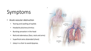
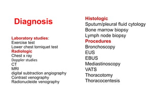
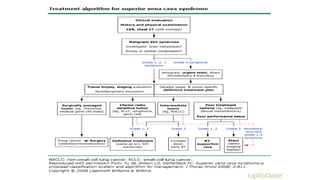
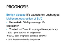
Superior vena cava obstruction (SVCO)
Anatomy SVC
o The confluence of the left and right
innominate(Brachiocephalic) veins at the level of
the cartilaginous portion of the first right rib gives
rise to the SVC.
o The left brachiocephalic vein > the right one
o The SVC has a diameter of 2 cm and an average
length of 7 cm.
Anatomy
o Left brachiocephalic vein crosses the anterosuperior
mediastinum from left to right posteriorly to the thymus or its
remnants
o SVC descends toward the right atrium laterally to the ascending
aorta and medially to the right mediastinal pleura and lung.
o Anatomical structures adjacent to the SVC are the
o Right paratracheal lymphatic chain.
o Right pulmonary artery, crossing the vessel posteriorly.
o Upper right pulmonary vein.
o Phrenic nerve.
o Thymus or its remnants with mediastinal fat.
Anatomy of the collateral SVC
routes:
1. Azygos venous system is the only direct path
into the SVC.
2. Internal thoracic vein is the collector between
SVC and inferior vena cava (IVC) via
epigastric and iliac veins.
3. Vertebral veins with intercostals, lumbar and
sacral veins, represent the posterior network
between SVC and IVC.
4. External thoracic vein system is the most
superficial and it is represented by axillary,
lateral thoracic and superficial epigastric
veins.
• The SVC obstruction (SVCO) was first described in 1757 by William
Hunter in a patient with a saccular syphilitic aortic aneurysm of the
ascending aorta.
• Almost two hundred years later, Stokes, in 1837, described SVC
obstruction related to a malignant tumor arising in the right lung; he
observed that the underlying clinical findings were the result of the
neoplastic compression on the vein and the progressive development of
collateral circulation.
History
In the pre-antibiotic era
◦ syphilitic thoracic aortic aneurysms,
• fibrosing mediastinitis,
• untreated infection were frequent causes of the SVC obstruction.
• More recently, the incidence of SVC obstruction due to thrombosis has
risen, largely because of increased use of intravascular devices such as
catheters and pacemakers.
 The first SVC bypass graft with an autologous superficial femoral vein was
described in 1951
 Subsequently in 1980, malignancy became the most common cause,
accounting for 90 % of cases.
 In 1987, the use of polytetrafluoroethylene (PTFE) grafts with proven
patency in patients with neoplastic involvement of the SVC.
History
Epidemiology
• 73 to 97% of the cases are secondary to malignancy
• Bronchogenic carcinoma is most frequent cause, accounting for 65 to 80%.
• 3% of the patients with lung cancer develop SVC involvement in which 10% of the with
right-sided tumors.
• Mediastinal tumors accounts for 20%.
• Metastatic lesions are responsible for 5% of malignant SVC obstructions.
Superior vena cava (SVC) obstruction:
• It is defined as “The symptoms resulting from compression or obstruction of the SVC
system at any level, from the left and right brachiocephalic veins to the right atrium.”.
Pathophysiology:
• The SVC due to the thin walls and inner low pressure it is easily obstructed by
I. External compression II. Invasion III. Constriction IV. thrombosis
due to hypercoagulation, intimal damage, and/or stasis may be involved.
• Severe obstruction dramatically increases the endovascular pressure up to 400–500
cmH2O.
Cont:
 Acute SVC obstruction, the collateral systems do not have time to accommodate the increased blood flow.
 Slow progression of obstruction, palliation from collateral circulation is more pronounced.
• when the azygous vein orifice is not involved, the collateral pathways are more efficient since this system easily accommodates
the shunted blood.
• when the azygous system can’t compensate due to the location of the tumor, the blood flow runs through the other venous
systems that are less efficient for the smaller caliber and major length of the pathway; for this reason symptoms more
pronounced.
Etiology
 Lung cancer
 Lymphomas
 Thymoma
 Mediastinal germ cell tumors
 Mediastinal metastases
 Mesothelioma
 Leiomyosarcoma
 Angiosarcoma
 Neoplastic thrombi
 Anaplastic thyroid cancer
Malignant
Etiology
Fibrosing mediastinitis
(idiopathic or radiation induced)
 Infectious diseases –
1. Tuberculosis,
2. Histoplasmosis,
3. Echinococcosis,
4. Syphilis,
5. Aspergillosis,
6. Blastomycosis,
7. Filariasis,
8. Nocardiosis.
Benign
Etiology
 Iatrogenic
1. Pacemaker and defibrillator
placement
2. Central venous catheters
 Thrombosis (non-neoplastic)
 Lymphadenopathies
1. sarcoidosis,
2. Behçet’s obstruction,
3. Castleman’s disease
 Aortic aneurysm
 Substernal goiter
 Pericardial, thymic, bronchogenic cysts
Benign
Superior vena cava (SVC)
Obstruction Anatomic
classification
Obstruction of the upper
SVC
proximal to the azygos
entry point
.  In this situation, there is no impediment to normal
blood flow through the azygos vein which opens
into the patent tract of the SVC.
 Venous drainage coming from the head neck,
shoulders and arms cannot directly reach the
right atrium.
 From the superior tract of the SVC, blood flow
is reversed and directed to the azygos, mainly
through the right superior intercostal vein.
Obstruction with
azygos involvement
In this case, the azygos vein cannot be used as
collateral pathway and the only viable blood return is
carried by minor vessels to IVC (cava-cava or
anazygotic circulation).
 From the internal thoracic veins, blood is forced to
the intercostal veins, then to azygos and hemiazygos
veins.
 The flow is thus reversed into the ascending
lumbar veins to the iliac veins.
 Direct anastomosis between the
azygos’ origin and the IVC and between
hemiazygos and left renal vein are also
active.
 In addition, the internal thoracic
veins can flow into the superior
epigastric veins.
 From the superior epigastric veins,
blood is carried to the inferior epigastric
veins across the superficial system of
the cutaneous abdominal veins and
finally to the iliac veins.
 Another course is between the
thoraco-epigastric vein (collateral of the
axillary vein) and the external iliac vein.
 In these conditions, the collateral circulation is
partly deep and partly superficial.
 Physical examination often reveals SVC
obstruction.
 The reversed circulation through the described
pathways, remains less efficient than the azygos
system and venous hypertension is usually more
severe.
 For this reason, this kind of SVC obstruction is
often related to important symptoms, dyspnea
and pleural effusion.
 The ensuing slow blood flow may be
responsible for superimposed thrombosis.
Obstruction of the lower
SVC
distal to the azygos entry
point
 In this condition, the obstruction is
just below the azygos arch.
 The blood flow is distributed from the
superior body into the azygos and
hemiazygos veins, in which the flow is
inverted, to the IVC tributaries.
Cont-
 In this type of obstruction, the superficial
collateral system is not always evident but the
azygos and hemiazygos congestion and
dilatation are usually important.
 The hemodynamic changes lead to edema
and cyanosis of the upper chest and pleural
effusion. Pleural effusion is often slowly-growing
and rightsided, probably due to anatomical
reasons:
 There is a wider anastomosis between
hemiazygos and IVC than between azygos and
IVC.
Classification of SVCO
There are three (03) main classification proposals
which follow different methods of categorization.
Doty and Standford’s classification (anatomical)
1. Type I: stenosis of up to
90% of the supraazygos
SVC
2. Type II: stenosis of more
than 90% of the supraazygos
SVC
3. Type III: complete
occlusion of SVC with
azygos reverse blood flow
4. Type IV: complete
occlusion of SVC with the
involvement of the major
tributaries and azygos vein
Yu’s classification (clinical)
1. Grade 0: asymptomatic (imaging evidence of SVC obstruction)
2. Grade 1: mild (plethora, cyanosis, head and neck edema)
3. Grade 2: moderate (grade 1 evidence + functional impairment)
4. Grade 3: severe (mild/moderate cerebral or laryngeal edema, limited cardiac reserve)
5. Grade 4: life-threatening (significant cerebral or laryngeal edema, cardiac failure)
6. Grade 5: fatal
Bigsby’s classification (operative risk)
1. Low risk
2. High risk
The low risk patients present with
o No dyspnea at rest,
o No facial cyanosis in the upright position,
o No change of dyspnea, No worsening of facial edema and Cyanosis during
the supine position.
The high risk patients present with
o Facial cyanosis or dyspnea at rest in the sitting position.
Symptoms
• Acute vascular obstruction
• Tearing and swelling of eyelids
• Headache,dizziness,tinnitus
• Bursting sensation in the head
• Red and edematous (face, neck and arms)
• Superficial veins distended (chest)
• sleep in a chair to avoid dyspnea.
Superior vena cava obstruction in a person with brochogenic carcinoma. Note the swelling
of his face first thing in the morning (left) and its resolution after being upright all day (right).
Sign
o Venous distension of neck-66%
o Venous distension of Chest-54%
o Edema upper half of the body-50%
o Paleness of lower half of the body-18%
o Engorged abdomen veins-12%
o Papilledema, stupor, and even coma.
o Cyanosis and edema are aggravated by
horizontal position and relived by upright
position
Life threatening complication of
venous HTN
• Cerebral edema, thrombosis, and hemorrhage
• Laryngeal and/or glossal edema.
Diagnosis
Diagnosis of SVCO can be made simply on physical
examination.
Laboratory studies:
Exercise test
Lower chest torniquet test
Radiologic
Chest x ray
Doppler studies
CT
MRI
digital subtraction angiography
Contrast venography
Radionucleide venography
Histologic
Sputum/pleural fluid cytology
Bone marrow biopsy
Lymph node biopsy
Procedures
Bronchoscopy
EUS
EBUS
Mediastinoscopy
VATS
Thoracotomy
Thoracocentesis
Diagnosis
Laboratory studies:
o Exercise test
o Lower chest torniquet
test
Laboratory studies:
Localizing Obstruction
 Pressure readings are taken from the ante-cubital vein with a 3-way stopcock spinal
manometer using 2.5 % citrate solution
Exercise test (Hussay et al)
 Patient opens and closes his fist forcefully for one minute while venous pressure readings are
being noted.
 In normal individuals, it remains constant
 In SVCO pressure will rise 10 cm. or more and then gradually recedes to normal.
Laboratory studies:
Lower chest tourniquet test
 in which a tourniquet constricts the superficial thoracic collaterals and raises the venous pressure if
obstruction is below the azygos.
Other Test
 Circulatory time is prolonged in SVCO
 Infra –red photography demonstrates superficial collaterals
 Phlebography
Chest x-ray
• The initial diagnostic test for
suspected SVCO.
• helpful in identifying the cause
of the disorder.
• Parish and colleagues in 1981 –
(16%) of the patients With
SVCO had normal Chest
radiography.
• Right sided findings are
common.
CT Scan
MRI
MRI images of case 3: a)
tight stenosis in the mid
SVC (white arrow);
Contrast venography
Venographic classification
1. Type I: stenosis of up to 90% of the
supra-azygos SVC
2. Type II: stenosis of more than 90%
of the supra-azygos SVC
3. Type III: complete occlusion of SVC
with azygos reverse blood flow
4. Type IV: complete occlusion of SVC
with the involvement of the major
tributaries and azygos vein
Digital Subtraction
Angiography DSA
DSA is gold standard to
assess upper extremities
thrombosis an widely
available in most Centre
Current guidelines stress the importance of accurate
histologic diagnosis prior to starting therapy, and the
upfront use of endovascular stents in severely
symptomatic patients to provide more rapid relief
than can be achieved using RT.
Kvale PA, Selecky PA, Prakash UB, American
College of Chest Physicians. Palliative care in lung
cancer: ACCP evidencebased clinical practice
guidelines (2nd edition). Chest 2007;132:368S.
TREATMENT OPTIONS
• The goals of treatment are to relieve symptoms and prevent further complications.
• Depending on the underlying condition, multiple treatment options are available for superior vena cava
obstruction. The primary treatment options include
1. Medical Care
2. Radiation
3. Chemotherapy
4. Thrombolytic therapy
5. Anticoagulation
6. Stents and balloon angioplasty and
7. Surgery..
Medical Care
 The goals of SVCO management are to relieve symptoms and
to attempt cure of the primary malignant
Conservative treatment -symptomatic improvement
 including elevation of the head end of the bed and supplemental
oxygen.
Emergency treatment (Corticosteroids and diuretics )
 For Brain edema,
 decreased cardiac output,
 or upper airway edema
Radiation therapy
Indications.
 The majority of cases of SVCO are caused by malignancy; thus, most patients
receive radiation treatment at some point in their illness.
Emergency radiation treatment
 To life-threatening cerebral or laryngeal edema prior to a tissue diagnosis of
malignancy.
 To relieve obstructive symptoms
 Inappropriate for the treatment of an underlying thrombosis or granulomatosis
causing the obstruction
Response to RT
3 to 4 days- Resolution of facial edema and venous distension
of the upper extremities .
1 to 3 weeks- Radiographic improvement .
Not effective -Thrombosis is cause for SVCO
 When RT successfully completed in pts of SVCO with
malignancies, 10% to 20% survive more than 2 years.
Chemotherapy
 Chemotherapy may be used as a primary therapy or as an
adjunct to radiotherapy
 treatment of choice for SVCO caused by Mediastinal
lymphoma is a combination of chemotherapy and radiotherapy.
Thrombolytic Therapy
 Peri-catheter thrombosis is seen in
approximately 50% of Non-anticoagulated patients with long
term Central vein Catheters
 Acute Cases- excellent results with thrombolytic therapy
• Thrombolytic = Urokinase
Anticoagulation
 Patients with SVCO are at increased risk for deep vein thrombosis and
pulmonary embolism.
 In patients for whom thrombosis is the cause of SVCO, anticoagulation
therapy should be administered after successful thrombolytic treatment.
 Once the symptoms subside after thrombolytic therapy, anticoagulation
should be maintained as long as the central venous catheter is present.
Anticoagulant = Heparin, Warfarin
(Coumarin)
Stents
 Recent advances in interventional radiology have contributed expandable
wire stents and balloon angioplasty.
 can be placed across the stenotic portion.
 stents have little thrombogenic potential
 After thrombolytic therapy, stent placement has been noted to be a more
successful approach.
 After stent, patients experience instantaneous relief of symptoms.
 The placement of stents is performed under local anesthesia.
 palliation of the symptoms
Balloon Angioplasty
 For localized lesions, balloon
angioplasty with or without
stenting has also been shown to
significantly reduce the symptoms
of SVCO.
Surgical Treatment
 Surgical bypass is an additional alternative to relieve SVCO.
 is usually recommended to benign disease and to only a few patients
with malignancy.
 Patients selected for surgery should have the Category-IV venographic
signs, i.e, total vena caval obstruction.
 Surgery in cases of fibrosing mediastinitis can be extremely
complicated, because of the extensive collateral circulation under high
venous pressure.
 Advantage is definitive removal of the obstruction and direct tissue
diagnosis.
 Long-term results after surgical bypass are lacking, because their life
expectancy is short.
Surgical Options
PROGNOSIS
Benign disease-life expectancy unchanged
Malignant obstruction of SVC
 Untreated - 30 days average life
expectancy
 Treated - < 7 month average life expectancy
- 20% 1-year survival for lung cancer
-NSCLC-poor prognosis, palliative care+RT
- 50% 2-year survival for lymphoma
supra vena cava obstruction (SVCO)

supra vena cava obstruction (SVCO)

  • 1.
    Superior vena cavaobstruction (SVCO)
  • 2.
    Anatomy SVC o Theconfluence of the left and right innominate(Brachiocephalic) veins at the level of the cartilaginous portion of the first right rib gives rise to the SVC. o The left brachiocephalic vein > the right one o The SVC has a diameter of 2 cm and an average length of 7 cm.
  • 3.
    Anatomy o Left brachiocephalicvein crosses the anterosuperior mediastinum from left to right posteriorly to the thymus or its remnants o SVC descends toward the right atrium laterally to the ascending aorta and medially to the right mediastinal pleura and lung. o Anatomical structures adjacent to the SVC are the o Right paratracheal lymphatic chain. o Right pulmonary artery, crossing the vessel posteriorly. o Upper right pulmonary vein. o Phrenic nerve. o Thymus or its remnants with mediastinal fat.
  • 4.
    Anatomy of thecollateral SVC routes: 1. Azygos venous system is the only direct path into the SVC. 2. Internal thoracic vein is the collector between SVC and inferior vena cava (IVC) via epigastric and iliac veins. 3. Vertebral veins with intercostals, lumbar and sacral veins, represent the posterior network between SVC and IVC. 4. External thoracic vein system is the most superficial and it is represented by axillary, lateral thoracic and superficial epigastric veins.
  • 5.
    • The SVCobstruction (SVCO) was first described in 1757 by William Hunter in a patient with a saccular syphilitic aortic aneurysm of the ascending aorta. • Almost two hundred years later, Stokes, in 1837, described SVC obstruction related to a malignant tumor arising in the right lung; he observed that the underlying clinical findings were the result of the neoplastic compression on the vein and the progressive development of collateral circulation. History
  • 6.
    In the pre-antibioticera ◦ syphilitic thoracic aortic aneurysms, • fibrosing mediastinitis, • untreated infection were frequent causes of the SVC obstruction. • More recently, the incidence of SVC obstruction due to thrombosis has risen, largely because of increased use of intravascular devices such as catheters and pacemakers.  The first SVC bypass graft with an autologous superficial femoral vein was described in 1951  Subsequently in 1980, malignancy became the most common cause, accounting for 90 % of cases.  In 1987, the use of polytetrafluoroethylene (PTFE) grafts with proven patency in patients with neoplastic involvement of the SVC. History
  • 7.
    Epidemiology • 73 to97% of the cases are secondary to malignancy • Bronchogenic carcinoma is most frequent cause, accounting for 65 to 80%. • 3% of the patients with lung cancer develop SVC involvement in which 10% of the with right-sided tumors. • Mediastinal tumors accounts for 20%. • Metastatic lesions are responsible for 5% of malignant SVC obstructions.
  • 8.
    Superior vena cava(SVC) obstruction: • It is defined as “The symptoms resulting from compression or obstruction of the SVC system at any level, from the left and right brachiocephalic veins to the right atrium.”.
  • 9.
    Pathophysiology: • The SVCdue to the thin walls and inner low pressure it is easily obstructed by I. External compression II. Invasion III. Constriction IV. thrombosis due to hypercoagulation, intimal damage, and/or stasis may be involved. • Severe obstruction dramatically increases the endovascular pressure up to 400–500 cmH2O.
  • 10.
    Cont:  Acute SVCobstruction, the collateral systems do not have time to accommodate the increased blood flow.  Slow progression of obstruction, palliation from collateral circulation is more pronounced. • when the azygous vein orifice is not involved, the collateral pathways are more efficient since this system easily accommodates the shunted blood. • when the azygous system can’t compensate due to the location of the tumor, the blood flow runs through the other venous systems that are less efficient for the smaller caliber and major length of the pathway; for this reason symptoms more pronounced.
  • 11.
    Etiology  Lung cancer Lymphomas  Thymoma  Mediastinal germ cell tumors  Mediastinal metastases  Mesothelioma  Leiomyosarcoma  Angiosarcoma  Neoplastic thrombi  Anaplastic thyroid cancer Malignant
  • 12.
    Etiology Fibrosing mediastinitis (idiopathic orradiation induced)  Infectious diseases – 1. Tuberculosis, 2. Histoplasmosis, 3. Echinococcosis, 4. Syphilis, 5. Aspergillosis, 6. Blastomycosis, 7. Filariasis, 8. Nocardiosis. Benign
  • 13.
    Etiology  Iatrogenic 1. Pacemakerand defibrillator placement 2. Central venous catheters  Thrombosis (non-neoplastic)  Lymphadenopathies 1. sarcoidosis, 2. Behçet’s obstruction, 3. Castleman’s disease  Aortic aneurysm  Substernal goiter  Pericardial, thymic, bronchogenic cysts Benign
  • 14.
    Superior vena cava(SVC) Obstruction Anatomic classification
  • 15.
    Obstruction of theupper SVC proximal to the azygos entry point .  In this situation, there is no impediment to normal blood flow through the azygos vein which opens into the patent tract of the SVC.  Venous drainage coming from the head neck, shoulders and arms cannot directly reach the right atrium.  From the superior tract of the SVC, blood flow is reversed and directed to the azygos, mainly through the right superior intercostal vein.
  • 16.
    Obstruction with azygos involvement Inthis case, the azygos vein cannot be used as collateral pathway and the only viable blood return is carried by minor vessels to IVC (cava-cava or anazygotic circulation).  From the internal thoracic veins, blood is forced to the intercostal veins, then to azygos and hemiazygos veins.  The flow is thus reversed into the ascending lumbar veins to the iliac veins.
  • 17.
     Direct anastomosisbetween the azygos’ origin and the IVC and between hemiazygos and left renal vein are also active.  In addition, the internal thoracic veins can flow into the superior epigastric veins.  From the superior epigastric veins, blood is carried to the inferior epigastric veins across the superficial system of the cutaneous abdominal veins and finally to the iliac veins.  Another course is between the thoraco-epigastric vein (collateral of the axillary vein) and the external iliac vein.
  • 18.
     In theseconditions, the collateral circulation is partly deep and partly superficial.  Physical examination often reveals SVC obstruction.  The reversed circulation through the described pathways, remains less efficient than the azygos system and venous hypertension is usually more severe.  For this reason, this kind of SVC obstruction is often related to important symptoms, dyspnea and pleural effusion.  The ensuing slow blood flow may be responsible for superimposed thrombosis.
  • 19.
    Obstruction of thelower SVC distal to the azygos entry point  In this condition, the obstruction is just below the azygos arch.  The blood flow is distributed from the superior body into the azygos and hemiazygos veins, in which the flow is inverted, to the IVC tributaries.
  • 20.
    Cont-  In thistype of obstruction, the superficial collateral system is not always evident but the azygos and hemiazygos congestion and dilatation are usually important.  The hemodynamic changes lead to edema and cyanosis of the upper chest and pleural effusion. Pleural effusion is often slowly-growing and rightsided, probably due to anatomical reasons:  There is a wider anastomosis between hemiazygos and IVC than between azygos and IVC.
  • 21.
  • 22.
    There are three(03) main classification proposals which follow different methods of categorization.
  • 23.
    Doty and Standford’sclassification (anatomical) 1. Type I: stenosis of up to 90% of the supraazygos SVC 2. Type II: stenosis of more than 90% of the supraazygos SVC 3. Type III: complete occlusion of SVC with azygos reverse blood flow 4. Type IV: complete occlusion of SVC with the involvement of the major tributaries and azygos vein
  • 24.
    Yu’s classification (clinical) 1.Grade 0: asymptomatic (imaging evidence of SVC obstruction) 2. Grade 1: mild (plethora, cyanosis, head and neck edema) 3. Grade 2: moderate (grade 1 evidence + functional impairment) 4. Grade 3: severe (mild/moderate cerebral or laryngeal edema, limited cardiac reserve) 5. Grade 4: life-threatening (significant cerebral or laryngeal edema, cardiac failure) 6. Grade 5: fatal
  • 25.
    Bigsby’s classification (operativerisk) 1. Low risk 2. High risk The low risk patients present with o No dyspnea at rest, o No facial cyanosis in the upright position, o No change of dyspnea, No worsening of facial edema and Cyanosis during the supine position. The high risk patients present with o Facial cyanosis or dyspnea at rest in the sitting position.
  • 26.
    Symptoms • Acute vascularobstruction • Tearing and swelling of eyelids • Headache,dizziness,tinnitus • Bursting sensation in the head • Red and edematous (face, neck and arms) • Superficial veins distended (chest) • sleep in a chair to avoid dyspnea.
  • 27.
    Superior vena cavaobstruction in a person with brochogenic carcinoma. Note the swelling of his face first thing in the morning (left) and its resolution after being upright all day (right).
  • 28.
    Sign o Venous distensionof neck-66% o Venous distension of Chest-54% o Edema upper half of the body-50% o Paleness of lower half of the body-18% o Engorged abdomen veins-12% o Papilledema, stupor, and even coma. o Cyanosis and edema are aggravated by horizontal position and relived by upright position
  • 29.
    Life threatening complicationof venous HTN • Cerebral edema, thrombosis, and hemorrhage • Laryngeal and/or glossal edema.
  • 30.
  • 31.
    Diagnosis of SVCOcan be made simply on physical examination.
  • 32.
    Laboratory studies: Exercise test Lowerchest torniquet test Radiologic Chest x ray Doppler studies CT MRI digital subtraction angiography Contrast venography Radionucleide venography Histologic Sputum/pleural fluid cytology Bone marrow biopsy Lymph node biopsy Procedures Bronchoscopy EUS EBUS Mediastinoscopy VATS Thoracotomy Thoracocentesis Diagnosis
  • 33.
    Laboratory studies: o Exercisetest o Lower chest torniquet test
  • 34.
    Laboratory studies: Localizing Obstruction Pressure readings are taken from the ante-cubital vein with a 3-way stopcock spinal manometer using 2.5 % citrate solution Exercise test (Hussay et al)  Patient opens and closes his fist forcefully for one minute while venous pressure readings are being noted.  In normal individuals, it remains constant  In SVCO pressure will rise 10 cm. or more and then gradually recedes to normal.
  • 35.
    Laboratory studies: Lower chesttourniquet test  in which a tourniquet constricts the superficial thoracic collaterals and raises the venous pressure if obstruction is below the azygos. Other Test  Circulatory time is prolonged in SVCO  Infra –red photography demonstrates superficial collaterals  Phlebography
  • 36.
    Chest x-ray • Theinitial diagnostic test for suspected SVCO. • helpful in identifying the cause of the disorder. • Parish and colleagues in 1981 – (16%) of the patients With SVCO had normal Chest radiography. • Right sided findings are common.
  • 37.
  • 38.
    MRI MRI images ofcase 3: a) tight stenosis in the mid SVC (white arrow);
  • 39.
    Contrast venography Venographic classification 1.Type I: stenosis of up to 90% of the supra-azygos SVC 2. Type II: stenosis of more than 90% of the supra-azygos SVC 3. Type III: complete occlusion of SVC with azygos reverse blood flow 4. Type IV: complete occlusion of SVC with the involvement of the major tributaries and azygos vein
  • 40.
    Digital Subtraction Angiography DSA DSAis gold standard to assess upper extremities thrombosis an widely available in most Centre
  • 41.
    Current guidelines stressthe importance of accurate histologic diagnosis prior to starting therapy, and the upfront use of endovascular stents in severely symptomatic patients to provide more rapid relief than can be achieved using RT. Kvale PA, Selecky PA, Prakash UB, American College of Chest Physicians. Palliative care in lung cancer: ACCP evidencebased clinical practice guidelines (2nd edition). Chest 2007;132:368S.
  • 42.
    TREATMENT OPTIONS • Thegoals of treatment are to relieve symptoms and prevent further complications. • Depending on the underlying condition, multiple treatment options are available for superior vena cava obstruction. The primary treatment options include 1. Medical Care 2. Radiation 3. Chemotherapy 4. Thrombolytic therapy 5. Anticoagulation 6. Stents and balloon angioplasty and 7. Surgery..
  • 43.
    Medical Care  Thegoals of SVCO management are to relieve symptoms and to attempt cure of the primary malignant Conservative treatment -symptomatic improvement  including elevation of the head end of the bed and supplemental oxygen. Emergency treatment (Corticosteroids and diuretics )  For Brain edema,  decreased cardiac output,  or upper airway edema
  • 44.
    Radiation therapy Indications.  Themajority of cases of SVCO are caused by malignancy; thus, most patients receive radiation treatment at some point in their illness. Emergency radiation treatment  To life-threatening cerebral or laryngeal edema prior to a tissue diagnosis of malignancy.  To relieve obstructive symptoms  Inappropriate for the treatment of an underlying thrombosis or granulomatosis causing the obstruction
  • 45.
    Response to RT 3to 4 days- Resolution of facial edema and venous distension of the upper extremities . 1 to 3 weeks- Radiographic improvement . Not effective -Thrombosis is cause for SVCO  When RT successfully completed in pts of SVCO with malignancies, 10% to 20% survive more than 2 years.
  • 46.
    Chemotherapy  Chemotherapy maybe used as a primary therapy or as an adjunct to radiotherapy  treatment of choice for SVCO caused by Mediastinal lymphoma is a combination of chemotherapy and radiotherapy.
  • 47.
    Thrombolytic Therapy  Peri-catheterthrombosis is seen in approximately 50% of Non-anticoagulated patients with long term Central vein Catheters  Acute Cases- excellent results with thrombolytic therapy • Thrombolytic = Urokinase
  • 48.
    Anticoagulation  Patients withSVCO are at increased risk for deep vein thrombosis and pulmonary embolism.  In patients for whom thrombosis is the cause of SVCO, anticoagulation therapy should be administered after successful thrombolytic treatment.  Once the symptoms subside after thrombolytic therapy, anticoagulation should be maintained as long as the central venous catheter is present. Anticoagulant = Heparin, Warfarin (Coumarin)
  • 49.
    Stents  Recent advancesin interventional radiology have contributed expandable wire stents and balloon angioplasty.  can be placed across the stenotic portion.  stents have little thrombogenic potential  After thrombolytic therapy, stent placement has been noted to be a more successful approach.  After stent, patients experience instantaneous relief of symptoms.  The placement of stents is performed under local anesthesia.  palliation of the symptoms
  • 50.
    Balloon Angioplasty  Forlocalized lesions, balloon angioplasty with or without stenting has also been shown to significantly reduce the symptoms of SVCO.
  • 51.
    Surgical Treatment  Surgicalbypass is an additional alternative to relieve SVCO.  is usually recommended to benign disease and to only a few patients with malignancy.  Patients selected for surgery should have the Category-IV venographic signs, i.e, total vena caval obstruction.  Surgery in cases of fibrosing mediastinitis can be extremely complicated, because of the extensive collateral circulation under high venous pressure.  Advantage is definitive removal of the obstruction and direct tissue diagnosis.  Long-term results after surgical bypass are lacking, because their life expectancy is short.
  • 52.
  • 54.
    PROGNOSIS Benign disease-life expectancyunchanged Malignant obstruction of SVC  Untreated - 30 days average life expectancy  Treated - < 7 month average life expectancy - 20% 1-year survival for lung cancer -NSCLC-poor prognosis, palliative care+RT - 50% 2-year survival for lymphoma

Editor's Notes

  • #33 , endoscopic ultrasound (EUS) or endobronchial ultrasound (EBUS) , mediastinoscopy , anterior mediastinotomy, median sternotomy, video-assisted thoracoscopy (VATS), and even thoracotomy might be indicated.
  • #37 Mediastinal widening Pleural effusion(s) Right hilar mass Cardiomegaly Calcified paratracheal lymph nodes- Granulomatous disease anterior mediastinal mass Aortic Nipple  Seen as a small soft-tissue density adjacent to the lateral border of the aortic knob on a frontal radiograph.  an aortic nipple is a radiological sign that represents the left superior intercostal vein as it runs around the aortic arch before joining the left brachiocephalic vein Conditions that can cause an aortic nipple are a) Normal variant is usually found in normal healthy patients in anywhere from 1.4-9.5% of people. b) Increase in venous flow such as  Recumbant position, or during expiration  Portal venous hypertension secondary to hepatofugal shunting from the liver,  congenital anomalies of the caval, azygos or hemiazygos circulation results in enlargement of the left superior intercostal vein. partial or total anomalous pulmonary venous drainage c) Caused by increased venous resistance as in  Congestive heart failure,  Budd Chiari sydrome  absence or obstruction of the inferior vena cava
  • #38 CT scanning provides 1) Anatomic details of the mediastinal and thoracic organs 2) Allows identification of the cause and extent of the obstruction, 3) Documents collateral circulation, 4) Provides guidance for Percutaneous biopsies 5) Guides the formulation for radiotherapy.
  • #39 MRI, by virtue of its multidimensional capabilities, shows the relationships of vessels, lymph nodes, and other mediastinal structures.
  • #40  The extent and site of obstruction.  The nature and degree of obstruction.  Patency of the superior vena cava.  Differentiation between intrinsic and extrinsic obstruction.  Assessment of collateral vessels  the degree of venous distension of the neck and arms
  • #44 Dexamethasone (Decadron, Dexasone) For symptomatic management in tumor associated edema.  8-40 mg IV once initially, followed by 4-6 mg IV/PO q6-8h
  • #45 Radiation Dosage  Initiated at high dose daily for the first few days. followed by conventional low daily doses. total dose is dependent on tumor histology.  Lymphomas (3000 to 4000 cGy,)  Carcinomas require (4000 to 5000 cGy or more) Lower doses of radiation treatment  When systemic disease is present and shortterm palliation is the goal.  Radiation to Heart and Spinal cord.  who are receiving chemotherapeutic agents such as doxorubicin, which can enhance radiation toxicity.
  • #46 Side effects of RT.  Persistent fever,  Bleeding or SVC perforation at the site of tumor invasion,  Nausea, Vomiting,  Anorexia,  Leukopenia,  Hemoptysis, Late Complications  Skin irritation;  Esophagitis;  Pulmonary or mediastinal fibrosis;
  • #48  Fast dissolution of emboli,  Quickened recovery,  Prevention of recurrent thrombus formation,  Rapid restoration of hemodynamic disturbances. Action:-  Converts plasminogen to plasmin, which degrades fibrin clots, fibrinogen, and other plasma proteins. Adult Dose: Loading dose: 4400 U/kg IV over 10 min and increase to 6000 U/kg/h Maintenance dose: 4400-6000 U/kg/h IV
  • #49 Action:- heparin  Inhibits thrombosis by inactivating activated factor X and inhibiting conversion of prothrombin to thrombin. Adult:-  5000 U IV bolus, then infusion to maintain aPTT 2-3 times the reference range Pediatric:- Initial dose: 50 U/kg IV Maintenance infusion: 15-25 U/kg/h IV Increase dose by 2-4 U/kg/h IV q6-8h using aPTT Action:-warfarin  Inhibits synthesis of vitamin K–dependent coagulation factors (factors II, VII, IX, X). Adult:-  Initial: 5-10 mg PO Maintenance: 2-10 mg PO qd to maintain INR of 2-3 Pediatric:-  0.05-0.34 mg/kg/d PO; adjust dose according to desired INR