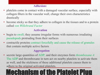
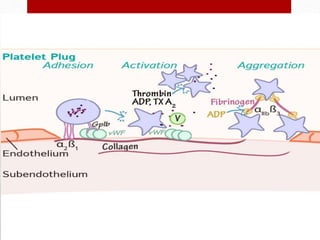
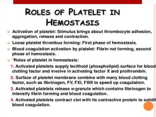
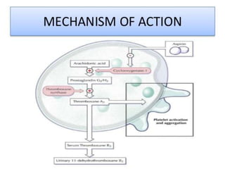
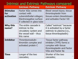
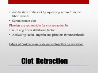
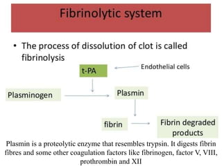
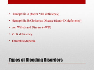
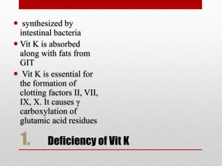
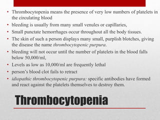
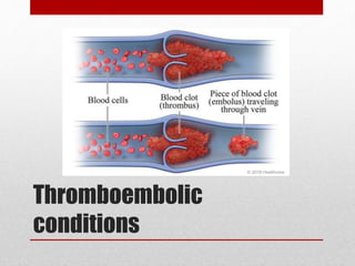
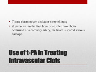
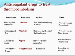
3. Steps of Hemostasis
• (1) vascular constriction,
• (2) formation of a platelet plug,
• (3) formation of a blood clot
• (4) Fibrous organization of the clot
4. Vascular Constriction
The spasm can last for many minutes or even hours, during
which time the processes of platelet plugging and blood
coagulation can take place. Mechanisms:
(1) local myogenic spasm,
(2) local autacoid factors from the traumatized tissues and
blood platelets eg thromboxane A2.
(3) nervous reflexes: initiated by pain nerve impulses or
other sensory impulses that originate from the traumatized
vessel or nearby tissues
5. Formation of the Platelet
Plug
• many small vascular holes develop throughout the body
each day that are often sealed by a platelet plug, rather
than by a blood clot.
6. • Platelets (also called thrombocytes) are minute discs 1 to
4 micrometers
• formed in the bone marrow from megakaryocytes
• fragment into the minute platelets either in the bone
marrow or soon after entering the blood,
• 150,000 and 300,000 per microliter.
• Half life in the blood of 8 to 12 days
• eliminated from the circulation mainly by the tissue
macrophage system
• do not have nuclei
7. (1) actin and myosin molecules, which
are contractile proteins thrombosthenin,
that can cause the platelets to contract;
(2) residuals of both the endoplasmic
reticulum and the Golgi apparatus that
synthesize various enzymes and store
large quantities of calcium ions;
(3) mitochondria
4) enzyme systems that are capable of
forming ATP and ADP
(4) enzyme systems that synthesize
prostaglandins, (COX) which are local
hormones that cause many vascular and
other local tissue reactions;
(5) fibrin-stabilizing factor,
(6) Platelet derived growth factor that
causes vascular endothelial cells,
vascular smooth muscle cells, and
fibroblasts to multiply and grow
8. • cell membrane coat of glycoproteins that repulses
adherence to normal endothelium and yet causes
adherence to injured endothelial cells and even more so to
exposed collagen
• platelet membrane contains large amounts of
phospholipids that activate multiple stages in the blood-
clotting process
9. Mechanism of the Platelet Plug
Adhesion:
• platelets come in contact with a damaged vascular surface, especially with
collagen fibers in the vascular wall, change their own characteristics
drastically
• become sticky so that they adhere to collagen in the tissues and to a protein
called von Willebrand Factor
Activation
• begin to swell; they assume irregular forms with numerous irradiating
pseudopods protruding from their surfaces
• contractile proteins contract forcefully and cause the release of granules
that contain multiple active factors
Aggregation
• secrete large quantities of ADP; and their enzymes form thromboxane A
The ADP and thromboxane in turn act on nearby platelets to activate them
as well, and the stickiness of these additional platelets causes them to
adhere to the original activated platelets.
10.
11.
12.
13.
14.
15.
16.
17.
18. Clot Retraction
• stabilization of the clot by squeezing serum from the
fibrin strands
• Serum cannot clot
Platelets are responsible for clot retraction by
• releasing fibrin stabilizing factor
• Activating actin , myosin and platelets thrombosthenin
Edges of broken vessels are pulled together by retraction
19. Plasmin is a proteolytic enzyme that resembles trypsin. It digests fibrin
fibres and some other coagulation factors like fibrinogen, factor V, VIII,
prothrombin and XII
20. Types of Bleeding Disorders
• Hemophilia A (factor VIII deficiency)
• Hemophilia B/Christmas Disease (factor IX deficiency)
• von Willebrand Disease (vWD)
• Vit K deficiency
• Thrombocytopenia
21. 1. Deficiency of Vit K
synthesized by
intestinal bacteria
Vit K is absorbed
along with fats from
GIT
Vit K is essential for
the formation of
clotting factors II, VII,
IX, X. It causes γ
carboxylation of
glutamic acid residues
22. Genetic basis of Hemophilia
• Hemophilia A and B are X-linked recessive disorders
• Hemophilia is typically expressed in males and carried by
females
23.
24. Von Willebrand Disease
• Von Willebrand factor (vWF) is a large multimeric
glycoprotein that performs two critical functions in
hemostasis:
• it acts as a bridging molecule at sites of vascular injury
for normal platelet adhesion
• vWF acts as a carrier for factor VIII in the circulation that
maintains the normal level of factor VIII by decreasing
the clearance of factor VIII fivefold.
• BT and CT both prolonged
25. Thrombocytopenia
• Thrombocytopenia means the presence of very low numbers of platelets in
the circulating blood
• bleeding is usually from many small venules or capillaries,
• Small punctate hemorrhages occur throughout all the body tissues.
• The skin of such a person displays many small, purplish blotches, giving
the disease the name thrombocytopenic purpura.
• bleeding will not occur until the number of platelets in the blood falls
below 50,000/ml,
• Levels as low as 10,000/ml are frequently lethal
• person’s blood clot fails to retract
• idiopathic thrombocytopenic purpura: specific antibodies have formed
and react against the platelets themselves to destroy them.
35. Causes of thromboembolic
conditions
(1) Any roughened endothelial surface of a vessel—as may
be caused by arteriosclerosis, infection, or trauma—is likely
to initiate the clotting process.
(2) Blood often clots when it flows very slowly through
blood vessels
36. 1. Thrombotic coronary occlusion- leading to myocardial infarction
2. Thrombotic cerebral occlusion- leading to stroke.
3. Deep vein thrombosis
• bed ridden patients, stasis of blood in lower extremities cause
intravascular throbosis in leg veins that may grow in length upto the
common iliac vein.
• Embolus may dislodge and block the pulmonary artery leading to sudden
death due to pulomanry embolism
3. Disseminated intravascular coagulation-
• clotting mechanism becomes activated in widespread areas of the
circulation
• results from the presence of large amounts of traumatized or dying tissue
in the body that releases great quantities of tissue factor into the blood.
• Septicemia, in which either circulating bacteria or bacterial toxins—
especially endotoxins—activate the clotting mechanisms.
• Patient may even begin to bleed due to consumption of clotting factors.
37. Use of t-PA in Treating
Intravascular Clots
• Tissue plasminogen activator-streptokinase
• if given within the first hour or so after thrombotic
occlusion of a coronary artery, the heart is spared serious
damage.
38.
39. Blood Coagulation tests
• Bleeding time (BT)
• Clotting time (CT)
• Prothrombin Time( PT)
• activated Partial Thromboplastin Time (aPTT)