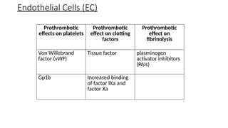
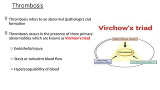
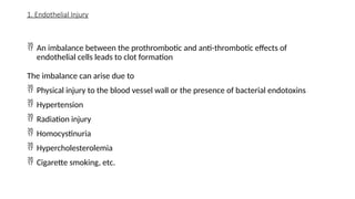
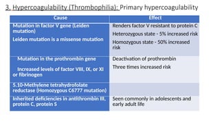
DR AMER ROHAID
MBBS MD
Assistant Professor Dept of Pathology
HEMOSTASIS & THROMBOSIS
• Define hemostasis
• Describe normal hemostasis
• Define a thrombus
• List the elements of a Virchow’s triad
• Describe the etiopathogenesis of thrombosis
• List the fates of a thrombus
• Describe the consequences of thrombosis
Definition
Hemostasis
Hemostasis is a precisely orchestrated process involving
platelets, clotting factors, and endothelium that occurs at
the site of vascular injury and culminates in the formation
of a blood clot, which serves to prevent or limit the extent
of bleeding.
Sequence of events
Primary hemostasis
• Formation of a platelet plug
Secondary hemostasis
• Deposition of fibrin
Clot stabilization and resorption
• Polymerized fibrin and platelet aggregates undergo contraction to form a solid permanent plug
Definition
Thrombosis
 The pathologic counterpart of hemostasis. It is the formation of a blood clot
(Thrombus) within intact vessels
Thrombus
• An aggregate of coagulated blood containing platelets, fibrin and entrapped
cellular elements, within a vascular lumen
Hemostasis and Thrombosis
 Hemostasis and thrombosis refer to the process of blood clot formation.
• Cells involved in hemostasis are endothelial cells and platelets which act in
conjunction with extracellular matrix
Haemostasis Thrombosis
Physiological Pathological
Platelets
 Platelets contain various type of granules which
secrete specific hemostatic substances
o α granules: Express adhesion molecule P-
selectin and contain fibrinogen, fibronectin,
factors V and VIII, platelet factor-4 (a heparin-
binding chemokine), platelet-derived growth
factor (PDGF), and transforming growth factor-
β (TGF-β)
o δ granules (Dense granules): Contain ADP and
ATP, calcium ions, histamine, serotonin,
epinephrine
• Platelets act through following steps: Adhesion,
Activation and Aggregation (3 As)
 Binding of vWF with platelet
surface receptor Gp1b
 Deficiency of vWF causes and
von Willebrand disease
 Deficiency of Gp1b causes
Bernard - Soulier disease
Platelet Adhesion
 Platelet activation triggered by thrombin and ADP. These also begets additional
rounds of platelet activation (Recruitment)
 Platelet activation begins by the release of granular components and once activated,
platelets also produce thromboxane A2 (TxA2)
• Platelets undergo a conformational change in the shape along the injured bv from
being spherical to long smooth discs with numerous spiky cell membrane extensions
 This change allows for increased surface area to react with coagulation factors via
negatively charged surface phospholipids and expression of GpIIb and GpIIIa proteins
 Deficiency of GpIIb and GpIIIa causes Glanzmann thrombasthenia
Platelet Activation
 Conformational change in GpIIb and GpIIIa allows binding of fibrinogen. The
initial wave of aggregation is reversible
 Concurrently, Thrombin activates the protease-activated receptor (PAR) which
stabilizes the platelet plug and initiates irreversible platelet contraction
 Thrombin also converts fibrinogen to fibrin
 This creates an irreversibly fused mass leading to secondary hemostatic plug
formation
• The balance between PGI2 and TxA2 helps in maintaining normal blood flow
versus hemostasis
Platelet Aggregation
 Is a series of amplifying enzymatic reactions that leads to the deposition of an insoluble
fibrin clot
 Extrinsic pathway, so-called because of its requirement of an exogenous trigger
 Intrinsic pathway, so-called because of its independence from exogenous factors and
dependency on Hageman’s factor which is endogenous
 Once the clot is organized and hemostasis is achieved it stimulates inhibitory effects of EC
on platelets and clotting factors
 Because of clot organization fibrinolytic cascade (plasmin) is activated which eventually
degrades the clot
Coagulation Cascade
I Fibrinogen
II Prothrombin
III Tissue factor
IV Calcium
V Proaccelerin, Labile factor
VI Unassigned – Old name of Factor
Va
VII Stable factor, proconvertin
VIII Antihemophilic factor A
IX Antihemophilic factor B or
Christmas factor
X Stuart-Prower factor
XI Plasma thromboplastin antecedent
XII Hageman factor
XIII Fibrin-stabilizing factor
Clinical tests to verify both arms of the coagulation cascade
Test Factors being
Tested
Normal
Value
Reduced in Increased in
Prothrombin
time (PT)
VII, X, II, V, and
fibrinogen
(Extrinsic
pathway)
11 to 13.5
seconds
Use of Estrogen
containing drugs
(OCP)
High dietary intake of
vitamin K
Use of blood thinners
(Coumadin, heparin)
Liver damage
Vitamin K deficiency
Partial
thromboplastin
time
XII, XI, IX, VIII, X,
V, II, and
fibrinogen
(Intrinsic
pathway)
28 to 35
seconds
- Hemophilia A or B
Pregnancy, miscarriage
Von Willebrand disease
Liver disease
leukaemia
Coagulation Cascade
Endothelial Cells (EC)
 Balance between anticoagulant and procoagulant activities of endothelium
 Anti-thrombotic properties of endothelium are activities directed against
(a) Platelets
(b) Coagulation factors
(c) Fibrinolysis
Endothelial Cells (EC)
 Inhibitory effects on platelets
o Prostaglandin I2 (Prostacyclin) and Nitrous Oxide (NO) produced by
endothelial cells impedes platelet adhesion
o PGI2 and NO also produce a vasodilator effect further impeding with platelet
aggregation
o EC also produce ADPase which reduces ADP also inhibiting platelet
aggregation (ADP is a potent activator of platelet aggregation).
 Inhibitory effects on coagulation factors: The following molecules are expressed on the
EC cell surface
o Heparin like molecule: Inhibits thrombin by activating antithrombin III
o Thrombomodulin: Modifies the activity of thrombin by binding to it.
o Endothelial protein C receptor: binds protein C
o Once bound to thrombomodulin, thrombin cleaves protein C (cofactor protein S).
o Activated protein C/protein S complex is a potent inhibitor of coagulation factors Va
and VIIIa.
Endothelial Cells (EC)
 Inhibitory effects on fibrinolysis
o Normal endothelial cells synthesize Tissue type plasminogen activator (t-PA)
o t-PA acts along tissue factor inhibitor pathway leading to fibrinolysis
Endothelial Cells (EC)
Inhibitory effects on
platelets
Inhibitory effects on
coagulation factors
Inhibitory effects on
fibrinolysis
Prostaglandin I2
(Prostacyclin)
Heparin like molecule Tissue type
plasminogen activator -
(T-PA)
Nitrous Oxide (NO) Thrombomodulin
ADPase Vit K
Endothelial Cells (EC)
 Prothrombotic effects on platelets
o Von Willebrand factor (vWF): Injury to endothelial cells brings platelets in
contact with the ECM which has upregulated vWF. It acts as a glue attaching
the platelets to the collagen in ECM
o Gp1b: A glycoprotein on the surface of platelets which interacts with vWF
Endothelial Cells (EC)
 Prothrombotic effect on clotting factors
o Tissue factor: Injured EC produce tissue factor which downregulates
thrombomodulin. Its production is also increased by TNF and IL1 which are
produced in response to bacterial toxins in circulation
o Increased binding of factor IXa and factor Xa
• The antifibrinolytic effect is shown by producing plasminogen activator inhibitors
(PAIs) which increase thrombosis
Endothelial Cells (EC)
Prothrombotic
effects on platelets
Prothrombotic
effect on clotting
factors
Prothrombotic
effect on
fibrinolysis
Von Willebrand
factor (vWF)
Tissue factor plasminogen
activator inhibitors
(PAIs)
Gp1b Increased binding
of factor IXa and
factor Xa
Endothelial Cells (EC)
Thrombosis
 Thrombosis refers to an abnormal (pathologic) clot
formation
 Thrombosis occurs in the presence of three primary
abnormalities which are known as Virchow’s triad
o Endothelial injury
o Stasis or turbulent blood flow
o Hypercoagulability of blood
1. Endothelial Injury
 An imbalance between the prothrombotic and anti-thrombotic effects of
endothelial cells leads to clot formation
The imbalance can arise due to
 Physical injury to the blood vessel wall or the presence of bacterial endotoxins
 Hypertension
 Radiation injury
 Homocystinuria
 Hypercholesterolemia
 Cigarette smoking, etc.
2. Abnormal Blood Flow
 Turbulence due to hypertension, endothelial injury, etc lead to formation of
countercurrents contributing to local pockets of stasis of blood which leads to
o Endothelial cell activation and increased prothrombotic activity
o Disrupt laminar flow and allow contact between platelets and endothelium
o Prevents washout of local clotting factors and inflow of clotting factor inhibitors
• Flow abnormalities can also occur due to focal dilation of vessels known as
aneurysms, certain hyperviscosity syndromes (polycythemia) and sickle cell anemia
3. Hypercoagulability (Thrombophilia): Primary hypercoagulability
Cause Effect
Mutation in factor V gene (Leiden
mutation)
Leiden mutation is a missense mutation
Renders factor V resistant to protein C
Heterozygous state - 5% increased risk
Homozygous state - 50% increased
risk
Mutation in the prothrombin gene
Increased levels of factor VIII, IX, or XI
or fibrinogen
Deactivation of prothrombin
Three times increased risk
5.10-Methylene tetrahydrofolate
reductase (Homozygous C6777 mutation)
Inherited deficiencies in antithrombin III,
protein C, protein S
Seen commonly in adolescents and
early adult life
 It is seen in cardiac failure, stasis, use of OCP and vascular injury
 Acquired hypercoagulability state
Condition Features
Heparin-induced thrombocytopenia (HIT)
syndrome
Seen in patients treated with
Unfractionated heparin
Antiphospholipid antibody syndrome Antibodies are produced
against cardiolipin
Hypercoagulability: Secondary (Acquired)
List of hypercoagulable states: High Risk of thrombosis
o Prolonged bed rest or immobilization
o Myocardial infarction
o Atrial fibrillation
o Tissue Injury (Surgery, #, burns)
o Cancer
o DIC
List of hypercoagulable states: Low Risk of thrombosis
o Abruptio placentae
o Nephrotic Syndrome
o OCP use
o Sickle cell anaemia
o Smoking
Thrombi
Thrombus: An aggregate of coagulated blood containing platelets, fibrin and
entrapped cellular elements, within a vascular lumen
 Location-specific thrombi
o Heart valves - Mural thrombi
o Venous thrombi - Phlebothrombosis
Thrombi
• Thrombi are focally attached to the underlying vascular surface at the point of
initiation
• Morphologically thrombi propagate towards the heart and hence arterial thrombi
grow against the direction of blood flow in a retrograde manner
• Venous thrombi grow in the direction of blood flow
• The propagating portion is poorly attached and can fragment and embolize
Arterial or Cardiac Venous
Thrombi on heart valves = Vegetations Phlebothrombosis
Usually begin at site of turbulence/endoth
injury
occur at sites of stasis
Frequently occlusive Almost INVARIABLY occlusive
Most common: carotid, cerebral, femoral Veins of lower extremities
White thrombi - Platelet rich Red / Stasis thrombi - Platelet poor and
RBC rich (because formed in sluggish
flow)
Thrombi
 Thrombi macroscopically and microscopically show laminations known as lines of Zahn which are
layers of pale platelet and compressed fibrin sandwiched between darker RBCs
 As these laminations are seen in clots formed in flowing blood, they differentiate between
antemortem and non-laminated post-mortem clots (Chicken fat clot)
Thrombi
Fate of thrombus
Thrombus
Propagation
Grows towards
heart
Vascular occlusion
Embolization
Dislodgement of
thrombus
Deposition and
vascular occlusion
elsewhere
Dissolution
Rapid action of
fibrinolytics results
in dissolution
Organization
and
recanalization
Long standing
thrombi undergo
recanalization
Restored patency of
the vessel
Clinical Implications of Thrombosis
 Clinical implications depend upon the location of thrombi with the most
common site being lower limb thrombosis
 Lower limb thrombosis is also referred to as deep vein thrombosis (DVT) and it
can occur above or below knee joint
 DVT occurring in popliteal, femoral, iliac veins has the potential of pulmonary
embolism and death
 Superficial venous thrombi typically occurs in the saphenous vein in the setting
of varicosities; prevent the milking action of veins
• Lower limb DVT is also associated with ↑risk of thromboembolism in
disseminated cancers i.e., migratory or Trousseau syndrome
SLOs
• Define haemostasis
• Describe normal haemostasis
• Define a thrombus
• List the elements of a Virchow’s triad
• Describe the etiopathogenesis of thrombosis
• List the fates of a thrombus
• Describe the consequences of thrombosis
THANK YOU

Thrombosis,embolism,infarctions,diagrams

  • 1.
    DR AMER ROHAID MBBSMD Assistant Professor Dept of Pathology HEMOSTASIS & THROMBOSIS
  • 2.
    • Define hemostasis •Describe normal hemostasis • Define a thrombus • List the elements of a Virchow’s triad • Describe the etiopathogenesis of thrombosis • List the fates of a thrombus • Describe the consequences of thrombosis
  • 3.
    Definition Hemostasis Hemostasis is aprecisely orchestrated process involving platelets, clotting factors, and endothelium that occurs at the site of vascular injury and culminates in the formation of a blood clot, which serves to prevent or limit the extent of bleeding. Sequence of events Primary hemostasis • Formation of a platelet plug Secondary hemostasis • Deposition of fibrin Clot stabilization and resorption • Polymerized fibrin and platelet aggregates undergo contraction to form a solid permanent plug
  • 4.
    Definition Thrombosis  The pathologiccounterpart of hemostasis. It is the formation of a blood clot (Thrombus) within intact vessels Thrombus • An aggregate of coagulated blood containing platelets, fibrin and entrapped cellular elements, within a vascular lumen
  • 5.
    Hemostasis and Thrombosis Hemostasis and thrombosis refer to the process of blood clot formation. • Cells involved in hemostasis are endothelial cells and platelets which act in conjunction with extracellular matrix Haemostasis Thrombosis Physiological Pathological
  • 6.
    Platelets  Platelets containvarious type of granules which secrete specific hemostatic substances o α granules: Express adhesion molecule P- selectin and contain fibrinogen, fibronectin, factors V and VIII, platelet factor-4 (a heparin- binding chemokine), platelet-derived growth factor (PDGF), and transforming growth factor- β (TGF-β) o δ granules (Dense granules): Contain ADP and ATP, calcium ions, histamine, serotonin, epinephrine • Platelets act through following steps: Adhesion, Activation and Aggregation (3 As)
  • 7.
     Binding ofvWF with platelet surface receptor Gp1b  Deficiency of vWF causes and von Willebrand disease  Deficiency of Gp1b causes Bernard - Soulier disease Platelet Adhesion
  • 8.
     Platelet activationtriggered by thrombin and ADP. These also begets additional rounds of platelet activation (Recruitment)  Platelet activation begins by the release of granular components and once activated, platelets also produce thromboxane A2 (TxA2) • Platelets undergo a conformational change in the shape along the injured bv from being spherical to long smooth discs with numerous spiky cell membrane extensions  This change allows for increased surface area to react with coagulation factors via negatively charged surface phospholipids and expression of GpIIb and GpIIIa proteins  Deficiency of GpIIb and GpIIIa causes Glanzmann thrombasthenia Platelet Activation
  • 10.
     Conformational changein GpIIb and GpIIIa allows binding of fibrinogen. The initial wave of aggregation is reversible  Concurrently, Thrombin activates the protease-activated receptor (PAR) which stabilizes the platelet plug and initiates irreversible platelet contraction  Thrombin also converts fibrinogen to fibrin  This creates an irreversibly fused mass leading to secondary hemostatic plug formation • The balance between PGI2 and TxA2 helps in maintaining normal blood flow versus hemostasis Platelet Aggregation
  • 12.
     Is aseries of amplifying enzymatic reactions that leads to the deposition of an insoluble fibrin clot  Extrinsic pathway, so-called because of its requirement of an exogenous trigger  Intrinsic pathway, so-called because of its independence from exogenous factors and dependency on Hageman’s factor which is endogenous  Once the clot is organized and hemostasis is achieved it stimulates inhibitory effects of EC on platelets and clotting factors  Because of clot organization fibrinolytic cascade (plasmin) is activated which eventually degrades the clot Coagulation Cascade
  • 13.
    I Fibrinogen II Prothrombin IIITissue factor IV Calcium V Proaccelerin, Labile factor VI Unassigned – Old name of Factor Va VII Stable factor, proconvertin VIII Antihemophilic factor A IX Antihemophilic factor B or Christmas factor X Stuart-Prower factor XI Plasma thromboplastin antecedent XII Hageman factor XIII Fibrin-stabilizing factor
  • 14.
    Clinical tests toverify both arms of the coagulation cascade Test Factors being Tested Normal Value Reduced in Increased in Prothrombin time (PT) VII, X, II, V, and fibrinogen (Extrinsic pathway) 11 to 13.5 seconds Use of Estrogen containing drugs (OCP) High dietary intake of vitamin K Use of blood thinners (Coumadin, heparin) Liver damage Vitamin K deficiency Partial thromboplastin time XII, XI, IX, VIII, X, V, II, and fibrinogen (Intrinsic pathway) 28 to 35 seconds - Hemophilia A or B Pregnancy, miscarriage Von Willebrand disease Liver disease leukaemia Coagulation Cascade
  • 15.
    Endothelial Cells (EC) Balance between anticoagulant and procoagulant activities of endothelium  Anti-thrombotic properties of endothelium are activities directed against (a) Platelets (b) Coagulation factors (c) Fibrinolysis
  • 16.
    Endothelial Cells (EC) Inhibitory effects on platelets o Prostaglandin I2 (Prostacyclin) and Nitrous Oxide (NO) produced by endothelial cells impedes platelet adhesion o PGI2 and NO also produce a vasodilator effect further impeding with platelet aggregation o EC also produce ADPase which reduces ADP also inhibiting platelet aggregation (ADP is a potent activator of platelet aggregation).
  • 17.
     Inhibitory effectson coagulation factors: The following molecules are expressed on the EC cell surface o Heparin like molecule: Inhibits thrombin by activating antithrombin III o Thrombomodulin: Modifies the activity of thrombin by binding to it. o Endothelial protein C receptor: binds protein C o Once bound to thrombomodulin, thrombin cleaves protein C (cofactor protein S). o Activated protein C/protein S complex is a potent inhibitor of coagulation factors Va and VIIIa. Endothelial Cells (EC)
  • 18.
     Inhibitory effectson fibrinolysis o Normal endothelial cells synthesize Tissue type plasminogen activator (t-PA) o t-PA acts along tissue factor inhibitor pathway leading to fibrinolysis Endothelial Cells (EC)
  • 19.
    Inhibitory effects on platelets Inhibitoryeffects on coagulation factors Inhibitory effects on fibrinolysis Prostaglandin I2 (Prostacyclin) Heparin like molecule Tissue type plasminogen activator - (T-PA) Nitrous Oxide (NO) Thrombomodulin ADPase Vit K Endothelial Cells (EC)
  • 20.
     Prothrombotic effectson platelets o Von Willebrand factor (vWF): Injury to endothelial cells brings platelets in contact with the ECM which has upregulated vWF. It acts as a glue attaching the platelets to the collagen in ECM o Gp1b: A glycoprotein on the surface of platelets which interacts with vWF Endothelial Cells (EC)
  • 21.
     Prothrombotic effecton clotting factors o Tissue factor: Injured EC produce tissue factor which downregulates thrombomodulin. Its production is also increased by TNF and IL1 which are produced in response to bacterial toxins in circulation o Increased binding of factor IXa and factor Xa • The antifibrinolytic effect is shown by producing plasminogen activator inhibitors (PAIs) which increase thrombosis Endothelial Cells (EC)
  • 22.
    Prothrombotic effects on platelets Prothrombotic effecton clotting factors Prothrombotic effect on fibrinolysis Von Willebrand factor (vWF) Tissue factor plasminogen activator inhibitors (PAIs) Gp1b Increased binding of factor IXa and factor Xa Endothelial Cells (EC)
  • 23.
    Thrombosis  Thrombosis refersto an abnormal (pathologic) clot formation  Thrombosis occurs in the presence of three primary abnormalities which are known as Virchow’s triad o Endothelial injury o Stasis or turbulent blood flow o Hypercoagulability of blood
  • 24.
    1. Endothelial Injury An imbalance between the prothrombotic and anti-thrombotic effects of endothelial cells leads to clot formation The imbalance can arise due to  Physical injury to the blood vessel wall or the presence of bacterial endotoxins  Hypertension  Radiation injury  Homocystinuria  Hypercholesterolemia  Cigarette smoking, etc.
  • 25.
    2. Abnormal BloodFlow  Turbulence due to hypertension, endothelial injury, etc lead to formation of countercurrents contributing to local pockets of stasis of blood which leads to o Endothelial cell activation and increased prothrombotic activity o Disrupt laminar flow and allow contact between platelets and endothelium o Prevents washout of local clotting factors and inflow of clotting factor inhibitors • Flow abnormalities can also occur due to focal dilation of vessels known as aneurysms, certain hyperviscosity syndromes (polycythemia) and sickle cell anemia
  • 26.
    3. Hypercoagulability (Thrombophilia):Primary hypercoagulability Cause Effect Mutation in factor V gene (Leiden mutation) Leiden mutation is a missense mutation Renders factor V resistant to protein C Heterozygous state - 5% increased risk Homozygous state - 50% increased risk Mutation in the prothrombin gene Increased levels of factor VIII, IX, or XI or fibrinogen Deactivation of prothrombin Three times increased risk 5.10-Methylene tetrahydrofolate reductase (Homozygous C6777 mutation) Inherited deficiencies in antithrombin III, protein C, protein S Seen commonly in adolescents and early adult life
  • 27.
     It isseen in cardiac failure, stasis, use of OCP and vascular injury  Acquired hypercoagulability state Condition Features Heparin-induced thrombocytopenia (HIT) syndrome Seen in patients treated with Unfractionated heparin Antiphospholipid antibody syndrome Antibodies are produced against cardiolipin Hypercoagulability: Secondary (Acquired)
  • 28.
    List of hypercoagulablestates: High Risk of thrombosis o Prolonged bed rest or immobilization o Myocardial infarction o Atrial fibrillation o Tissue Injury (Surgery, #, burns) o Cancer o DIC
  • 29.
    List of hypercoagulablestates: Low Risk of thrombosis o Abruptio placentae o Nephrotic Syndrome o OCP use o Sickle cell anaemia o Smoking
  • 30.
    Thrombi Thrombus: An aggregateof coagulated blood containing platelets, fibrin and entrapped cellular elements, within a vascular lumen  Location-specific thrombi o Heart valves - Mural thrombi o Venous thrombi - Phlebothrombosis
  • 32.
    Thrombi • Thrombi arefocally attached to the underlying vascular surface at the point of initiation • Morphologically thrombi propagate towards the heart and hence arterial thrombi grow against the direction of blood flow in a retrograde manner • Venous thrombi grow in the direction of blood flow • The propagating portion is poorly attached and can fragment and embolize
  • 33.
    Arterial or CardiacVenous Thrombi on heart valves = Vegetations Phlebothrombosis Usually begin at site of turbulence/endoth injury occur at sites of stasis Frequently occlusive Almost INVARIABLY occlusive Most common: carotid, cerebral, femoral Veins of lower extremities White thrombi - Platelet rich Red / Stasis thrombi - Platelet poor and RBC rich (because formed in sluggish flow) Thrombi
  • 34.
     Thrombi macroscopicallyand microscopically show laminations known as lines of Zahn which are layers of pale platelet and compressed fibrin sandwiched between darker RBCs  As these laminations are seen in clots formed in flowing blood, they differentiate between antemortem and non-laminated post-mortem clots (Chicken fat clot) Thrombi
  • 35.
    Fate of thrombus Thrombus Propagation Growstowards heart Vascular occlusion Embolization Dislodgement of thrombus Deposition and vascular occlusion elsewhere Dissolution Rapid action of fibrinolytics results in dissolution Organization and recanalization Long standing thrombi undergo recanalization Restored patency of the vessel
  • 36.
    Clinical Implications ofThrombosis  Clinical implications depend upon the location of thrombi with the most common site being lower limb thrombosis  Lower limb thrombosis is also referred to as deep vein thrombosis (DVT) and it can occur above or below knee joint  DVT occurring in popliteal, femoral, iliac veins has the potential of pulmonary embolism and death  Superficial venous thrombi typically occurs in the saphenous vein in the setting of varicosities; prevent the milking action of veins • Lower limb DVT is also associated with ↑risk of thromboembolism in disseminated cancers i.e., migratory or Trousseau syndrome
  • 37.
    SLOs • Define haemostasis •Describe normal haemostasis • Define a thrombus • List the elements of a Virchow’s triad • Describe the etiopathogenesis of thrombosis • List the fates of a thrombus • Describe the consequences of thrombosis
  • 38.

Editor's Notes

  • #1 Hemostasis can be defined simply as the process by which blood clots form at sites of vascular injury
  • #8 TXA2 s a potent inducer of platelet aggregation
  • #16 TXA2 – potent vasoconstrictor vs. PGI2 - vasodilator
  • #17 Thrombomodulin is produced by all EC except for brain microcirculation; Vitamin K mediates γ carboxyglutamate carboxylation in protein C and protein S
  • #18 These inhibitory effects of endothelial cells on platelets prevent abnormal platelet aggregation and maintain the normal flow of blood under physiologic conditions
  • #24 Homocystinuria (HCU)[2] is an inherited disorder of the metabolism of the amino acid methionine due to a deficiency of cystathionine beta synthase or methionine synthase.[
  • #26 Common (> 1% of the population: Factor V mutation (G169IA mutation. factor V Leiden) Prothrombin mutation (G20210A variant); 5.10-Methylene tetrahydrofolate reductase (Homozygous C6777 mutation); Increased levels of factor VIII, IX, or XI or fibrinogen Rare: Antithrombin III deficiency ; Protein C deficiency; Protein S deficiency; Very Rare Fibrinolysis defects; Homozygous homocystinuria (Deficiency of cystathionine beta synthetase)
  • #34  arterial thrombi (so called “white clots”) are platelet-rich, venous thrombi (“red clots”) are high in RBC and fibrin content