
![31511 Cytopathology of Head and Neck Lesions
of individual cells or groups of cells, which, with or without some stromal substance,
are taken out from their tissue context and dispersed on glass slides. Therefore, the
cytological evaluation relies less on the architectural pattern – although the relation-
ship between the cells and the stroma remains important – and focuses more on the
features of individual cells. For example, there are characteristic cytological features
of malignant cells, which include increased nuclear to cytoplasmic ratio, abnormally
dark (hyperchromatic) or light (hypochromatic) nuclear staining, irregular nuclear
membranes and chromatin pattern, marked pleiomorphism (variation of nuclear size
and shape), and abnormal cell-to-cell interactions (crowded, overlapping nuclei; cell
within another cell; dissociation of cells when they are expected to be cohesive).
The acceptance of diagnostic cytology as a valid discipline of medicine is mostly
due to the work of Papanicolaou (1883–1962) who studied cervicovaginal cells for
hormonal evaluation and in the early 1940s concluded that this cytological method
was of value in the diagnosis of uterine cervical cancer.
By now several methods of cell collection have been developed to reach virtually
any site of the body. For example, sputum can be evaluated for lower airway cells,
urine can be examined for altered bladder or kidney cells, fluids from body cavities
can be aspirated and examined for abnormal floating cells, inner body surfaces –
airways, bowels, etc. – can be washed and brushed to sample the lining cells, and
fine needle aspiration (FNA) may be able to reach more hidden sites.
In the head and neck (H&N) area FNA has become the most commonly used cell
collection method, hence it is discussed in detail in this chapter. Aspiration cytology
began to gain popularity in the United States in the 1970s. H&N specialists were
among the first to recognize the value of aspiration cytology in the study of thyroid
nodules [1] and in the diagnosis of metastatic malignancy in cervical lymph nodes
[2, 3]. Of all the potential FNA sites, the H&N area is probably the most complex
since it includes numerous different structures from which both benign and malignant
tumors may arise. This chapter focuses on the discussion of the cytopathology of the
most common, mainly neoplastic, diseases of the three most frequently aspirated
H&N sites: salivary glands, thyroid, and cervical lymph nodes. Important mimics of
neoplastic lesions are also included to help prevent rendering an incorrect diagnosis.
11.2 FNA Method
11.2.1 Technique of Sampling
The target lesion/nodule can be localized and the aspiration can be guided by
palpation or ultrasound. With ultrasound guidance, the aspirator is able to perform
FNAs of smaller, non-palpable lesions and target the solid portion of complex
(solid and cystic) lesions with accuracy, thus achieving a better outcome. A 25-gauge
(0.02 in./0.51 mm outer diameter), or less frequently a 23-gauge (0.025 in./0.64 mm
outer diameter) “fine” needle is attached to a syringe and inserted into a syringe](https://image.slidesharecdn.com/headneckcancercurrentperspectivesadvancesandchallenges-130605015223-phpapp01/85/Head-neck-cancer-current-perspectives-advances-and-challenges-3-320.jpg)
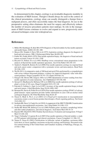
![316 G. Tarjan
holder. The needle quickly penetrates the overlying skin – local anesthesia is
unnecessary – and is directed into the lesion (Fig. 11.1). A few rapid short strokes
are made with the needle tip within the lesion while 2–3 cc of vacuum is applied
with the syringe. Before the needle is pulled out of the lesion, the vacuum is
released. (Rarely, the procedure is performed using only the needle without a
syringe and without creating vacuum; the so-called French technique.) Once the
needle is withdrawn, the aspirated material can be expelled from the needle onto
glass slides to make direct smears, or flushed into a liquid to be used for the liquid-
based cytology method (LBC), cell block, flow cytometry or molecular studies
(discussed in the following section).
11.2.2 Processing of the Aspirated Sample
The aspirated sample (the “aspirate”) may be directly smeared on glass slides and
air-dried or alcohol fixed for staining with Romanowsky-type (Diff-Quik, Wright-
Giemsa, Wright) stain or Papanicolaou stain, respectively. The preference of stain
depends on the tissue/lesion aspirated. For example, Romanowsky stained slides are
preferred for assessing hematopoetic elements (cells originating from bone marrow
and lymphoid organs), because variations in cell size are accentuated on air-dried
smears and immaturity of the chromatin (the intranuclear DNA-protein complex)
and the cytoplasmic characteristics are better appreciated.
The application of LBC to thyroid FNA was first suggested in the 1990s [4].
Contrary to direct smears, LBC methods (ThinPrep, SurePath, Cytospin prepara-
tions) require special instrumentation for preparing a monolayer of cells after align-
ing them on a membrane. Advocates of LBC prefer this method for the ability to
screen fewer, consistently well-fixed, and less blood-containing slides.
Fig. 11.1 Instruments prepared for FNA procedure: syringe holder with syringe and needle](https://image.slidesharecdn.com/headneckcancercurrentperspectivesadvancesandchallenges-130605015223-phpapp01/85/Head-neck-cancer-current-perspectives-advances-and-challenges-4-320.jpg)
![31711 Cytopathology of Head and Neck Lesions
Cell blocks can be prepared from the centrifuged sediment of suspended cells in
the needle rinse fluid. The sediment is congealed into a solid mass with the help of
plasma/thrombin, then fixed in formalin and embedded in paraffin. Most impor-
tantly, immunostains can be performed on thin sections of this cell block to charac-
terize the cells by their specific binding of a labeled antibody.
FNA can provide sample for flow cytometry which is used primarily to immuno-
phenotype hematopoetic malignancies. Since this method requires about one million
cells, two FNA passes (two courses of aspiration) should provide a sufficient num-
ber of cells. Using this method, numerous cells can be analyzed for the coexpression
of two or three different cell surface markers simultaneously, facilitating the detection
of even a small monoclonal/neoplastic cell population.
If an infectious lesion is suspected, the aspirate from a separate designated “pass” can
be sent to the microbiology laboratory for microbiologic examination (Fig. 11.5d).
Molecular diagnostic methods are also available for samples obtained using
FNA. These include conventional karyotype cytogenetics (analysis of total chromo-
somes), polymerase chain reaction (a method of DNA/RNA amplification used for
analyzing short segments of the genetic material), and in situ hybridization. In situ
hybridization is an “on-slide” technique that permits microscopic visualization of
specific chromosomal segments using nucleic acid probes usually linked to a
fluorescent label (FISH). It also allows the detection of numerical and/or structural
abnormalities of chromosomes in cytological preparations where, conveniently, the
cells are already disaggregated in a single layer on the slide.
11.2.3 Advantages and Potential Complications of FNA
Among its advantages it should be mentioned that FNA can be performed as an
outpatient procedure, does not require anesthesia, is minimally discomforting to the
patient, is cost effective, and has rapid turnaround time. In the hands of experienced
individuals, FNA is highly sensitive (negative FNA result can safely predict if the
final diagnosis will be negative) and specific (positive FNA result predicts the pres-
ence of malignancy with high probability). Positive and negative FNA test results
prove to be correct predicting the final diagnosis in a high proportion of cases (FNA
has high positive and negative predictive values). Review and meta-analysis of 3459
FNAs from all head and neck sites in 30 studies revealed 89.6% sensitivity, 96.5%
specificity, 96.2% positive predictive value, 90.3% negative predictive value, and
93.1% accuracy [5]. Diagnostic errors are more likely to occur when the cytopathol-
ogist attempts to interpret the cytological findings without being fully informed of
the exact location of the aspirated lesion and the patient’s medical history, symp-
toms, and physical and radiological findings. As an example, note the similarities in
the aspirates of a cystic SCCA metastasis, a branchial cleft cyst, an epidermal inclu-
sion cyst, and a pilomatricoma (Figs. 11.26a, 11.32a, 11.33a, and 11.34a, respec-
tively), making it obvious that being uninformed of the history and the exact location
of the lesion may result in incorrect diagnosis. In the hands of a fully informed](https://image.slidesharecdn.com/headneckcancercurrentperspectivesadvancesandchallenges-130605015223-phpapp01/85/Head-neck-cancer-current-perspectives-advances-and-challenges-5-320.jpg)
![318 G. Tarjan
cytopathologist, 100% sensitivity, 86% specificity, 97% positive predictive value,
100% negative predictive value, and 97% accuracy could be achieved performing
H&N FNA with ultrasound guidance [6].
Potential complications of FNA are bleeding, infection, and tumor cell seeding.
In the absence of a bleeding diathesis, which is a contraindication to FNA, the occa-
sional local hemorrhage can usually be controlled with local pressure. Infection is
very rare if the skin is thoroughly cleaned with 95% ethanol before FNA. Although
occasional implantation of tumor cells along the needle track cannot be completely
excluded, its clinical significance is questionable [7].
11.3 Cytopathology of Salivary Gland Lesions
FNA is a useful technique for evaluating masses both in the major – parotid,
submandibular, and sublingual – salivary glands and in the numerous tiny minor
salivary glands found throughout the oropharynx. Preoperative identification of
non-neoplastic lesions (e.g. the benign gland enlargement called sialadenosis,
inflammations, cysts, or intraparotid lymph nodes) can negate the need for a surgi-
cal procedure. Malignant neoplasms, carcinomas and lymphomas, are found in less
than 10% of salivary gland FNAs. When FNA reveals neoplastic tissue, the distinc-
tion of benign from malignant neoplasms and the exact classification of malignant
neoplasms may dictate the urgency and extent of the subsequent operation.
Unfortunately, some salivary gland neoplasms are difficult to diagnose on FNA.
Perhaps the main reason is that the majority of these tumors arise from the same
epithelial and/or myoepithelial cell lines, rendering them alike. In addition, these
cells are able to undergo squamous, mucinous, oncocytic, and sebaceous metaplas-
tic changes hindering the identification of the lesion. (Metaplasia is the non-neo-
plastic transformation of one adult cell type to another.) Hence, although the
sensitivity of salivary gland FNA is approximately 90% and its specificity is about
95% in expert hands [8, 9], specific classification of malignant neoplasms can be
achieved in only about 85% of salivary gland FNAs. Following a non-diagnostic
FNA, the sensitivity and specificity of a repeat FNA was reported to be 84 and 93%,
respectively, in distinguishing benign from malignant tumors [10].
The salivary glands are complex structures composed of acinar, ductal, and mes-
enchymal tissues. Therefore, the FNA of a normal salivary gland will yield ductal
cells and glandular elements composed of serous and/or mucinous acinar cells.
Mature fat may also be present. Cytologically, the acinar cells have granular or
finely vacuolated cytoplasm. They appear in globular/ball-like cohesive groups
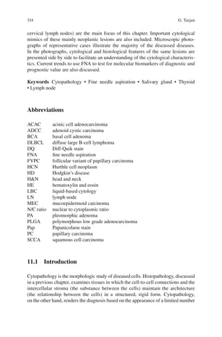
which may aggregate in grapelike clusters connected by ducts (Fig. 11.2). Occasional
attached elongated myoepithelial cells may be present. The cohesive ductal cells
appear in orderly sheets or tubules.
Chronic sialadenitis (chronic inflammation of the salivary gland), which can
mimic a neoplasm, is one of the most often aspirated lesions of the major salivary
glands. The smears show small groups of acinar cells and ductal cells admixed with
lymphocytes (Fig. 11.3). As the sialadenitis progresses toward the atrophic phase](https://image.slidesharecdn.com/headneckcancercurrentperspectivesadvancesandchallenges-130605015223-phpapp01/85/Head-neck-cancer-current-perspectives-advances-and-challenges-6-320.jpg)
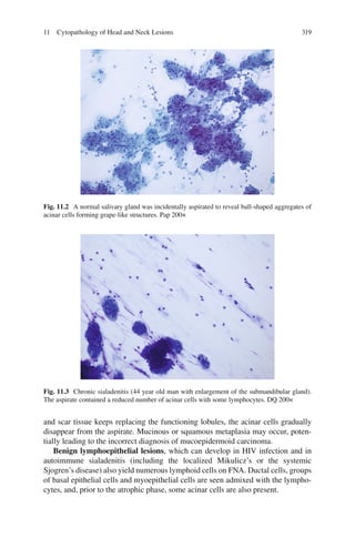
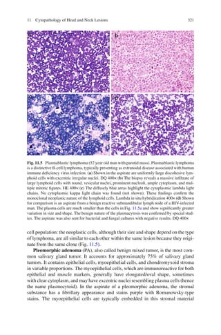
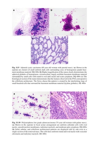
![32711 Cytopathology of Head and Neck Lesions
presence of intermediate cells is specific to MEC. At the other end of the spectrum, in
high grade MEC, there are few mucocytes and the predominance of the squamous
cell-like epidermoid cells yields a similar appearance to squamous cell carcinoma
(SCCA). However, as with mucinous adenocarcinoma, SCCA contains no intermedi-
ate cells. Moreover, in MEC the epidermoid cells rarely show obvious keratinization,
which is a key feature of the malignant squamous cells in SCCA (Fig. 11.26a). The
presence of intermediate cells and the lack of keratinization also help to distinguish
MEC from the more aggressive adenosquamous carcinoma which contains malig-
nant glandular elements in addition to malignant squamous cells. In the future, molec-
ular studies of fine-needle aspirates may assist in rendering the diagnosis of MEC
(Fig. 11.13) and serve as prognostic indicators. The t(11;19) translocation that under-
lies MEC has been cloned [11]. The resulting MECT1/MAML2 translocation is
highly specific for MEC and imparts a better prognosis [12].
Acinic cell adenocarcinoma (ACAC) occurs in both major and minor salivary
glands. The cells aspirated from well differentiated ACAC bear a striking resem-
blance to the normal acinar cells from which they derive. However, in contrast to
FNA of normal salivary gland tissue, FNA of ACAC yields a more cellular aspirate
in which the tumor cells do not appear in balls but rather in sheets or cords, and
other components of a normal salivary gland (ductal cells, fat) are not present
(Fig. 11.14). Because of their ample cytoplasm and central nucleolus, the cells of
ACAC may resemble cells of an oncocytic tumor but the granular or finely vacuo-
lated cytoplasm is different from the uniformly granular cytoplasm of oncocytes.
The very aggressive salivary duct carcinoma, being histologically similar to the
comedocarcinoma of the breast, will yield sheets of obviously malignant cells with
high nuclear to cytoplasmic ratio and pleiomorphic nuclei in a necrotic background
(Fig. 11.7a).
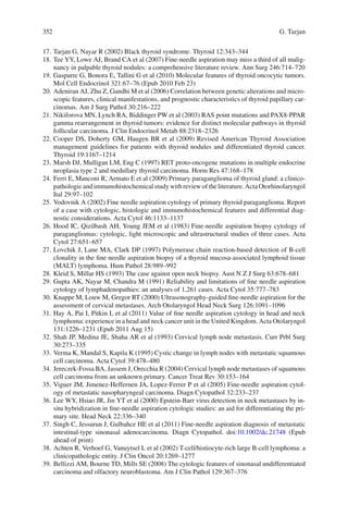
Fig. 11.13 Clear cell-rich mucoepidermoid carcinoma (56 year old woman with parotid mass).
(a) Shown in the aspirate is a cohesive group of relatively large cells with clear/vacuolated
cytoplasm. DQ 400× (b) Biopsy of the mass revealed an infiltrating carcinoma with clear cell
change. HE 400× The positive MAML2 rearrangement by FISH supported MEC, while the
negative EWS translocation and negative human papillomavirus DNA effectively excluded a
hyalinizing clear cell carcinoma and a clear cell non-keratinizing SCCA, respectively](https://image.slidesharecdn.com/headneckcancercurrentperspectivesadvancesandchallenges-130605015223-phpapp01/85/Head-neck-cancer-current-perspectives-advances-and-challenges-15-320.jpg)
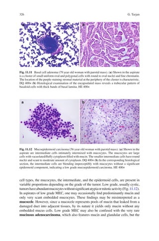
![328 G. Tarjan
The cytologic appearance of small cell carcinoma of the salivary gland is similar
to that of the undifferentiated small cell carcinoma of the lung. In the highly cellular
aspirates, the tumor cells have high nuclear to cytoplasmic ratio (N/C ratio), irregular
fragile nuclear membranes with nuclear molding and frequent smearing artifact.
11.4 Cytopathology of Thyroid Lesions
Thyroid FNA is a safe, accurate, and cost-effective method for guiding the clinical
management of thyroid nodules. Given that 4–7% of the adult population has pal-
pable thyroid lesions and even more people have non-palpable nodules detected by
ultrasound [13, 14], it is no wonder that thyroid FNA is one of the most commonly
performed FNAs – over 350,000 thyroid FNAs are done annually in the USA. In
expert hands, the diagnostic accuracy of thyroid FNA to distinguish benign lesions
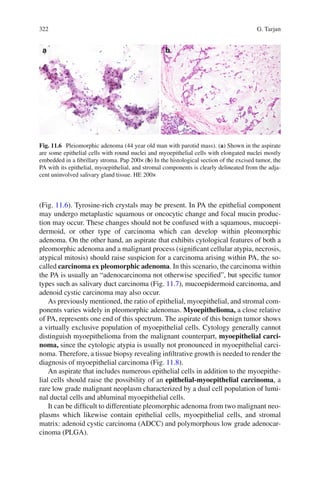
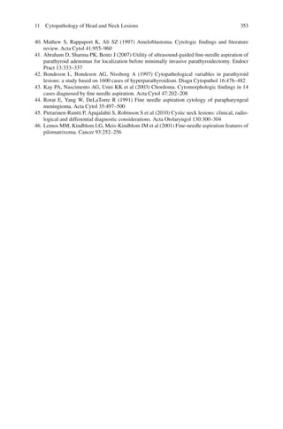
from malignant ones (which represent only a small fraction of the nodules) ranges
Fig. 11.14 Acinic cell carcinoma (56 year old woman with parotid mass). (a) Shown in the aspi-
rate is a sheet of large cells with vacuolated cytoplasm. The nuclei are uniformly round and excen-
trically located. Pap 400× (b) In other smears, the Romanowsky-type stain highlights fine granules
in the tumor cells’ ample cytoplasm. DQ 1000× oil (c) The vacuolated cells can also be clearly
identified in the histologic section of the tumor. HE 400× (d) However, other sections may show
non-vacuolated tumor cells in a follicular pattern resembling thyroid tissue. HE 400×](https://image.slidesharecdn.com/headneckcancercurrentperspectivesadvancesandchallenges-130605015223-phpapp01/85/Head-neck-cancer-current-perspectives-advances-and-challenges-16-320.jpg)
![32911 Cytopathology of Head and Neck Lesions
from 80 to over 95% [15]. This accuracy refers only to adequate specimens. The
criteria for adequacy include the presence of a sufficient number of well-visualized
thyroid follicular cells in the aspirate (six groups, each containing 10 follicular cells
or 180–320 follicular cells using LBC method). The rare exceptions to this require-
ment are mentioned in the discussion of colloid nodule and chronic thyroiditis. The
presence of even a large number of macrophages (members of the body’s defense
mechanism that engulf and digest cellular debris and pathogens) does not render a
thyroid aspirate adequate in the absence of follicular cells. The presence of mac-
rophages usually indicates cyst formation which is generally benign but may also
occur in papillary carcinomas.
The two main components of a normal thyroid aspirate are hormone-producing
follicular cells and colloid, the storage site of thyroid hormone. Scant lymphocytes
may be seen but their presence is generally associated with inflammatory condi-
tions. The neuroendocrine parafollicular C cells are too few to be recognized in
normal thyroid FNA. The follicular cells are uniform, cuboidal-shaped cells with
round granular and slightly hyperchromatic nuclei, smooth nuclear membranes, and
scant pale cytoplasm (Fig. 11.15b). Individually scattered follicular cells may lose
Fig. 11.15 (a) A sheet of follicular cells and colloid is seen in the aspirate of a hyperplastic thy-
roid nodule. Note the honeycomb arrangement of the cells and the wrinkled appearance of the
colloid. Pap 100× (b) A thyroid microfollicle and a colloid “droplet” is shown in the aspirate of a
follicular adenoma. Thinprep Pap 1000× oil (c) Accidentally aspirated skeletal muscle may resem-
ble colloid. However, the cross-striation in the center is specific for skeletal muscle. DQ 200×](https://image.slidesharecdn.com/headneckcancercurrentperspectivesadvancesandchallenges-130605015223-phpapp01/85/Head-neck-cancer-current-perspectives-advances-and-challenges-17-320.jpg)
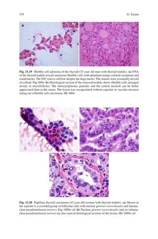
![330 G. Tarjan
their cytoplasm and resemble lymphocytes. More often, however, benign follicular
cells form flat, cohesive sheets with evenly distanced nuclei, yielding a honeycomb
pattern. Normal follicles appear in the aspirate as macrofollicles, comprised of
numerous follicular cells surrounding and/or surrounded by colloid (Fig. 11.15a).
The appearance of colloid depends on its functional state, varying from watery to
dense. Watery colloid has a thin membrane coating appearance in direct smears and
a tissue-paper appearance in LBC preparations. Thick colloid can show linear crack-
ing artifact in direct smears and appear as dense orange-blue droplets in LBC prepa-
rations (Fig. 11.15b). Especially when highlighted under polarized light, calcium
oxalate crystals may occasionally be detected in thyroid aspirates of adults. These
crystals can be found in any colloid-containing thyroid tissue, regardless if it is
benign or malignant (Fig. 11.16) [16]. When follicular cells undergo reactive/repar-
ative change, they may reveal enlarged nuclei with prominent nucleoli. However,
they maintain their cohesiveness and their orderly macrofollicular arrangement.
Follicular cells may undergo oncocytic change, manifesting abundant finely
granular cytoplasm, enlarged nuclei, and prominent nucleoli similar to the onco-
cytes described in the salivary glands (Fig. 11.19a). The cytoplasm of oncocytes is
green or orange with Papanicolaou stain, blue or grey-pink with Romanowsky-type
stains, and pink with hematoxylin-eosin stain. In the thyroid these cells are gener-
ally called Hurthle cells, despite the fact that their correct description was provided
by Askanazy in 1898, and the cells described by Hurthle in 1894 are now believed
to represent the parafollicular C cells.
A brown pigment consisting of neuromelanin and/or lipofuscin may accumulate
in the cytoplasm of follicular cells due to the administration of tetracycline or mino-
cycline resulting in the “black thyroid syndrome”. Interestingly, the pigment does
not deposit in autonomously functioning nodules; for example, a follicular adenoma
would stand out from the otherwise uniformly black thyroid [17].
Fig. 11.16 Calcium oxalate crystals (44 year old woman with thyroid nodule). (a) Shown in the
aspirate of a follicular variant of papillary carcinoma are crystals embedded in the colloid. DQ
400× (b) Polarized light highlights the crystals. DQ 400×](https://image.slidesharecdn.com/headneckcancercurrentperspectivesadvancesandchallenges-130605015223-phpapp01/85/Head-neck-cancer-current-perspectives-advances-and-challenges-18-320.jpg)
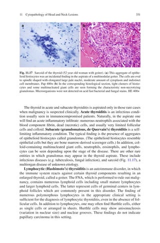
![332 G. Tarjan
In the case of the rare Riedel’s thyroiditis, which results in progressive fibrosis
of the tissues within and next to the thyroid, FNA usually yields non-diagnostic
hypocellular aspirate.
In Graves’ disease (or toxic diffuse hyperplasia), an autoimmune stimulus
causes increased benign proliferation of follicular cells (hyperplasia), leading to
diffuse enlargement of the thyroid associated with toxic thyroid hormone overpro-
duction. FNA, though rarely performed in this condition, may show the morpho-
logic signs of hyperactivity: the distinctive “flame cells”, which are characterized by
marginal cytoplasmic vacuoles and red frayed edges on Romanowsky stains sec-
ondary to the hyperfunctioning endoplasmic reticulum. The other findings, the vari-
able number of follicular cells arranged in honeycomb sheets and the abundant
colloid, are non-specific and are also seen in aspirates of benign hyperplastic nod-
ules of a nodular goiter. The aspirate of Graves’ disease treated with radioactive
iodine or carbimazole may include crowded follicular cells with atypia. This repre-
sents treatment effect and knowledge of the clinical history helps to avoid over-
interpreting them as signs of malignancy.
Nodule(s) of a (multi)nodular goiter are the most common lesions targeted in
thyroid FNA. Nodular goiter is the result of repeated cycles of hyperplasia and invo-
lution, usually due to iodine deficiency. The resulting hyperplastic nodules show
cytological features similar to those in Graves’ disease: abundant colloid and vari-
able number of follicular cells in monolayer orderly sheets (Fig. 11.15a). Hurthle
cell change is frequently encountered. Features of involution may be seen: mac-
rophages in cystic degeneration; hemosiderin-laden macrophages in old hemor-
rhages; spindle cells or fibrous tissue fragments in fibrosis. Sometimes, the colloid
content is so prominent in a nodular goiter that the FNA yields only abundant,
essentially acellular (cell-free) colloid. In this case if there is no clinical or radio-
logical sign of malignancy, the sample is considered adequate despite the absence
of follicular cells, and the diagnosis of “benign colloid nodule” is rendered.
The previously discussed lesions (subacute thyroiditis, lymphocytic thyroiditis,
Graves’ disease, hyperplastic/colloid nodule) are reported as “Category: Benign fol-
licular lesion” according to the 2010 Bethesda System for Reporting Thyroid
Cytopathology. In this category, the false-negative rate (when a malignant lesion is
incorrectly diagnosed as benign) is 1–10% [18]. Therefore, patients with benign
cytology are generally followed for up to 3–5 years with ultrasonography and repeat
FNA if clinically indicated (for significant growth of the nodule, presence of
microcalcifications, etc.).
An aspirate, in which the number of follicular cells is significantly increased while
the amount of colloid is decreased, raises suspicion for a follicular neoplasm.
A follicular neoplasm, contrary to the hyperplastic nodule, is an unregulated indepen-
dently functioning neoplastic lesion. It needs to be surgically excised because cytol-
ogy cannot distinguish between a benign follicular adenoma and a malignant follicular
carcinoma. (Only histological evaluation is able to disclose the diagnostic feature of
malignancy: capsular and/or vascular invasion.) Therefore, it is important to make the
often difficult distinction between a cellular hyperplastic nodule and a follicular neo-
plasm. The increased cellularity and scant colloid alone are not sufficient for the diagnosis](https://image.slidesharecdn.com/headneckcancercurrentperspectivesadvancesandchallenges-130605015223-phpapp01/85/Head-neck-cancer-current-perspectives-advances-and-challenges-20-320.jpg)
![33311 Cytopathology of Head and Neck Lesions
of follicular neoplasm. To render this diagnosis, one has to see follicular cell crowding,
overlapping, and microfollicle formation affecting the majority of the follicular cells
(Fig. 11.18). The definition of a microfollicle is 6–12 (generally accepted as less than
15) follicular cells arranged in a ring, with or without a small amount of central colloid
(Fig. 11.15b). The follicular cells may also form crowded ribbons/trabeculae and
three-dimensional groups. The nuclei are round, slightly hyperchromatic, and in none
of these lesions are the nuclear features of papillary carcinoma present.
Whether Hurthle cell neoplasms are separate entities or only oncocytic variants
of follicular neoplasms is still being debated [19]. The typical aspirates are highly
cellular and consist of a virtually exclusive population of Hurthle cells dispersed as
isolated cells or forming crowded clusters and sheets. Marked variation in cell and
nuclear size is common (Fig. 11.19).
Papillary carcinoma (PC) accounts for up to 80% of malignant thyroid lesions.
FNA is well-suited to diagnose these common thyroid neoplasms because the
diagnosis is mostly based on cytologic features: numerous oval nuclei with irregular
nuclear membranes, finely granular (powdery) chromatin with several small
marginally placed nucleoli, nuclear grooves imparting a coffee bean appearance,
dense “squamoid” cytoplasm, and intranuclear pseudoinclusions. These pseudoin-
clusions are created by the invagination of the cytoplasm into the nucleus, therefore
they are characterized by sharply defined edges and cytoplasmic staining qualities
(Fig. 11.20). Certain cytological features reflect the characteristic histological features
of papillary carcinoma: there may be papillae with or without fibrovascular cores in
the aspirate of a conventional PC, and follicular structures in the aspirate of a folli-
cular variant of papillary carcinoma (FVPC). Other possible findings include viscous
stringy “bubble gum” colloid, multinucleated giant cells, and psammoma bodies.
The latter are concentrically laminated calcifications believed to represent tips of
papillae that became calcified.
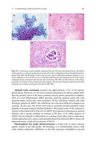
Fig. 11.18 Follicular thyroid carcinoma (25 year old woman with thyroid nodule). (a) In the cel-
lular aspirate, the follicular cells are primarily arranged in microfollicles suggesting a follicular
neoplasm. Pap 400× (b) Histological section of the lesion reveals an encapsulated lesion with a
predominantly microfollicular pattern. The capsular invasion (arrowhead) is diagnostic for follicu-
lar carcinoma. HE 40×](https://image.slidesharecdn.com/headneckcancercurrentperspectivesadvancesandchallenges-130605015223-phpapp01/85/Head-neck-cancer-current-perspectives-advances-and-challenges-21-320.jpg)
![33511 Cytopathology of Head and Neck Lesions
None of these criteria alone is specific for the diagnosis of PC. (For example,
nuclear grooves may be seen in chronic thyroiditis, hyperplastic nodules, follicu-
lar neoplasms, and intranuclear pseudoinclusions may be found in medullary
carcinoma.) The more features of PC, the more certain the diagnosis of PC. False
negative diagnoses, which occur in approximately 5% of cases, are predomi-
nantly due to PC variants. In one of these, in the FVPC, the tumor is composed
of small to medium-sized follicles instead of papillae, and nuclear grooves and
intranuclear pseudoinclusions are less common than in conventional PC
(Fig. 11.16a). Furthermore, the abundant colloid in the aspirate of the macrofol-
licular variant of PC can closely mimic a benign follicular lesion. About 10% of
PCs become predominantly cystic. In this case, the aspirate contains thin fluid,
abundant histiocytes, and often scant carcinoma cells. Many times even these
scant carcinoma cells are less typical than in conventional PC. The tumor cells
have more abundant vacuolated cytoplasm and less prominent finely granular
chromatin. The typical nuclear features of PC are also less prominent in the
aggressive tall cell and columnar cell variants, which are characterized by tall
cells and crowded clusters of columnar cells, respectively. In the oncocytic vari-
ant the carcinoma cells undergo oncocytic change, resembling a Hurthle cell
neoplasm. In the Warthin-like variant of PC, the numerous lymphocytes and the
oncocytic tumor cells together resemble Warthin’s tumor of the salivary gland.
Thus, careful attention to nuclear features is essential for all thyroid aspirates to
avoid overlooking a PC.
Molecular testing of thyroid FNA samples may improve the diagnostic accu-
racy and help to reduce the number of cases falling into the “indeterminate” or
“atypia of undetermined significance” category. Point mutations of the BRAF
and RAS genes or RET/PTC rearrangements are found in more than 70% of
papillary thyroid carcinomas [20]. RAS mutations or PAX8/PPAR gamma rear-
rangements are identified in about 75% of follicular carcinomas [21]. The data
on these markers are fast accumulating. The guidelines of the American Thyroid
Association already recommend the use of these molecular markers for indeter-
minate cytology to assist in guiding patient management [22]. In the future, aspirates
of patients with PC may be tested for some markers, such as BRAF or RAS, to
select the radioactive-iodine-resistant carcinomas and to apply molecular targeted
therapy.
Poorly differentiated thyroid carcinoma, including its classic form, the insular
carcinoma, may arise from either papillary carcinoma or follicular carcinoma.
However, the bulk of the tumor no longer shows the features of either of those. Rather,
it is composed of cellular islands of tumor cells separated by thin fibrous bands.
Therefore, the FNA will usually reveal numerous crowded nests/three-dimensional
clusters of relatively uniform, small follicular cells with high nuclear to cytoplasmic
ratio. Apoptotic and mitotic forms are often present with necrotic debris in the back-
ground, reflecting the high turn-over of this aggressive tumor. A subset of poorly dif-
ferentiated thyroid carcinomas shows predominantly isolated cells with coarsely
granular chromatin. In these cases, the positive thyroglobulin and negative calcitonin
and CEA immunostains help to distinguish it from medullary carcinoma.](https://image.slidesharecdn.com/headneckcancercurrentperspectivesadvancesandchallenges-130605015223-phpapp01/85/Head-neck-cancer-current-perspectives-advances-and-challenges-23-320.jpg)
![336 G. Tarjan
The markedly aggressive undifferentiated/anaplastic carcinoma carries the
poorest prognosis among primary thyroid carcinomas. The FNA yields cellular
smears showing round to polygonal and/or spindle-shaped cells with significant
size-variation, arranged in groups or dispersed individually in the background of
acute inflammatory cells, fibrin, blood, necrotic cells, and debris (the so-called
tumor diathesis). Sometimes bizarre and multinucleated tumor cells, plasmocytoid
and rhabdoid cells, and osteoclast-like giant cells are present. There are often abnor-
mal ones among the numerous mitotic figures (Fig. 11.21). Along with the loss of
differentiation, this tumor loses its immunoreactivity for the usual follicular cell
markers: TTF-1 and thyroglobulin. The majority still retains immunoreactivity for
pan-keratin, which helps to differentiate the spindle cell-rich form of anaplastic
carcinoma from the very rare primary thyroid sarcoma. Clinical correlation and
imaging studies showing a thyroid-centered lesion are needed to exclude a meta-
static malignancy.
Medullary carcinoma (MC) is a malignant neoplasm arising from the neuroen-
docrine parafollicular C cells of the thyroid. It accounts for 5–7% of thyroid carci-
nomas. While 25% of MCs are hereditary and can be the manifestation of a “multiple
endocrine neoplasia syndrome”, the great majority of MCs are sporadic. The hered-
itary MC is associated with germline point mutations in the RET proto-oncogene.
In sporadic MCs various somatic RET mutations have been identified [23].
Cytologically, MC is characterized by anastomosing clusters or a mostly dispersed
population of relatively uniform tumor cells. These can be spindled, plasmocytoid
with excentric nuclei, or Hurthle cell-like with finely granular cytoplasm. The cyto-
plasmic granules appear red with a Romanowsky-type stain. The nuclei typically
show the neuroendocrine-type coarsely granular “salt-and-pepper” chromatin with
inconspicuous nucleoli. Intranuclear pseudoinclusions, also common in PC, are
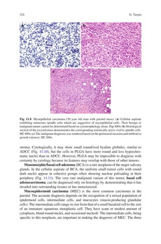
Fig. 11.21 Anaplastic thyroid carcinoma (46 year old woman with rapidly enlarging thyroid
mass). (a) FNA reveals numerous highly atypical discohesive epithelioid cells with frequent
mitotic figures (arrowheads). DQ 200× (b) Histological section of the lesion shows an infiltrating
neoplasm composed of large epithelioid tumor cells with high grade features (arrow). These can
be contrasted with the residual benign thyroid follicles (arrowhead). The tumor cells were cytokeratin
positive by immunohistochemistry. HE 200×](https://image.slidesharecdn.com/headneckcancercurrentperspectivesadvancesandchallenges-130605015223-phpapp01/85/Head-neck-cancer-current-perspectives-advances-and-challenges-24-320.jpg)
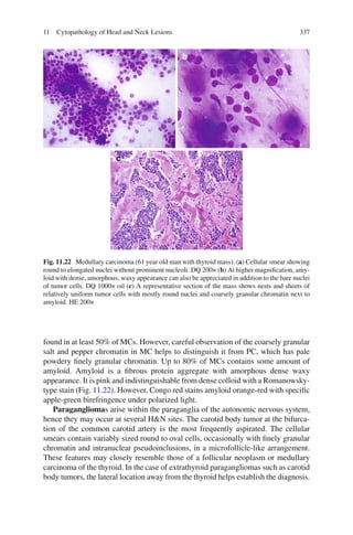
![338 G. Tarjan
However, primary paraganglioma of the thyroid has also been described [24] and
this rare neuroendocrine tumor may occasionally be aspirated [25], causing
significant diagnostic challenge. Negative immunocytochemical testing for thyro-
globulin and calcitonin is helpful to distinguish a paraganglioma of the thyroid from
a follicular neoplasm or medullary carcinoma, respectively. Separation of benign
from malignant paraganglioma is difficult but the tumor diathesis, the more promi-
nent nucleoli, and the mitotic figures favor a malignant process [26] (Fig. 11.23).
Primary thyroid malignant lymphomas, mostly non-Hodgkin’s lymphomas,
account for less than 5% of thyroid malignances. Secondary involvement of the
thyroid by lymphoma is more frequent than primary thyroid lymphoma; about 20%
Fig. 11.23 Malignant paraganglioma of the thyroid (54 year old woman with thyroid mass). (a)
Cellularity varies widely in aspirates of paragangliomas. Sometimes the smears contain mostly
blood components and only rare tumor cells or bare nuclei with occasional pseudoinclusions, as in
this case. The usual location of a paraganglioma at the carotid bifurcation helps to distinguish it
from a papillary or medullary thyroid carcinoma, which must be entertained in the differential. Pap
40× (b) However, in this case, the tumor (arrow) was arising from the thyroid (arrowhead). On
immunohistochemical studies, the tumor was found to be reactive with synaptophysin and chro-
mogranin and non-reactive with calcitonin and keratins. S-100 highlighted sustentacular cells.
Diagnosis of malignant paraganglioma was rendered based on these features as well as on tumor
invasion into skeletal muscle and cricoid cartilage. HE 40× (c) A representative section of the mass
shows the characteristic “Zellballen” pattern composed of nests of large polygonal cells, separated
by richly vascular septa. HE 200×](https://image.slidesharecdn.com/headneckcancercurrentperspectivesadvancesandchallenges-130605015223-phpapp01/85/Head-neck-cancer-current-perspectives-advances-and-challenges-26-320.jpg)
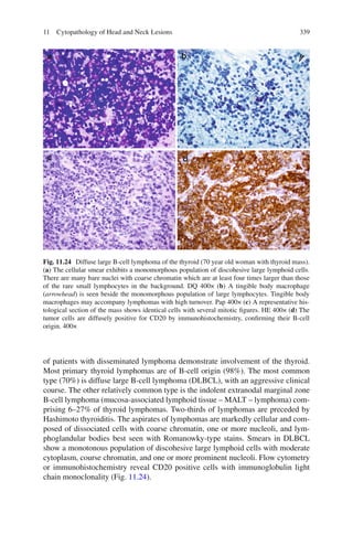
![340 G. Tarjan
In MALT-lymphoma, the abnormal B cells appear in three forms: the small to
intermediate-sized marginal zone B-cells with irregular nuclei, moderately dis-
persed chromatin and inconspicuous nucleoli; the intermediate to large monocy-
toid B-cells with open chromatin and abundant pale staining cytoplasm, and the
sometimes dominating plasmacytoid cells. The distinction of this low-grade lym-
phoma from lymphocytic thyroiditis is sometimes problematic. Definitive diag-
nosis of lymphoma in cytological specimens may be facilitated by the
documentation of a clonal lymphoid proliferation by flow cytometric immuno-
phenotyping or immunocytochemistry, or by molecular techniques such as the
polymerase chain reaction-based assay for immunoglobulin heavy chain gene
rearrangement [27].
Metastatic tumors to the thyroid are rare, the reported incidence varying from
2.7 to 4%. The most frequent ones are renal, colorectal, lung, breast, melanoma,
lymphoma, and H&N carcinoma metastases. The possible diagnosis of metastatic
tumor should be entertained when the cytological features of the aspirated malig-
nant cells are different from those of the previously discussed primary thyroid
neoplasms. Information about a malignant tumor elsewhere in the body is of
utmost importance. Especially when the features of the metastatic tumor are simi-
lar to those of a primary thyroid neoplasm: cytological features of metastatic renal
cell and breast carcinoma can mimic those of a follicular neoplasm; metastatic
papillary lung carcinoma can resemble papillary thyroid carcinoma; metastatic
melanoma may be misinterpreted as medullary carcinoma. Negative immunocy-
tochemistry for thyroglobulin and thyroid transcription factor-1 can assist in iden-
tifying a metastatic tumor.
11.5 Cytopathology of Cervical Lymph Node Lesions
Cervical lymphadenopathy is not a rare phenomenon. Palpable enlarged lymph
nodes (LN) in the neck are often associated with infections but can also be the first
sign of cancer. Cervical lymphadenopathy is present in over 30% of patients with
H&N cancer when they first seek medical care [28]. FNA has become the primary
method for the initial evaluation of LNs when are they suspicious for malignancy
[29]. Ultrasound-guided FNA of cervical LNs has a sensitivity of 89%, specificity
of 98%, and accuracy of 94.5% to diagnose the most common malignancy, meta-
static squamous cell carcinoma [30]. Although previously regarded as limited in its
use for diagnosing primary lymphoid malignancies, FNA in combination with
immunophenotyping and molecular studies is gaining respect in providing accurate
diagnosis of lymphoma. FNA combined with immunocytochemistry/flow cytome-
try has reached a sensitivity of 95.5% and a positive predictive value of 96.8% in the
investigation of lymphoma presenting in the neck [31].
When evaluating the aspirate from a suspected LN, the first step is to confirm
that, in fact, a LN was aspirated. The characteristic cytological features of a LN
include lymphoid cells, which are discohesive cells with scant to moderate cytoplasm,](https://image.slidesharecdn.com/headneckcancercurrentperspectivesadvancesandchallenges-130605015223-phpapp01/85/Head-neck-cancer-current-perspectives-advances-and-challenges-28-320.jpg)
![34111 Cytopathology of Head and Neck Lesions
and lymphoglandular bodies (Fig. 11.25). The latter represent fragments of lymphocyte
cytoplasms measuring up to the size of a red blood cell. Lymphoglandular bodies
are best appreciated on Romanowsky-type stain as round pale blue bodies with lacy
internal structure. The presence of these cells in the background are important in the
identification of lymphoid tissue, as non-lymphoid “small round blue cell tumors”
such as Ewing’s sarcoma or small cell carcinoma may resemble lymphocytes.
On the other hand, FNA of a LN, that is completely replaced by metastasis or
becomes fibrotic/necrotic due to treatment effect, may not yield sufficient lympho-
cytes to be recognized as a LN.
Once it is established that the aspirate is derived from a lymph node, a metastatic
neoplasm needs to be excluded. In general, FNA of LNs with metastatic neoplasms
shows non-hematopoetic (carcinoma, sarcoma, or melanoma) cells in the back-
ground of polymorphous lymphoid cells. Most metastatic carcinomas exhibit
cohesive groups of round-polygonal tumor cells with well-defined cytoplasm.
However, metastatic melanoma and high grade SCCA may display a single cell pat-
tern resembling a lymphoproliferative disorder.
SCCA is the most common neoplasm (90%) to metastasize to LNs in the H&E
region [32]. The aspirate of metastatic squamous cell carcinoma is usually highly
cellular. The hyperchromatic tumor cells are arranged in sheets and groups which
have irregular borders with loosely attached cells hanging from their edges. The
amount of scattered, independent neoplastic cells depends on the differentiation and
cohesiveness of the tumor. The single cells often exhibit bizarre forms, including
tadpole and spindle shapes. The keratinization is best demonstrated by Papanicolaou
stain: the keratotic cells show dense orange color (orangeophilia) due to the affinity
Fig. 11.25 The aspirate from a benign reactive cervical lymph node exhibits a polymorphous
population of discohesive lymphoid cells and numerous lymphoglandular bodies (see arrowhead).
DQ 400×](https://image.slidesharecdn.com/headneckcancercurrentperspectivesadvancesandchallenges-130605015223-phpapp01/85/Head-neck-cancer-current-perspectives-advances-and-challenges-29-320.jpg)
![342 G. Tarjan
of keratinized squamous cells for one of the components of the Papanicolaou stain
(Fig. 11.26). Cystic SCCA metastasis can present a significant diagnostic pitfall: if
the metastasis and eventually the entire LN undergo cystic change, the often poorly
cellular aspirates will show mostly dirty necrotic content and inflammatory cells
with scant anucleate or mildly atypical squamous cells. These findings can easily be
confused with an inflammatory process or a branchial cleft cyst. The possibility of
cystic SCCA must always be kept in mind since the frequency of cystic SCCA
metastasis varies from 9.1 to 21.5% depending on the site of the primary tumor [33].
Knowing that the patient has a primary SCCA, especially if that is in the H&N
region, helps to avoid this mistake. However, in about 2–5% of cervical SCCA
metastases, the location of the primary carcinoma is never discovered [34].
Some unique carcinoma types may also present diagnostic difficulties when they
metastasize to LNs. Metastasis of the Epstein-Barr virus-related undifferentiated
nasopharyngeal carcinoma may exhibit only scattered large carcinoma cells
accompanied by abundant lymphocytes, some eosinophils, and occasionally granu-
lomas [35]. Thus, it can be confused with lymphoma, especially Hodgkin’s lymphoma,
Fig. 11.26 Cervical lymph node with SCCA metastasis (50 year old man with enlarged neck LN).
(a) The aspirate contains many large atypical cells, including a tadpole-like cell, with orange-col-
ored cytoplasm and hyperchromatic nuclei. Pap 200× (b) Another smear shows the polymorphous
lymphoid population, reactive to the metastatic orange-colored squamous cells. Pap 200× (c) The
characteristic whorled pattern of SCCA and focal keratinization can be appreciated in the corre-
sponding histological section. HE 200×](https://image.slidesharecdn.com/headneckcancercurrentperspectivesadvancesandchallenges-130605015223-phpapp01/85/Head-neck-cancer-current-perspectives-advances-and-challenges-30-320.jpg)
![34311 Cytopathology of Head and Neck Lesions
if particular attention is not paid to the scant epithelial cells that mark for cytokeratin
on immunocytochemistry. Epstein-Barr virus detection by in-situ hybridization in
FNA specimens can be used as a supplemental tool to render this diagnosis [36].
Basaloid squamous cell carcinomas and basaloid type salivary gland carcinomas
occasionally exhibit numerous single cells with high nuclear to cytoplasmic ratio
when they metastasize. They can be differentiated from lymphoma if the focal cohe-
sive groups are not missed and their reactivity for cytokeratin is confirmed by immu-
nocytochemistry. Rare reports of FNA of metastatic sinonasal adenocarcinoma,
intestinal-type, describe moderately cellular smears with numerous naked, atypical
nuclei, and rare signet ring-like cells in the background of abundant mucin [37].
If only lymphoid cells are present in a cervical LN FNA, it must be determined
whether these lymphoid cells represent a benign lymphoid process or a malignant
lymphoma.
Benign lymphoid hyperplasia due primarily to viral or bacterial infections is a
common cause of cervical lymph node enlargement. The hallmark of a reactive LN is
the polymorphous nature of the lymphoid cells. The mixed population displays a
broad size spectrum ranging from small mature cells to large immunoblasts. The small
mature lymphocytes with dark chromatin and scant cytoplasm always dominate
(Fig. 11.25). Numerous tingible body macrophages can be seen. These are large cells
with engulfed particles in their ample cytoplasm. Though tingible macrophages are
most commonly found in reactive LNs, they may also be present in any high turnover
lymphomas such as Burkitt’s lymphoma or large cell lymphoma (Fig. 11.24b).
Certain specific reactive conditions can be distinguished by their unique features:
in infectious mononucleosis some atypical large lymphoid cells, often with eccen-
tric nuclei and pale open chromatin, are observed. In cytomegalovirus infection,
numerous immunoblasts and characteristic intranuclear inclusions may be seen.
Some benign conditions exhibit an increased number of epithelioid histiocytes
that aggregate providing a granulomatous appearance. These granulomatous
lymphadenopathies include: infectious diseases (tuberculosis, fungi, toxoplasmo-
sis, cat-scratch disease) with or without necrosis; dermatopathic lymphadenopathy
associated with skin rash; Langerhans cell histiocytosis with eosinophils and
grooved nuclei; sarcoidosis with non-necrotizing tight granulomas; Rosai-Dorfman
disease with large histiocytes containing intact lymphocytes in their cytoplasm. The
histiocytic aggregates should not be confused with the cohesive groups of atypical
epithelial cells seen in metastatic carcinoma. An exception to the non-neoplastic
nature of the granulomatous conditions is the granulomatous reaction associated
with Hodgkin’s disease.
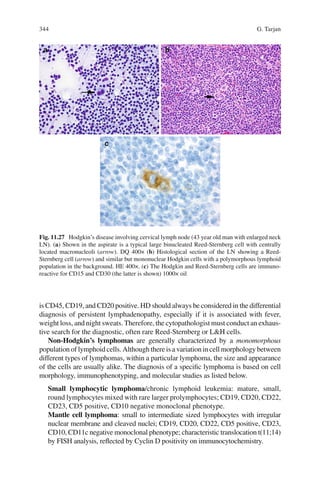
Classic Hodgkin’s disease (HD) is also an exception to the general guideline that
polymorphous lymphoid population equals a benign condition. In HD the smears
reveal a mixed background population, including lymphocytes, eosinophils, plasma
cells, and histiocytes. However, Reed-Sternberg cells or their variants are the hall-
mark cytological findings in this condition. Reed-Sternberg cells are large cells with
several irregular hyperchromatic nuclei and macronucleoli. They are CD15 and
CD30 positive by immunochemistry (Fig. 11.27). A variant, the popcorn-shaped
L&H cell in the lymphocyte predominant HD, has a different immunophenotype: it](https://image.slidesharecdn.com/headneckcancercurrentperspectivesadvancesandchallenges-130605015223-phpapp01/85/Head-neck-cancer-current-perspectives-advances-and-challenges-31-320.jpg)
![34511 Cytopathology of Head and Neck Lesions
Follicular lymphoma: small to intermediate sized lymphocytes with irregular
nuclear membrane and notched/cleaved nuclei; CD19, CD20, CD22 positive,
CD5 negative monoclonal phenotype, CD 10 positive in 60% of cases; t(14;18)
translocation and IGH/bcl-2 fusion by molecular studies.
Marginal zone lymphoma: small to intermediate sized lymphocytes with round
nuclei, small nucleoli, broad rim of lightly blue cytoplasm; CD19, CD20, CD22
positive, CD5, CD10 negative monoclonal phenotype.
Lymphoplasmocytic lymphoma: small plasmocytoid cells and plasma cells;
CD23, CD5, CD10 negative, surface IgM positive, CD20 variable phenotype.
Lymphoblastic lymphoma: medium sized lymphoid cells with blast-type chro-
matin and scant cytoplasm; CD1, CD10, CD19, TdT, CD4/CD8 positive.
Burkitt’s lymphoma: medium sized round lymphoid cells with nucleoli, narrow
rim of basophilic cytoplasm with vacuoles; CD19, CD22 positive, TdT negative,
CD10 variable monoclonal phenotype.
Diffuse large B-cell lymphoma: uniformly large atypical cells with irregular
nuclear membranes and prominent nucleoli; more often positive for B-cell mark-
ers and shows monoclonality if surface immunoglobulin is not lost.
There are rare lymphomas in which the broad size-variation of the lymphocytes
resembles a reactive process:
T-cell/histiocyte-rich B-cell lymphoma: an uncommon morphologic variant of
diffuse large B-cell lymphoma with less than 10% malignant large B cells scat-
tered amidst a majority population of reactive T lymphocytes and histiocytes.
Accurate diagnosis of this entity is difficult and rests with careful immunohis-
tochemical analysis of the tumor cells [38].
In other rare lymphomas the pleiomorphism of the lymphoid cells mimics a non-
lymphoid neoplasm:
Anaplastic large cell lymphoma: pleiomorphic large highly atypical cells with fre-
quent multinucleation, horseshoe shaped nuclei, abundant cytoplasm; majority are of
T-cell, minority are of B-cell or null-cell origin with variable phenotype; characteris-
tically CD30 positive, may be EMA positive; majority harbors the translocation of
t(2:5) resulting in ALK protein dysregulation detectable by immunocytochemistry.
11.6 Cytopathology of Other Lesions of the H&N Region
The cytological features of sinonasal salivary gland-type adenocarcinomas are iden-
tical to those described in the section of salivary glands. The cytological features of
intestinal-type sinonasal adenocarcinoma, SCCA, undifferentiated nasopharyngeal
carcinoma, and paraganglioma were also discussed previously.
FNA of sinonasal melanomas reveals cells identical to those in aspirates from
cutaneous melanomas. Namely, FNA yields a highly cellular smear of poorly cohe-
sive plasmocytoid to elongated, occasionally binucleated cells with macronucleoli,
intranuclear pseudoinclusions, and frequent melanin pigment that appears as fine,
dusty cytoplasmic granules.](https://image.slidesharecdn.com/headneckcancercurrentperspectivesadvancesandchallenges-130605015223-phpapp01/85/Head-neck-cancer-current-perspectives-advances-and-challenges-33-320.jpg)
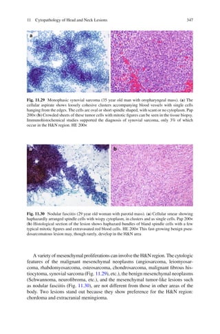
![346 G. Tarjan
There are two malignant neoplasms unique to the sinonasal tract: the aggressive
sinonasal undifferentiated carcinoma and the olfactory neuroblastoma, an uncom-
mon neuroectodermal tumor of the superior nasal cavity. These tumors are rarely
aspirated. According to scarce reports, sinonasal undifferentiated carcinoma
yields hypercellular smears with a single-cell-predominant pattern and a back-
ground of naked nuclei and nuclear debris. The cells are medium-sized with irregu-
lar nuclear contours, small nucleoli, and mitotic figures. Olfactory neuroblastoma
exhibits similar cellularity, cellular arrangement, and chromatin. In contrast, olfac-
tory neuroblastoma demonstrates smooth nuclear contours, fibrillary cytoplasm,
rosette formation, and absent mitotic figures [39].
Jaw lesions, including odontogenic tumors, are infrequently aspirated and FNA
experience is limited. The cytological features of ameloblastoma are probably the
most described. The hypercellular smears have two distinct cell populations: small,
hyperchromatic, basaloid-type cells with peripheral palisading, and scattered larger
cells with more open chromatin. Occasional fragments of mesenchymal cells with
more elongated nuclei and ample, clear cytoplasm are also present. The rare malig-
nant cases exhibit prominent cytologic pleomorphism, cellular crowding, molding
and a high mitotic/karyorrhectic index [40].
Parathyroid cysts are rare, representing less than 1% of all neck swellings. They
can present clinically as low neck masses and mimic a thyroid nodule. However,
FNA can assist in formulating a correct preoperative diagnosis. The aspirate typi-
cally reveals clear, colorless and watery fluid that has an extremely high concentra-
tion of parathyroid hormone. Ultrasound-guided FNA combined with parathyroid
hormone measurement has also been described in the preoperative localization of
parathyroid adenomas, with a specificity of 95% and sensitivity of 91% [41].
Cytological features of parathyroid lesions include discohesive cells or bare nuclei,
about the size of red blood cells, which are mostly uniform but occasionally show
marked variation in size (Fig. 11.28). In addition, some cells may be arranged in a
nested pattern associated with capillaries [42].
Fig. 11.28 Parathyroid gland (49 year old woman). (a) The smear reveals relatively uniform cells
with evenly spaced round nuclei and indistinct nucleoli. DQ 200× (b) Identical cells are seen in the
histological section. HE 200×](https://image.slidesharecdn.com/headneckcancercurrentperspectivesadvancesandchallenges-130605015223-phpapp01/85/Head-neck-cancer-current-perspectives-advances-and-challenges-34-320.jpg)
![348 G. Tarjan
Chordoma is a malignant tumor that frequently arises from the base of the skull
and may extend into the nasal cavity, maxillary antrum, or nasopharynx. The aspi-
rate shows abundant myxoid-chondroid stroma, which has bright purple color with
Romanowsky-type stains. Embedded in this stroma are small ovoid cells and large
cells with vacuolated clear cytoplasm representing the diagnostic physaliphorous
cells [43] (Fig. 11.31).
The rare extracranial meningioma has been reported in various H&N sites. The
aspirate reveals many oval to spindle shaped cells, either by themselves or in larger
groups. The cells have round nuclei, evenly dispersed fine chromatin, and small
distinct nucleoli. Spindle-shaped cells in concentric whorles, and psammoma bod-
ies characteristic of meningioma have also been described [44].
Finally, it is essential to discuss lesions which, by cytological examination, can
be difficult to be distinguished from a cystic carcinoma metastasis:
Branchial cleft cysts, a result of developmental abnormalities of the branchial
apparatus, usually arise at the anterior border of the sternomastoid muscle. They are
lined predominantly by squamous epithelium, but minor columnar epithelial cell
Fig. 11.31 Chordoma (62 year old man with vertebral column mass). (a) Shown in the smear
preparation is sparse cellularity with prominent myxoid background. DQ 100× (b) The cells have
rather uniform round-oval nuclei with distinct nucleoli and vacuolated cytoplasm. Occasional
binucleate cells are seen. DQ 400× (c) Corresponding histologic section shows the vacuolated cells
and the myxoid stroma. HE 400×](https://image.slidesharecdn.com/headneckcancercurrentperspectivesadvancesandchallenges-130605015223-phpapp01/85/Head-neck-cancer-current-perspectives-advances-and-challenges-36-320.jpg)
![34911 Cytopathology of Head and Neck Lesions
lining may be present. FNAs of branchial cleft cysts yield thick yellow fluid which
containing many anucleate squames, some squamous cells, and occasional colum-
nar cells and lymphocytes. Amorphous debris with neutrophils is commonly seen in
the background (Fig. 11.32).
Thyroglossal duct cysts show similar cytological features. These are cystic rem-
nants of the embryonic thyroglossal duct and therefore are typically located in the
midline of the neck near the hyoid bone. Although many thyroglossal duct cysts
contain thyroid follicles adjacent to the squamous and ciliated columnar cell lining,
follicular epithelium or colloid may rarely appear in the aspirate.
Epidermal inclusion cysts are superficial dermal cystic lesions of hair follicular
and/or traumatic origin. They are lined by squamous epithelium and contain kerati-
nous debris. When these cysts are ruptured, numerous neutrophils and foreign
body-type multinucleate giant cells can be present in the smears, in addition to the
anucleate squames and mature squamous cells (Fig. 11.33).
In markedly inflamed branchial cleft cysts, thyroglossal duct cysts, and epider-
mal inclusion cysts, the benign squamous epithelial cells may exhibit significant
reactive atypia. In this case, the distinction from a well-differentiated SCCA that
shows little nuclear atypia, may be difficult. The patient’s clinical history and the
lesion’s exact location may provide some guidance. Particular attention should be
paid to make sure that features of SCCA such as increased N/C ratio, nuclear hyper-
chromasia, or nuclear membrane irregularity are not present. On the other hand,
clinically benign-appearing cervical cystic lesions may turn out to be malignant. In
a study aimed to investigate the incidence of unsuspected carcinoma in routinely
excised cervical cysts at a tertiary care teaching hospital, the incidence of unsus-
pected carcinoma in 196 consecutive cystic neck lesions initially diagnosed as bran-
chial cleft cysts was 3.6% (6 metastatic squamous cell carcinoma and 1 metastatic
papillary thyroid carcinoma were found) [45].
Fig. 11.32 Branchial cleft cyst (25 year old man with neck lesion). (a) The aspirated fluid contains
mature squamous cells and anucleate squames. No hyperchromasia or atypical nuclei are seen. Pap
100× (b) Histological section of the cyst wall shows benign squamous epithelial lining rimmed by
benign lymphoid tissue. HE 100×](https://image.slidesharecdn.com/headneckcancercurrentperspectivesadvancesandchallenges-130605015223-phpapp01/85/Head-neck-cancer-current-perspectives-advances-and-challenges-37-320.jpg)
![350 G. Tarjan
Pilomatricoma (pilomarixoma, calcifying epithelioma of Malherbe) is a benign
skin adnexal tumor that can be clinically mistaken for a cyst. It is composed of small
basaloid cells, keratinous debris associated with foreign body-type giant cells, and
ghost cells with abundant pink cytoplasm and an empty space that once contained
the now absent nucleus. Therefore, in the aspirate of pilomatricoma, the cytologic
features include a pink fibrillary material enveloping clusters of basaloid cells with
high N/C ratio and evenly dispersed chromatin, multinucleated giant cells, and
sheets of ghost cells which can resemble SCCA (Fig. 11.34). The recognition of the
unique constellation of cytological features in the appropriate clinical context helps
to distinguish pilomatricoma from carcinoma [46].
Fig. 11.33 Epidermal inclusion cyst (36 year old man with neck lesion). (a) Predominantly anu-
cleate squames are present in the aspirate. Pap 200× (b) Histological section demonstrates the
squames as they become detached from the surface of the cyst’s benign keratinizing squamous
epithelial lining. HE 200×
Fig. 11.34 Pilomatricoma (22 year old woman with facial mass superficial to the parotid). (a) The
aspirate reveals basaloid cells, multinucleated giant cell, and keratinous debris. Pap 200× (b)
Histological section of the lesion contains the characteristic elements: basaloid cells, ghost cells,
keratinous debris, and multinucleated giant cells. HE 200×](https://image.slidesharecdn.com/headneckcancercurrentperspectivesadvancesandchallenges-130605015223-phpapp01/85/Head-neck-cancer-current-perspectives-advances-and-challenges-38-320.jpg)

This document discusses cytopathology of head and neck lesions. It begins by introducing cytopathology as the microscopic study of diseased cells dispersed on glass slides, as opposed to histopathology which examines tissues maintaining cellular architecture. Fine needle aspiration is described as the main cell collection method for head and neck lesions, being minimally invasive and allowing rapid diagnosis. The document focuses on the cytopathology of common lesions in salivary glands, thyroid, and cervical lymph nodes. It provides details on FNA technique and processing, highlights advantages like sensitivity and specificity, and notes limitations such as difficulty distinguishing some salivary gland tumors. Representative cytology images are included to illustrate normal and diseased states.


