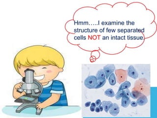
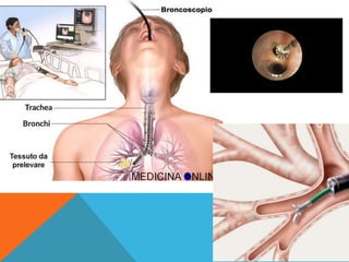
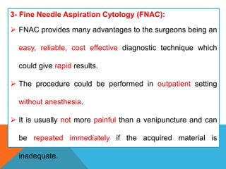
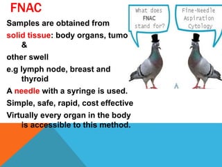
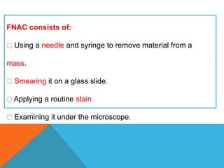
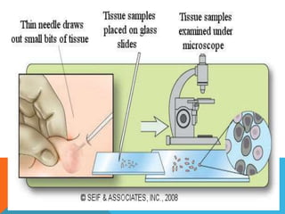
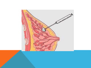
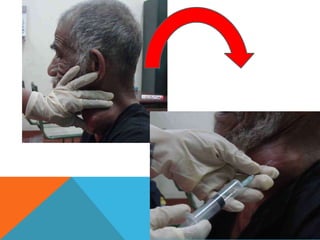
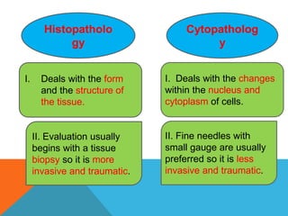
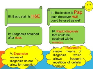
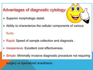
2. outlines:
1- Definition of cytopathology.
2- Differences between cytopathology and histopathology.
3- Advantage and disadvantage of cytopathology.
4- Sampling techniques.
5- Indications of cytopathology.
3. DEFINITION
Scientific study of cells obtained from
tissues or body secretions to identify
disease.
The individual cells reflect the normal
and abnormal morphology of the
tissue from which they are derived.
5. TYPES
Based on sampling techniques,
cytology is classified into the
following:
1. Exfoliative Cytology.
2. Abrasive Cytology.
3. Aspiration Cytology.
6. Sampling techniques
The collection of exfoliated cells
‘’Exfoliative Cytology’’.
Brushing or similar abrasive techniques
“Abrasive Cytology”
Aspiration biopsy
7. EXFOLIATIVE CYTOLOGY(NATURAL SPONTANEOUS
EXFOLIATION )
Based on spontaneous shedding of cells derived
from the lining of an organ into a cavity.
• It is the simplest of the three sampling
techniques.
• Contents of the sample are derived from several
sources.
Examples: vaginal smear, sputum, urine, effusion.
The material is collected spontaneously or by a
syringe or a cotton swab.
8.
9. ABRASIVE CYTOLOGY(ARTIFICIAL ENHANCED
EXFOLIATION)
• Cells are obtained directly from the surface of the target
of interest.
• Samples are taken by :
scraping, : from cervix( pap smear), vagina, oral
cavity(Buccal mucosal smear), and skin lesions.
brushing, washing. and lavage: bronchi, GIT, and urinary
tract
Examples: cervical scraper, endoscopy, and gastric lavage.
Samples can be obtained from superficial or deep lesions.
10. The purpose of this procedure is to dislodge cells…. enrich
the sample with cells obtained directly from the surface of the
target of interest.
11.
12.
13.
14. 3- Fine Needle Aspiration Cytology (FNAC):
FNAC provides many advantages to the surgeons being an
easy, reliable, cost effective diagnostic technique which
could give rapid results.
The procedure could be performed in outpatient setting
without anesthesia.
It is usually not more painful than a venipuncture and can
be repeated immediately if the acquired material is
inadequate.
15. FNAC
Samples are obtained from
solid tissue: body organs, tumors
&
other swell
e.g lymph node, breast and
thyroid
A needle with a syringe is used.
Simple, safe, rapid, cost effective
Virtually every organ in the body
is accessible to this method.
16. FNAC consists of:
Using a needle and syringe to remove material from a
mass.
Smearing it on a glass slide.
Applying a routine stain.
Examining it under the microscope.
17.
18.
19.
20. Fixatives used in Cytopathology:
1- 95% ethyl alcohol (for Pap
smear & FNA).
2- Spray fixatives (does not result in
lysis of RBC & better reserved of
nuclear details).
3-Carnoy’s fixative (lysis of RBC).
4-Other (Formaline)
21. Stains used in cytopathology:
1. Papnicolaou stain:
Mainly used in Exfoliative cytopathology.
Good & better demonstration of nuclear
details.
2. Hematoxyline & Eosin (H& E):
Mainly used in FNA.
3. Leishman & Giemsa stains:
Mainly in FNA.
22. Characteristics of ideal stain in cytopathology:
1-Evaluation of architectural pattern of tissue
fragments.
2- Proper evaluation of nuclear morphology (most
important characteristic).
3- Proper evaluation of cytoplasmic morphology.
4- Evaluation of background (blood, secretions,
mucin…….)
23. Staining methods in Cytopathology:
1.Air – dried Giemsa staining:
Air drying follows by staining with Giemsa).
Well demonstrated cytoplasmic details.
Exaggerated cells & nuclear size.
Poorly seen individual cells.
2. Wet –fixed Pap staining:
Alcohol fixation follows by staining with Pap or H &
E.
Excellent demonstration of nuclear details.
Normal size of cell & nucleus.
Clearly seen individual cells.
24. .Air – dried
Giemsa
staining:
Wet –fixed
Pap
staining:
Air drying Alcohol fixation
staining with
Giemsa).
staining with Pap or H & E
Well demonstrated
cytoplasmic details
Excellent demonstration of
nuclear details
Exaggerated cells &
nuclear size.
Normal size of cell & nucleus.
.
25. Differences between cytopathology and
histopathology
It differs importantly from histopathology in that cytology
examines only discrete cells or small groups of cells, but
histopathology examines intact tissues.
26. Histopatholo
gy
Cytopatholog
y
I. Deals with the form
and the structure of
the tissue.
I. Deals with the changes
within the nucleus and
cytoplasm of cells.
II. Evaluation usually
begins with a tissue
biopsy so it is more
invasive and traumatic.
II. Fine needles with
small gauge are usually
preferred so it is less
invasive and traumatic.
27. III. Basic stain is H&E
IV. Diagnosis obtained
after days.
IV. Rapid diagnosis
that could be
obtained within
minutes.
V. Inexpensive
simple means of
diagnosis which
allows frequent
repetition of cellular
sampling
V. Expensive
means of
diagnosis do not
allow for repetition
III. Basic stain is Pap
stain (however H&E
could be used as well)
28. Advantages of diagnostic cytology:
Superior morphologic detail.
Ability to characterize the cellular components of various
fluids.
Rapid: Speed of sample collection and diagnosis .
Inexpensive: Excellent cost effectiveness.
Simple :Minimally invasive diagnostic procedure not requiring
surgery or specialized anesthesia.
29. Advantages of diagnostic cytology:
It is better in evaluating the infectious diseases.
No injury to tissue allowing repeated sampling
It is better for hormonal assay.
Cytopathological smear cover a wider surface than
that involved in surgical biopsy.
30. The disadvantage of diagnostic cytology:
Tumors cannot be graded.
Margins cannot be evaluated.
Interpretation of the morphological cellular changes is based
only on individual cell observation.
Limited information about structural arrangement
(architecture) of the cells within the lesion.
Less definitive diagnosis as to the specific tumor type or the
distribution of an inflammatory infiltrate.
31. Very bad negative test:
A problem related to the small sample size and "blind"
nature of many common collection techniques, is the
possibility that the specimen may not represent the
primary lesion.
32. The following points should be kept in mind when starting a
cytological evaluation:
1) Cytology is an aid to clinical diagnosis, and results must be
interpreted in the context of the overall case. The cytologic
diagnosis must be based on the entire clinical evidence
available, rather than on changes in individual cells.
2) Cytology can be but is not always a substitute for
histopathology.
34. Indications for Cytopathology:
1- Differentiation between benign and malignant lesions.
2- Diagnosis of malignancy and the identification of the
neoplastic cells in primary, metastatic (secondary).
3- Diagnosis of premalignant diseases.
4- Detection of inflammation and certain types of pathogenic
agents.
35. 5- Study of the hormonal patterns through the examination of
the squamous cells in vaginal smears; which are under the
influence of ovarian hormones.
6- Follow-up and monitoring of response to chemotherapy and
irradiation, the latter producing certain cellular features which
could be diagnostic on cytological examination.
36. 7- The identification of sex chromosome: if a newborn presents
with ambiguous genitalia, one can not tell whether the sex is male
or female.
The presence of a dark dot attached to the nuclear membrane
from inside (Barr body +ve) indicates that a sex chromosome is
present, i.e., the genotype of the baby is XX (♀).
38. Cytopathology can be further
subdivided into:
Gynecological Cytopathology, include
cervicovaginal
cytopathology………..etc.
Non gynecological Cytopathology
include cytopathology of all other
organs.
Fine Needle Aspiration (FNA): include
FNA of breast, FNA of thyroid…….etc.
39. Gynecological cytopathology:
Two types of epithelia are present in female genital
organs
.1
Columnar epithelium: lined uterus & endocervix.
.2
Squamous epithelium: lined ectocervix & vagina.
Squamous Metaplasia of endocervical epithelium
(columnar) into Squamous epithelium is usual
process occurring in all women, as a result of
hormonal effects. (Changes more in cytoplasm of
cells more than nuclei).
For reporting of cervico- vaginal cytology, Bethesda
system (1991) is currently used.
46. characteristics Normal Squamous cells Metaplatic Squamous
cells
Malignant Squamous
cells
Cell arrangement Exfoliated singly Cohesive sheets or
groups
Singly or dyscohesive
sheets.
Variable size & shape of
cells.
Cytoplasmic
characteristics
Abundant
cytoplasm
Well defined cell
borders
Either pale to dense
cytoplasm.
Cytoplasmic processes.
Poor defined cell
borders.
Scant cytoplasm
Nuclear characteristics Centrally located
nucleus.
Absent nucleoli.
Low N/C ratio.
Larger nuclei than
normal.
With or without
nucleoli.
Variable size & shape.
High N/c ratio.
Prominent nucleoli.
51. Non-Gynecological cytopathology:
Include cytological examination of all other organs (e.g.
respiratory system cytopathology, urine
cytopathology……….)
1. Serous effusions:
Accumulation of fluid in serous cavities (e.g. pleura,
peritoneum )is abnormal & can results from many causes
(inflammation, cancers….).
Cytological examination of serous effusions is performed
mainly to establish the presence or absence of malignancy
(either primary mesothelioma or metastatic carcinoma to
these sites) .
Metastatic carcinomas to the serous surfaces are from
lung, breast, colon, stomach,
& ovary
Better to differentiate between mesothelioma & metastatic
carcinoma by using special stains & immunohistochemistry.
52. Characteristic reactive mesothelial cells Malignant mesothelioma Adenocarcinomas
(metastatic)
Fluid characteristics Clear, turbid Always hemorrhagic Hemorrhagic
Cells arrangement Singly mainly, less as tissue
fragments.
Ill defined cell borders.
Small to large complex
sheets of cells.
Well defined cell borders.
Acinar (gland like
formation).
Well defined cell borders.
Nuclear characteristics Variable in number & shape.
Normal N/C ratio
Multiple micronucleoli.
Same
Increase N/C ratio
Single, macronucleoli,
coarse chromatin.
Increase N/C ratio
Cytoplasmic characteristics Moderate to abundant
cytoplasm.
Scant to moderate amount. Scant to moderate amount.
Presence of cytoplasmic
vacuoles & signet ring cells.
56. 2. Sputum Cytology:
Offers highly diagnostic values for diagnosis of
centrally located lung cancer (Squamous, small
cell cancers of lung).
3 to 5 consecutive daily sputum examinations is
advisable to increase the rate of detection of
cancers.
Sputum may spontaneously coughed or induced.
Sputum sample is fixed with Saccomanno
fixative(2% polyethylene glycol With 50% ethyl
alcohol).
Sputum sample is considered satisfactory when it
contains alveolar macrophages.
58. FNA 0f Breast:
FNA is by far the most popular , simple, cost effective,
reliable, & diagnostic procedure in cytological
diagnosis of breast lesions.
FNA is important part in triple screening test for
early detection of beast carcinoma :
1- (physical examination a thorough physical examination of the whole
breast area, including both breasts, nipples, armpits .
2-Imaging tests
Imaging tests for breast cancer may involve:
•
mammogram: a way of examining the breasts using low-dose X-rays
•
ultrasound:
•
The tests used will depend on the woman’s age. For women aged 35
years or older, both a mammogram and an ultrasound will be used.
•
For women younger than 35 years, an ultrasound is used first and a
mammogram may also be used if more information is needed.
3- FNA).
59. Characteristics Benign pattern Malignant pattern
Cellularity Almost low cellularity High cellularity
Cells arrangements Sheets of uniform ductal cells (cohesive) Single, variable size malignant
cells.
(poorly cohesive)
Single bare nuclei FREQUENT ABSENT
Nuclear characteristics Uniform size Enlarged, atypical nuclei.