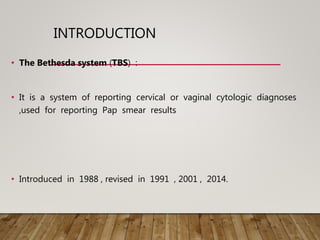
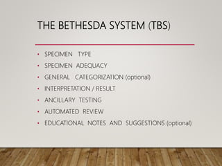
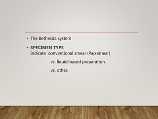
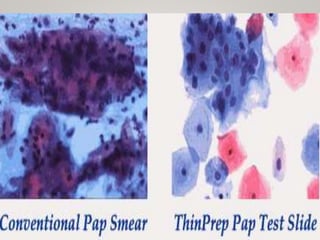
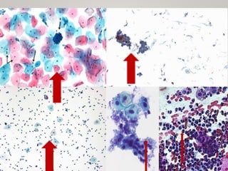
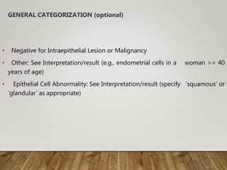
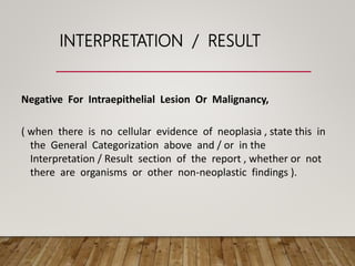
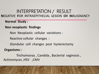
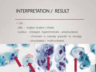
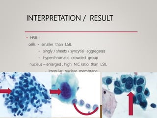
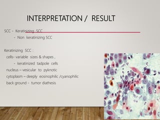
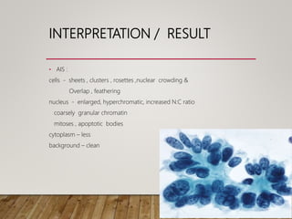
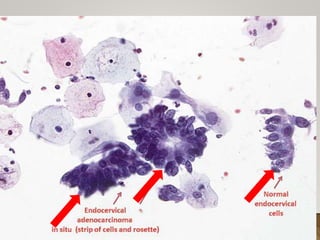
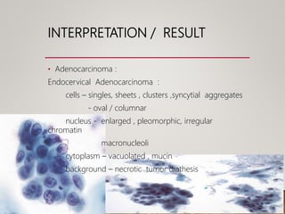
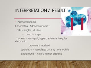
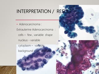
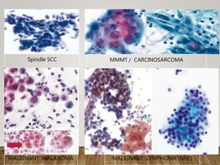
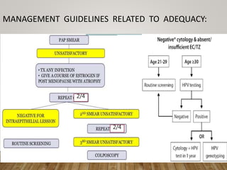
THE BETHESDA SYSTEM
DR.ABHINAV GOLLA
MEDICURE DIAGNOSTICS AND RESEARCH CENTER
VIJAYANAGAR COLONY
HYDERABAD , TELANGANA
INTRODUCTION
• The Bethesda system (TBS) :
• It is a system of reporting cervical or vaginal cytologic diagnoses
,used for reporting Pap smear results
• Introduced in 1988 , revised in 1991 , 2001 , 2014.
THE BETHESDA SYSTEM (TBS)
• SPECIMEN TYPE
• SPECIMEN ADEQUACY
• GENERAL CATEGORIZATION (optional)
• INTERPRETATION / RESULT
• ANCILLARY TESTING
• AUTOMATED REVIEW
• EDUCATIONAL NOTES AND SUGGESTIONS (optional)
• The Bethesda system
• SPECIMEN TYPE
Indicate conventional smear (Pap smear)
vs. liquid-based preparation
vs. other.
• Conventional Pap
In a conventional Pap smear , samples are smeared directly onto a
microscope slide after collection.
• Liquid based cytology
 The sample of (epithelial) cells is taken from the Transitional Zone
 Liquid-based cytology uses an arrow-shaped brush
 The cells taken are suspended in a bottle of preservative &
transported to the laboratory .
SPECIMEN ADEQUACY
• Satisfactory for evaluation
• Unsatisfactory for evaluation …(specify reason)
• Specimen rejected/not processed (specify reason)
• Specimen processed and examined, but unsatisfactory for evaluation of
epithelial abnormality because of (specify reason)
• Satisfactory :
 Presence / absence of Endocervical / transformation zone
component .
 Appropriate labeling & identifying information .
 Relevant clinical information .
• Unsatisfactory :
Rejected specimen
Fully evaluated , unsatisfactory
• Minimum squamous cellularity criteria :
5000 well visualized / well preserved squamous cells in liquid based
preparation .
8000 – 12000 cells in conventional preparation .
• Endocervical / Transformation Zone component :
10 well preserved endocervical / squamous metaplastic cells singly / in
clusters .
GENERAL CATEGORIZATION (optional)
• Negative for Intraepithelial Lesion or Malignancy
• Other: See Interpretation/result (e.g., endometrial cells in a woman >= 40
years of age)
• Epithelial Cell Abnormality: See Interpretation/result (specify ‘squamous’ or
‘glandular’ as appropriate)
INTERPRETATION / RESULT
Negative For Intraepithelial Lesion Or Malignancy,
( when there is no cellular evidence of neoplasia , state this in
the General Categorization above and / or in the
Interpretation / Result section of the report , whether or not
there are organisms or other non-neoplastic findings ).
INTERPRETATION / RESULT
NEGATIVE FOR INTRAEPITHELIAL LESION OR MALIGNANCY
Normal Study :
Non neoplastic findings
Non Neoplastic cellular variations :
Reactive cellular changes :
Glandular cell changes post hysterectomy
Organisms :
Trichomonas , Candida , Bacterial vaginosis ,
Actinomyces ,HSV ,CMV
NORMAL
• Squamous cell
• Endocervical cells
• Endometrial cells
• Superficial squamous cell : Mature ,polygonal
cytoplasm – mostly Eosinophilic
nucleus - pyknotic
cells show relatively prominent cellular outline .
• Intermediate squamous cell : Mature
cytoplasm - Cyanophilic
nucleus - non pyknotic ,vesicular nucleus
large oval / rounded nuclei with groove
granular chromatin
• Parabasal / Basal squamous cells :
small , oval , round
immature
cytoplasm - cyanophilic / eosinophilic
nucleus – oval, fine chromatin
• Endocervical cells :
 Columnar in shape & contain mucin
 In cervical smears endocervical cells arranged singly in
layers & in sheets forming a palisade .
 Honey comb / picket fence appearance .
 nucleus is large ,rounded, placed at basal portion
nucleus is granular evenly distributed chromatin
 cytoplasm - eosinophilic / basophilic
Endometrial cells :
 in smears appear as rounded clusters
 central core of connective tissue with elongated cells
(stromal)surrounded by round- oval cells(glandular) - Exodus
 cytoplasm - scanty , vacuolated
 nucleus - size similar to intermediate cells
- karyorrhexis
 cell border - ill defined
 histiocytes
• In liquid based preparation , exfoliated endometrial
cells may be slightly larger , with more easily
visible nucleoli and enhanced chromatin details
compared to conventional smear preparations .
EXODUS
NILM- NON NEOPLASTIC
FINDINGS
• Non neoplastic cellular variations
• Squamous Metaplasia :
 nucleus - round to oval
 evenly distributed chromatin
 in conventional smears – spider cells
NILM- NON NEOPLASTIC
FINDINGS
• Non neoplastic cellular variations
Keratotic cellular changes
 Keratosis
 Hyperkeratosis
 Parakeratosis
 Dyskeratosis
KERATOHYALINE GRANULES
PARAKERATOSIS
• Squamous cells with dense orangeophilic or
eosinophilic cytoplasm .
• Cells- isolated ,in sheets, in whorls
• Cell shape – round / polygonal / spindle shaped
• Nucleus - pyknotic
HYPERKERATOSIS
• Anucleate squamous cells with ghost like nuclear
holes .
NILM- NON NEOPLASTIC
FINDINGS
• Non neoplastic cellular changes :
• TUBAL METAPLASIA :
 columnar ciliated / pseudo stratified
 nucleus - round-oval , enlarged , pleomorphic ,
hyperchromatic &
 N:C ratio – high
 Cytoplasm - vacuoles / goblet cell change
NILM- NON NEOPLASTIC
FINDINGS
• Non neoplastic cellular changes :
ATROPHY
 flat monolayer sheets of parabasal like cells
 nucleus enlargement , N:C ratio, regular contour
 abundant inflammatory exudate,
 basophilic granular backgroud
 blue blobs – conventional preparations
NILM- NON NEOPLASTIC
FINDINGS• Non neoplastic cellular changes :
Pregnancy related cellular changes
 navicular cells- boat shaped intermediate cells
- abundant basophilic clear cytoplasm
- nucleus- vesicular , delicate chromatin
 decidua
 cytotrophoblast & syncytiotrophoblast
 Arias – Stella reaction
NILM- NON NEOPLASTIC
FINDINGS• Reactive cellular changes :
• 1. Inflammation :
 reactive squamous cells- mild nuclear enlargement without
significant chromatin abnormality .
 reactive endocervical cells – variation in nuclear size, prominent
nucleoli , fine chromatin, intra cytoplasmic PML’s
 perinuclear halos
NILM- NON NEOPLASTIC
FINDINGS• Reactive cellular changes :
• 2. Lymphocytic (Follicular) cervicitis:
NILM- NON NEOPLASTIC
FINDINGS• Reactive cellular changes :
• 3. Radiation :
cell size ,bizarre shape
nucleus size / N , Binucleation / Multinucleation
cytoplasm - vacuolated polychromatic , intra cytoplasmic
PML’s
NILM- NON NEOPLASTIC
FINDINGS• Reactive cellular changes :
• 4. IUCD :
Glandular cells - singly / clusters in clean background
signet ring appearance
NILM- NON NEOPLASTIC
FINDINGS
Glandular cell changes Post hysterectomy :
benign appearing glandular cells
NILM- ORGANISMS
1. Trichomonas vaginalis :
Pear shaped cyanophilic
nucleus – pale , vesicular , eccintrically
cytoplasm – eosinophilic granules
flagella
leptothrix
squamous cell TRICH change , polyballs
NILM- ORGANISMS
2. Candida :
budding yeast &/ pseudo hyphae
in liquid based preparation : spearing of epithelial cells/ shish
kebab effect
NILM- ORGANISMS
3. Bacterial vaginosis
clue cells
LACTOBACILLI
NILM- ORGANISMS
4. Actinomyces :
cotton ball clusters
NILM- ORGANISMS
5. HSV :
nucleus – ground glass appearance
- dense eosinophilic intranuclear inclusions
( Cowdry ) surrounded by a halo
large multinucleated / mononucleate squamous cells
NILM- ORGANISMS
6. CMV :
Endocervical glandular cells affected
cells & nucleus – enlarged
large eosinophilic intranuclear viral inclusions with
halo
INTERPRETATION / RESULT
OTHERS :
Endometrial cells (in a woman >= 40 years of
age)
(Specify if ‘negative for squamous intraepithelial
lesion’)
INTERPRETATION / RESULT
EPITHELIAL CELL ABNORMALITIES
SQUAMOUS CELL
 Atypical squamous cells
- of undetermined significance (ASC-US)
- cannot exclude HSIL (ASC-H)
 Low grade squamous intraepithelial lesion (LSIL)
(encompassing: HPV/mild dysplasia/CIN 1)
 High grade squamous intraepithelial lesion (HSIL)
(encompassing: moderate and severe dysplasia, CIS,
CIN 2 and CIN 3)
- with features suspicious for invasion (if invasion is
INTERPRETATION / RESULT
EPITHELIAL CELL ABNORMALITIES
GLANDULAR CELL
ATYPICAL
 endocervical cells (not otherwise specified (NOS) or specify in comments),
 endometrial cells (NOS or specify in comments),
 glandular cells (NOS or specify in comments)
ATYPICAL
 endocervical cells, favor neoplastic
glandular cells, favor neoplastic
ENDOCERVICAL ADENOCARCINOMA IN SITU
ADENOCARCINOMA:
 Endocervical
 Endometrial
 extrauterine
 not otherwise specified (NOS)
INTERPRETATION / RESULT
• OTHER MALIGNANT NEOPLASMS: (specify)
1. Uncommon primary tumors of cervix & uterine corpus:
 Carcinomas ( spindle SCC, Poorly diff SCC ,)
 Neuroendocrine ( small cell ,large cell, carcinoid ,glassy
,mucinous , MMMT ,Clear cell, Sarcoma, )
2. secondary / metastatic tumors :
 Extra uterine carcinomas (Breast , Stomach, Ovary ,Colon
, Kidney ,Bladder)
INTERPRETATION / RESULT
• ASC
• ASC-US:
changes that are suggestive of LSIL but are
insufficient for a definitive interpretation
ASC – 1. Squamous differentiation
2. N:C ratio
3. nucleus – size enlarged hyperchromasia ,
clumped chromatin ,irregular , multinucleated
Seen in atypical parakeratosis , atypical repair , atypia
in postmenopausal woman
ATYPICAL
PARAKERATOSIS
INTERPRETATION / RESULT
• ASC
• ASC-H:
Atypical immature metaplasia
Crowded sheet pattern
INTERPRETATION / RESULT
• LSIL :
cells - singles/ clusters / sheets
nucleus - enlarged , hyperchromatic , anisonucleosis
- chromatin is coarsely granular to smudgy
- binucleated / multinucleated
koilocytes & increased keratinization
INTERPRETATION / RESULT
• HSIL :
cells - smaller than LSIL
- singly / sheets / syncytial aggregates
- hyperchromatic crowded group
nucleus – enlarged , high N:C ratio than LSIL
- irregular nuclear membrane
INTERPRETATION / RESULT
• HSIL with invasion
INTERPRETATION / RESULT
SCC - Keratinizing SCC
- Non keratinizing SCC
Keratinizing SCC :
cells- variable sizes & shapes ,
- keratinized tadpole cells
nucleus – vesicular to pyknotic
cytoplasm – deeply eosinophilic /cyanophilic
back ground - tumor diathesis
INTERPRETATION / RESULT
SCC - Keratinizing SCC
- Non keratinizing SCC
Non Keratinizing SCC :
cells- singly / syncytial aggregates
nucleus – irregular coarsely clumped chromatin
- nucleoli prominent
- features of HSIL
back ground - tumor diathesis
INTERPRETATION / RESULT
• Atypical Endocervical cells: NOS
cells - sheets , cell crowding
nucleus - overlapping , enlarged ,pleomorphism
- nucleoli
- increased N:C ratio
INTERPRETATION / RESULT
• Atypical Endocervical cells , favour Neoplastic
cells - sheets , strips ,rosettes / feathering
- ill-defined border
nucleus - crowding ,overlap, pseudostratification
- enlarged
- coarse chromatin , increased N:C ratio
INTERPRETATION / RESULT
• Atypical Endometrial cells :
cells - small groups (5-10 cells)
- ill-defined border
nucleus – enlarged
mild hyperchromasia , chromatin heterogenecity
nucleoli
cytoplasm - vacuolated & scanty
INTERPRETATION / RESULT
• AIS :
cells - sheets , clusters , rosettes ,nuclear crowding &
Overlap , feathering
nucleus - enlarged, hyperchromatic, increased N:C ratio
coarsely granular chromatin
mitoses , apoptotic bodies
cytoplasm – less
background – clean
INTERPRETATION / RESULT
• Adenocarcinoma :
Endocervical Adenocarcinoma
Endometrial Adenocarcinoma
Extrauterine Adenocarcinoma
INTERPRETATION / RESULT
• Adenocarcinoma :
Endocervical Adenocarcinoma :
cells – singles, sheets , clusters ,syncytial aggregates
- oval / columnar
nucleus - enlarged , pleomorphic, irregular
chromatin
macronucleoli
cytoplasm – vacuolated , mucin
background – necrotic tumor diathesis
INTERPRETATION / RESULT
• Adenocarcinoma :
Endometrial Adenocarcinoma :
cells – singles, clusters ,
- round in shape
nucleus - enlarged , hyperchromasia, irregular
chromatin
prominent nucleoli
cytoplasm – vacuolated , scanty , cyanophilic
background – watery tumor diathesis
INTERPRETATION / RESULT
• Adenocarcinoma :
Extrauterine Adenocarcinoma :
cells – few , variable shape
nucleus - variable
cytoplasm – variable
background – no diathesis
Spindle SCC MMMT / CARCINOSARCOMA
MALIGNANT MALANOMA MALIGNANT LYMPHOMA (NHL)
ANCILLARY TESTING
• Provide a brief description of the test method(s) and report the result so
that it is easily understood by the clinician.
AUTOMATED REVIEW
If case examined by automated device, specify device and result.
EDUCATIONAL NOTES AND SUGGESTIONS
(optional)
Suggestions should be concise and consistent with clinical follow-up guidelines
published by professional organizations (references to relevant publications may
be included).
MANAGEMENT GUIDELINES RELATED TO ADEQUACY:
2/4
2/4
• Colposcopy is recommended (if LSIL)
• Loop electrosurgical excision or colposcopy for HSIL.
• ASCUS….. (HPV testing/ Repeat cytology in 1 year
If HPV negative - cotesting - cotesting at 3 years.
If HPV positive - colposcopy.
If 1 year cytology is negative - routine screening
• NILM – Repeat cotesting in 3 years is preferred.
• NILM – after 65 years – No further screening is required if
prior negative screening.
TAKE HOME MESSAGE
The 2014 BETHESDA SYSTEM FOR REPORTING CERVICAL
CYTOLOGY
SPECIMEN TYPE:
Indicate conventional smear (Pap smear) vs. liquid-based preparation vs. other
SPECIMEN ADEQUACY :
• Satisfactory for evaluation ( describe presence or absence of endocervical/transformation
zone component and any other quality indicators, e.g., partially
obscuring blood, infl ammation, etc. )
• Unsatisfactory for evaluation . . . ( specify reason )
– Specimen rejected/not processed (s pecify reason )
– Specimen processed and examined, but unsatisfactory for evaluation of epithelial
- abnormality because of ( specify reason )
GENERAL CATEGORIZATION ( optional )
• Negative for Intraepithelial Lesion or Malignancy
• Other: See Interpretation/Result ( e.g., endometrial cells in a woman ≥45 years of
age )
• Epithelial Cell Abnormality: See Interpretation/Result ( specify ‘squamous’ or
‘glandular’ as appropriate )
INTERPRETATION/RESULT
NEGATIVE FOR INTRAEPITHELIAL LESION OR MALIGNANCY
(When there is no cellular evidence of neoplasia, state this in the General
Categorization above and/or in the Interpretation/Result section of the report --
whether or not there are organisms or other non-neoplastic findings )
NON-NEOPLASTIC FINDINGS ( optional to report optional to report; list not
inclusive )
• Non-neoplastic cellular variations
– Squamous metaplasia
– Keratotic changes
– Tubal metaplasia
– Atrophy
– Pregnancy-associated changes
Reactive cellular changes associated with:
– Infl ammation (includes typical repair)
• Lymphocytic (follicular) cervicitis
– Radiation
– Intrauterine contraceptive device (IUD)
• Glandular cells status post hysterectomy
ORGANISMS
• Trichomonas vaginalis
• Fungal organisms morphologically consistent with Candida spp.
• Shift in flora suggestive of bacterial vaginosis
• Bacteria morphologically consistent with Actinomyces spp.
• Cellular changes consistent with herpes simplex virus
• Cellular changes consistent with cytomegalovirus
OTHER
• Endometrial cells ( in a woman ≥45 years of age )
( Specify if “negative for squamous intraepithelial lesion” )
EPITHELIAL CELL ABNORMALITIES
SQUAMOUS CELL
• Atypical squamous cells
– of undetermined signifi cance (ASC-US)
– cannot exclude HSIL (ASC-H)
• Low-grade squamous intraepithelial lesion (LSIL)
( encompassing: HPV/mild dysplasia/CIN 1 )
• High-grade squamous intraepithelial lesion (HSIL)
( encompassing: moderate and severe dysplasia, CIS; CIN 2 and CIN 3 )
– with features suspicious for invasion (i f invasion is suspected )
• Squamous cell carcinoma
GLANDULAR CELL
• Atypical
– endocervical cells (NOS o r specify in comments )
– endometrial cells (NOS o r specify in comments )
– glandular cells (NOS o r specify in comments )
• Atypical– endocervical cells, favor neoplastic
– glandular cells, favor neoplastic
• Endocervical adenocarcinoma in situ
• Adenocarcinoma– endocervical– endometrial– extrauterine– not otherwise specifi
ed (NOS)
OTHER MALIGNANT NEOPLASMS: (specify)
ADJUNCTIVE TESTING
Provide a brief description of the test method(s) and report the result so that it is
easily understood by the clinician.
COMPUTER-ASSISTED INTERPRETATION OF CERVICAL CYTOLOGY
If case examined by an automated device, specify device and result.
EDUCATIONAL NOTES AND COMMENTS APPENDED TO CYTOLOGY
REPORTS ( optional )
Suggestions should be concise and consistent with clinical follow-up guidelines
published by professional organizations (references to relevant publications may be
included).
REFERRENCES
• The Bethesda System for Reporting Cervical Cytology
– 3 rd edition – Ritu Nayar , David C.Wilbur
• Gynecological Cytopathology Cervix- 2 nd edition –
Suresh Bhambhani
• Comprehensive Cytopathology – 2 nd edition –
Marluce Bibbo
THANK YOU

Bethesda 180521085301

  • 1.
    THE BETHESDA SYSTEM DR.ABHINAVGOLLA MEDICURE DIAGNOSTICS AND RESEARCH CENTER VIJAYANAGAR COLONY HYDERABAD , TELANGANA
  • 2.
    INTRODUCTION • The Bethesdasystem (TBS) : • It is a system of reporting cervical or vaginal cytologic diagnoses ,used for reporting Pap smear results • Introduced in 1988 , revised in 1991 , 2001 , 2014.
  • 3.
    THE BETHESDA SYSTEM(TBS) • SPECIMEN TYPE • SPECIMEN ADEQUACY • GENERAL CATEGORIZATION (optional) • INTERPRETATION / RESULT • ANCILLARY TESTING • AUTOMATED REVIEW • EDUCATIONAL NOTES AND SUGGESTIONS (optional)
  • 4.
    • The Bethesdasystem • SPECIMEN TYPE Indicate conventional smear (Pap smear) vs. liquid-based preparation vs. other.
  • 5.
    • Conventional Pap Ina conventional Pap smear , samples are smeared directly onto a microscope slide after collection. • Liquid based cytology  The sample of (epithelial) cells is taken from the Transitional Zone  Liquid-based cytology uses an arrow-shaped brush  The cells taken are suspended in a bottle of preservative & transported to the laboratory .
  • 7.
    SPECIMEN ADEQUACY • Satisfactoryfor evaluation • Unsatisfactory for evaluation …(specify reason) • Specimen rejected/not processed (specify reason) • Specimen processed and examined, but unsatisfactory for evaluation of epithelial abnormality because of (specify reason)
  • 8.
    • Satisfactory : Presence / absence of Endocervical / transformation zone component .  Appropriate labeling & identifying information .  Relevant clinical information . • Unsatisfactory : Rejected specimen Fully evaluated , unsatisfactory
  • 9.
    • Minimum squamouscellularity criteria : 5000 well visualized / well preserved squamous cells in liquid based preparation . 8000 – 12000 cells in conventional preparation . • Endocervical / Transformation Zone component : 10 well preserved endocervical / squamous metaplastic cells singly / in clusters .
  • 11.
    GENERAL CATEGORIZATION (optional) •Negative for Intraepithelial Lesion or Malignancy • Other: See Interpretation/result (e.g., endometrial cells in a woman >= 40 years of age) • Epithelial Cell Abnormality: See Interpretation/result (specify ‘squamous’ or ‘glandular’ as appropriate)
  • 12.
    INTERPRETATION / RESULT NegativeFor Intraepithelial Lesion Or Malignancy, ( when there is no cellular evidence of neoplasia , state this in the General Categorization above and / or in the Interpretation / Result section of the report , whether or not there are organisms or other non-neoplastic findings ).
  • 13.
    INTERPRETATION / RESULT NEGATIVEFOR INTRAEPITHELIAL LESION OR MALIGNANCY Normal Study : Non neoplastic findings Non Neoplastic cellular variations : Reactive cellular changes : Glandular cell changes post hysterectomy Organisms : Trichomonas , Candida , Bacterial vaginosis , Actinomyces ,HSV ,CMV
  • 14.
    NORMAL • Squamous cell •Endocervical cells • Endometrial cells
  • 15.
    • Superficial squamouscell : Mature ,polygonal cytoplasm – mostly Eosinophilic nucleus - pyknotic cells show relatively prominent cellular outline .
  • 16.
    • Intermediate squamouscell : Mature cytoplasm - Cyanophilic nucleus - non pyknotic ,vesicular nucleus large oval / rounded nuclei with groove granular chromatin
  • 17.
    • Parabasal /Basal squamous cells : small , oval , round immature cytoplasm - cyanophilic / eosinophilic nucleus – oval, fine chromatin
  • 19.
    • Endocervical cells:  Columnar in shape & contain mucin  In cervical smears endocervical cells arranged singly in layers & in sheets forming a palisade .  Honey comb / picket fence appearance .  nucleus is large ,rounded, placed at basal portion nucleus is granular evenly distributed chromatin  cytoplasm - eosinophilic / basophilic
  • 21.
    Endometrial cells : in smears appear as rounded clusters  central core of connective tissue with elongated cells (stromal)surrounded by round- oval cells(glandular) - Exodus  cytoplasm - scanty , vacuolated  nucleus - size similar to intermediate cells - karyorrhexis  cell border - ill defined  histiocytes
  • 22.
    • In liquidbased preparation , exfoliated endometrial cells may be slightly larger , with more easily visible nucleoli and enhanced chromatin details compared to conventional smear preparations . EXODUS
  • 23.
    NILM- NON NEOPLASTIC FINDINGS •Non neoplastic cellular variations • Squamous Metaplasia :  nucleus - round to oval  evenly distributed chromatin  in conventional smears – spider cells
  • 25.
    NILM- NON NEOPLASTIC FINDINGS •Non neoplastic cellular variations Keratotic cellular changes  Keratosis  Hyperkeratosis  Parakeratosis  Dyskeratosis
  • 26.
  • 27.
    PARAKERATOSIS • Squamous cellswith dense orangeophilic or eosinophilic cytoplasm . • Cells- isolated ,in sheets, in whorls • Cell shape – round / polygonal / spindle shaped • Nucleus - pyknotic
  • 28.
    HYPERKERATOSIS • Anucleate squamouscells with ghost like nuclear holes .
  • 29.
    NILM- NON NEOPLASTIC FINDINGS •Non neoplastic cellular changes : • TUBAL METAPLASIA :  columnar ciliated / pseudo stratified  nucleus - round-oval , enlarged , pleomorphic , hyperchromatic &  N:C ratio – high  Cytoplasm - vacuoles / goblet cell change
  • 30.
    NILM- NON NEOPLASTIC FINDINGS •Non neoplastic cellular changes : ATROPHY  flat monolayer sheets of parabasal like cells  nucleus enlargement , N:C ratio, regular contour  abundant inflammatory exudate,  basophilic granular backgroud  blue blobs – conventional preparations
  • 31.
    NILM- NON NEOPLASTIC FINDINGS•Non neoplastic cellular changes : Pregnancy related cellular changes  navicular cells- boat shaped intermediate cells - abundant basophilic clear cytoplasm - nucleus- vesicular , delicate chromatin  decidua  cytotrophoblast & syncytiotrophoblast  Arias – Stella reaction
  • 33.
    NILM- NON NEOPLASTIC FINDINGS•Reactive cellular changes : • 1. Inflammation :  reactive squamous cells- mild nuclear enlargement without significant chromatin abnormality .  reactive endocervical cells – variation in nuclear size, prominent nucleoli , fine chromatin, intra cytoplasmic PML’s  perinuclear halos
  • 34.
    NILM- NON NEOPLASTIC FINDINGS•Reactive cellular changes : • 2. Lymphocytic (Follicular) cervicitis:
  • 35.
    NILM- NON NEOPLASTIC FINDINGS•Reactive cellular changes : • 3. Radiation : cell size ,bizarre shape nucleus size / N , Binucleation / Multinucleation cytoplasm - vacuolated polychromatic , intra cytoplasmic PML’s
  • 36.
    NILM- NON NEOPLASTIC FINDINGS•Reactive cellular changes : • 4. IUCD : Glandular cells - singly / clusters in clean background signet ring appearance
  • 37.
    NILM- NON NEOPLASTIC FINDINGS Glandularcell changes Post hysterectomy : benign appearing glandular cells
  • 38.
    NILM- ORGANISMS 1. Trichomonasvaginalis : Pear shaped cyanophilic nucleus – pale , vesicular , eccintrically cytoplasm – eosinophilic granules flagella leptothrix squamous cell TRICH change , polyballs
  • 39.
    NILM- ORGANISMS 2. Candida: budding yeast &/ pseudo hyphae in liquid based preparation : spearing of epithelial cells/ shish kebab effect
  • 40.
    NILM- ORGANISMS 3. Bacterialvaginosis clue cells LACTOBACILLI
  • 41.
    NILM- ORGANISMS 4. Actinomyces: cotton ball clusters
  • 42.
    NILM- ORGANISMS 5. HSV: nucleus – ground glass appearance - dense eosinophilic intranuclear inclusions ( Cowdry ) surrounded by a halo large multinucleated / mononucleate squamous cells
  • 43.
    NILM- ORGANISMS 6. CMV: Endocervical glandular cells affected cells & nucleus – enlarged large eosinophilic intranuclear viral inclusions with halo
  • 44.
    INTERPRETATION / RESULT OTHERS: Endometrial cells (in a woman >= 40 years of age) (Specify if ‘negative for squamous intraepithelial lesion’)
  • 45.
    INTERPRETATION / RESULT EPITHELIALCELL ABNORMALITIES SQUAMOUS CELL  Atypical squamous cells - of undetermined significance (ASC-US) - cannot exclude HSIL (ASC-H)  Low grade squamous intraepithelial lesion (LSIL) (encompassing: HPV/mild dysplasia/CIN 1)  High grade squamous intraepithelial lesion (HSIL) (encompassing: moderate and severe dysplasia, CIS, CIN 2 and CIN 3) - with features suspicious for invasion (if invasion is
  • 46.
    INTERPRETATION / RESULT EPITHELIALCELL ABNORMALITIES GLANDULAR CELL ATYPICAL  endocervical cells (not otherwise specified (NOS) or specify in comments),  endometrial cells (NOS or specify in comments),  glandular cells (NOS or specify in comments) ATYPICAL  endocervical cells, favor neoplastic glandular cells, favor neoplastic ENDOCERVICAL ADENOCARCINOMA IN SITU ADENOCARCINOMA:  Endocervical  Endometrial  extrauterine  not otherwise specified (NOS)
  • 47.
    INTERPRETATION / RESULT •OTHER MALIGNANT NEOPLASMS: (specify) 1. Uncommon primary tumors of cervix & uterine corpus:  Carcinomas ( spindle SCC, Poorly diff SCC ,)  Neuroendocrine ( small cell ,large cell, carcinoid ,glassy ,mucinous , MMMT ,Clear cell, Sarcoma, ) 2. secondary / metastatic tumors :  Extra uterine carcinomas (Breast , Stomach, Ovary ,Colon , Kidney ,Bladder)
  • 48.
    INTERPRETATION / RESULT •ASC • ASC-US: changes that are suggestive of LSIL but are insufficient for a definitive interpretation ASC – 1. Squamous differentiation 2. N:C ratio 3. nucleus – size enlarged hyperchromasia , clumped chromatin ,irregular , multinucleated Seen in atypical parakeratosis , atypical repair , atypia in postmenopausal woman
  • 49.
  • 50.
    INTERPRETATION / RESULT •ASC • ASC-H: Atypical immature metaplasia Crowded sheet pattern
  • 51.
    INTERPRETATION / RESULT •LSIL : cells - singles/ clusters / sheets nucleus - enlarged , hyperchromatic , anisonucleosis - chromatin is coarsely granular to smudgy - binucleated / multinucleated koilocytes & increased keratinization
  • 52.
    INTERPRETATION / RESULT •HSIL : cells - smaller than LSIL - singly / sheets / syncytial aggregates - hyperchromatic crowded group nucleus – enlarged , high N:C ratio than LSIL - irregular nuclear membrane
  • 53.
    INTERPRETATION / RESULT •HSIL with invasion
  • 54.
    INTERPRETATION / RESULT SCC- Keratinizing SCC - Non keratinizing SCC Keratinizing SCC : cells- variable sizes & shapes , - keratinized tadpole cells nucleus – vesicular to pyknotic cytoplasm – deeply eosinophilic /cyanophilic back ground - tumor diathesis
  • 55.
    INTERPRETATION / RESULT SCC- Keratinizing SCC - Non keratinizing SCC Non Keratinizing SCC : cells- singly / syncytial aggregates nucleus – irregular coarsely clumped chromatin - nucleoli prominent - features of HSIL back ground - tumor diathesis
  • 56.
    INTERPRETATION / RESULT •Atypical Endocervical cells: NOS cells - sheets , cell crowding nucleus - overlapping , enlarged ,pleomorphism - nucleoli - increased N:C ratio
  • 57.
    INTERPRETATION / RESULT •Atypical Endocervical cells , favour Neoplastic cells - sheets , strips ,rosettes / feathering - ill-defined border nucleus - crowding ,overlap, pseudostratification - enlarged - coarse chromatin , increased N:C ratio
  • 58.
    INTERPRETATION / RESULT •Atypical Endometrial cells : cells - small groups (5-10 cells) - ill-defined border nucleus – enlarged mild hyperchromasia , chromatin heterogenecity nucleoli cytoplasm - vacuolated & scanty
  • 59.
    INTERPRETATION / RESULT •AIS : cells - sheets , clusters , rosettes ,nuclear crowding & Overlap , feathering nucleus - enlarged, hyperchromatic, increased N:C ratio coarsely granular chromatin mitoses , apoptotic bodies cytoplasm – less background – clean
  • 61.
    INTERPRETATION / RESULT •Adenocarcinoma : Endocervical Adenocarcinoma Endometrial Adenocarcinoma Extrauterine Adenocarcinoma
  • 62.
    INTERPRETATION / RESULT •Adenocarcinoma : Endocervical Adenocarcinoma : cells – singles, sheets , clusters ,syncytial aggregates - oval / columnar nucleus - enlarged , pleomorphic, irregular chromatin macronucleoli cytoplasm – vacuolated , mucin background – necrotic tumor diathesis
  • 63.
    INTERPRETATION / RESULT •Adenocarcinoma : Endometrial Adenocarcinoma : cells – singles, clusters , - round in shape nucleus - enlarged , hyperchromasia, irregular chromatin prominent nucleoli cytoplasm – vacuolated , scanty , cyanophilic background – watery tumor diathesis
  • 64.
    INTERPRETATION / RESULT •Adenocarcinoma : Extrauterine Adenocarcinoma : cells – few , variable shape nucleus - variable cytoplasm – variable background – no diathesis
  • 65.
    Spindle SCC MMMT/ CARCINOSARCOMA MALIGNANT MALANOMA MALIGNANT LYMPHOMA (NHL)
  • 66.
    ANCILLARY TESTING • Providea brief description of the test method(s) and report the result so that it is easily understood by the clinician. AUTOMATED REVIEW If case examined by automated device, specify device and result. EDUCATIONAL NOTES AND SUGGESTIONS (optional) Suggestions should be concise and consistent with clinical follow-up guidelines published by professional organizations (references to relevant publications may be included).
  • 67.
    MANAGEMENT GUIDELINES RELATEDTO ADEQUACY: 2/4 2/4
  • 68.
    • Colposcopy isrecommended (if LSIL) • Loop electrosurgical excision or colposcopy for HSIL. • ASCUS….. (HPV testing/ Repeat cytology in 1 year If HPV negative - cotesting - cotesting at 3 years. If HPV positive - colposcopy. If 1 year cytology is negative - routine screening • NILM – Repeat cotesting in 3 years is preferred. • NILM – after 65 years – No further screening is required if prior negative screening.
  • 69.
    TAKE HOME MESSAGE The2014 BETHESDA SYSTEM FOR REPORTING CERVICAL CYTOLOGY SPECIMEN TYPE: Indicate conventional smear (Pap smear) vs. liquid-based preparation vs. other SPECIMEN ADEQUACY : • Satisfactory for evaluation ( describe presence or absence of endocervical/transformation zone component and any other quality indicators, e.g., partially obscuring blood, infl ammation, etc. ) • Unsatisfactory for evaluation . . . ( specify reason ) – Specimen rejected/not processed (s pecify reason ) – Specimen processed and examined, but unsatisfactory for evaluation of epithelial - abnormality because of ( specify reason )
  • 70.
    GENERAL CATEGORIZATION (optional ) • Negative for Intraepithelial Lesion or Malignancy • Other: See Interpretation/Result ( e.g., endometrial cells in a woman ≥45 years of age ) • Epithelial Cell Abnormality: See Interpretation/Result ( specify ‘squamous’ or ‘glandular’ as appropriate ) INTERPRETATION/RESULT NEGATIVE FOR INTRAEPITHELIAL LESION OR MALIGNANCY (When there is no cellular evidence of neoplasia, state this in the General Categorization above and/or in the Interpretation/Result section of the report -- whether or not there are organisms or other non-neoplastic findings ) NON-NEOPLASTIC FINDINGS ( optional to report optional to report; list not inclusive ) • Non-neoplastic cellular variations – Squamous metaplasia – Keratotic changes – Tubal metaplasia – Atrophy – Pregnancy-associated changes
  • 71.
    Reactive cellular changesassociated with: – Infl ammation (includes typical repair) • Lymphocytic (follicular) cervicitis – Radiation – Intrauterine contraceptive device (IUD) • Glandular cells status post hysterectomy ORGANISMS • Trichomonas vaginalis • Fungal organisms morphologically consistent with Candida spp. • Shift in flora suggestive of bacterial vaginosis • Bacteria morphologically consistent with Actinomyces spp. • Cellular changes consistent with herpes simplex virus • Cellular changes consistent with cytomegalovirus OTHER • Endometrial cells ( in a woman ≥45 years of age ) ( Specify if “negative for squamous intraepithelial lesion” )
  • 72.
    EPITHELIAL CELL ABNORMALITIES SQUAMOUSCELL • Atypical squamous cells – of undetermined signifi cance (ASC-US) – cannot exclude HSIL (ASC-H) • Low-grade squamous intraepithelial lesion (LSIL) ( encompassing: HPV/mild dysplasia/CIN 1 ) • High-grade squamous intraepithelial lesion (HSIL) ( encompassing: moderate and severe dysplasia, CIS; CIN 2 and CIN 3 ) – with features suspicious for invasion (i f invasion is suspected ) • Squamous cell carcinoma GLANDULAR CELL • Atypical – endocervical cells (NOS o r specify in comments ) – endometrial cells (NOS o r specify in comments ) – glandular cells (NOS o r specify in comments ) • Atypical– endocervical cells, favor neoplastic – glandular cells, favor neoplastic • Endocervical adenocarcinoma in situ • Adenocarcinoma– endocervical– endometrial– extrauterine– not otherwise specifi ed (NOS)
  • 73.
    OTHER MALIGNANT NEOPLASMS:(specify) ADJUNCTIVE TESTING Provide a brief description of the test method(s) and report the result so that it is easily understood by the clinician. COMPUTER-ASSISTED INTERPRETATION OF CERVICAL CYTOLOGY If case examined by an automated device, specify device and result. EDUCATIONAL NOTES AND COMMENTS APPENDED TO CYTOLOGY REPORTS ( optional ) Suggestions should be concise and consistent with clinical follow-up guidelines published by professional organizations (references to relevant publications may be included).
  • 74.
    REFERRENCES • The BethesdaSystem for Reporting Cervical Cytology – 3 rd edition – Ritu Nayar , David C.Wilbur • Gynecological Cytopathology Cervix- 2 nd edition – Suresh Bhambhani • Comprehensive Cytopathology – 2 nd edition – Marluce Bibbo THANK YOU