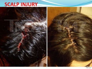
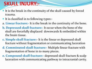
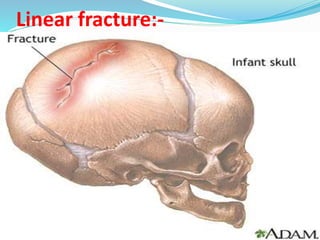
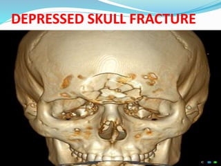
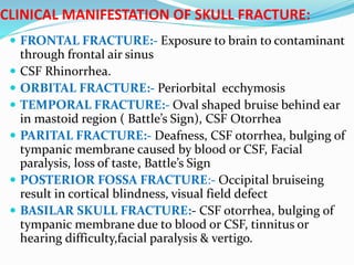
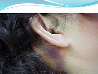
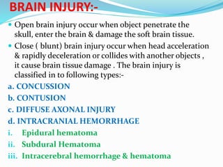
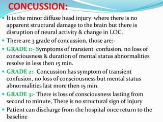
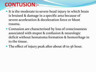
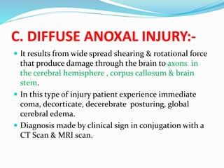
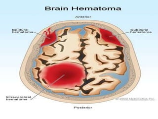
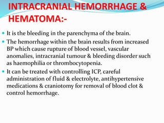
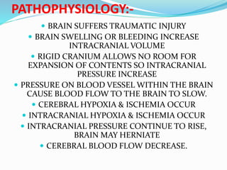
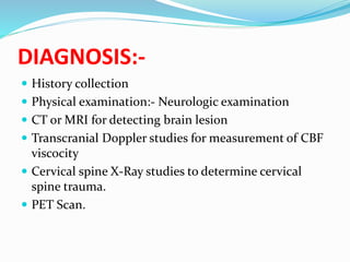
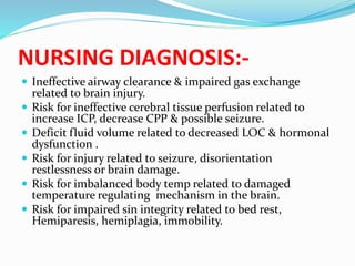
INTRODUCTION:-
 Head injury is a broad classification that include the
injury to the scalp, skull or brain.
 A serious form of head injury is traumatic brain
injury(TBI)
 In India about 1.6 million sustain TBI & seek medical
Aid & out of these 10% dies.
ETIOLOGY:-
 Motor vehicle collisions & Fall
 Fire
 Assaults
 Sports related trauma
 Recreational injuries
 War related injury
TYPE OF HEAD INJURIES:-
1. Scalp injury
2. Skull fracture:-
Skull fracture is
again 5 types, those are:-
a. Linear skull fracture
b. Depressed skull
fracture
c. Simple skull fracture
d. Comminuted skull
fracture
e. Compound skull
fracture
3. Brain injury
a. Concussion
b. Contusion
c. Diffuse axonal injury
d. Intracranial hemorrhage
i. Epidural hematoma
ii. Sub dural hematoma
iii.Intra cerebral hematoma
SCALP LACERATION:-
 It is classified as minor head injury.
 Scalp contain many blood vessels which has poor
constricting ability so when there is scalp injury it bleed
profusely which can be detected from out side.
 The scalp trauma may results in an abrasion ( brush wound),
contusion, laceration or hematoma beneath the layer of the
tissue of the scalp( subgaleal hematoma)
 Scalp injury may cause portal of entry of microorganisms
which can cause intra cranial infection
 It can be diagnosed by physical examination. Inspection,
palpation.
SCALP INJURY
SKULL INJURY:-
 It is the break in the continuity of the skull caused by forced
trauma.
 It is classified in to following types:-
a. Linear fracture:- It is the break in the continuity of the bone.
b. Depressed skull fracture:- It occur when the bone of the
skull are forcefully displaced downwards & embedded within
the brain tissue.
c. Simple skull fracture:- It is the linear or depressed skull
fracture without fragmentation or communicating laceration
d. Comminuted skull fracture:- Multiple linear fracture with
fragmentation of bone in to many pieces.
e. Compound skull fracture:- depressed skull fracture & scalp
laceration with communicating pathway to intracranial cavity.
Linear fracture:-
DEPRESSED SKULL FRACTURE
Comminuted skull fracture
COMPOUND SKULL FRACTURE
CLINICAL MANIFESTATION OF SKULL FRACTURE:
 FRONTAL FRACTURE:- Exposure to brain to contaminant
through frontal air sinus
 CSF Rhinorrhea.
 ORBITAL FRACTURE:- Periorbital ecchymosis
 TEMPORAL FRACTURE:- Oval shaped bruise behind ear
in mastoid region ( Battle’s Sign), CSF Otorrhea
 PARITAL FRACTURE:- Deafness, CSF otorrhea, bulging of
tympanic membrane caused by blood or CSF, Facial
paralysis, loss of taste, Battle’s Sign
 POSTERIOR FOSSA FRACTURE:- Occipital bruiseing
result in cortical blindness, visual field defect
 BASILAR SKULL FRACTURE:- CSF otorrhea, bulging of
tympanic membrane due to blood or CSF, tinnitus or
hearing difficulty,facial paralysis & vertigo.
Battle’s Sign
BRAIN INJURY:-
 Open brain injury occur when object penetrate the
skull, enter the brain & damage the soft brain tissue.
 Close ( blunt) brain injury occur when head acceleration
& rapidly deceleration or collides with another objects ,
it cause brain tissue damage . The brain injury is
classified in to following types:-
a. CONCUSSION
b. CONTUSION
c. DIFFUSE AXONAL INJURY
d. INTRACRANIAL HEMORRHAGE
i. Epidural hematoma
ii. Subdural Hematoma
iii. Intracerebral hemorrhage & hematoma
CONCUSSION:
 It is the minor diffuse head injury where there is no
apparent structural damage to the brain but there is
disruption of neural activity & change in LOC.
 There are 3 grade of concussion, those are:-
 GRADE 1:- Symptoms of transient confusion, no loss of
consciousness & duration of mental status abnormalities
resolve in less then 15 min.
 GRADE 2:- Concussion has symptom of transient
confusion, no loss of consciousness but mental status
abnormalities last more then 15 min.
 GRADE 3:- There is loss of consciousness lasting from
second to minute, There is no structural sign of injury
 Patient can discharge from the hospital once return to the
baseline
CONTUSION:-
 It is the moderate to severe head injury in which brain
is bruised & damage in a specific area because of
severe acceleration & deceleration force or blunt
trauma.
 Contusion are characterised by loss of consciousness
associated with stupor & confusion & neurologic
deficit without hematoma formation & hemorrhage in
to the tissue.
 The effect of injury peak after about 18 to 36 hour.
C. DIFFUSE ANOXAL INJURY:-
 It results from wide spread shearing & rotational force
that produce damage through the brain to axons in
the cerebral hemisphere , corpus callosum & brain
stem.
 In this type of injury patient experience immediate
coma, decorticate, decerebrate posturing, global
cerebral edema.
 Diagnosis made by clinical sign in conjugation with a
CT Scan & MRI scan.
D. INTRACRANIAL HEMORRHAGE:-
 In this there is collection of blood in the brain in
epidural (above the durameter), sub dural ( below the
dura) or intracerebral ( within the brain).
 Symptoms are delay until the hematoma is large
enough to cause the distoration of brain & increase
ICP.
 Intracranial hemorrhage is three types, those are:-
i. Epidural hematoma
ii. Subdural hematoma
iii. Intracerebral hemorrhage & hematoma.
EPIDURAL HEMATOMA:-
 It is the head injury and collection of blood in the epidural
space between skull & durameter.
 Bleeding & hematoma occur due to rupture of middle
meningial artery.
 The sign & symptoms includes brief loss of consciousness
followed by a lucid interval in which the patient is a wake &
conversant.
 If compensation mechanism fail to maintain ICP the volume
of the blood clot increase in the patient goes towards the
COMA.
SUBDURAL HEMATOMA:-
 It is the collection of blood in sub dural space..
 The cause of hematoma is trauma, rupture of aneurysm
or rupture of small blood vessels that bridge the
subdural space.
 Acute & subdural hematoma sign & symptoms includes
Loss Of Consciousness, increase BP, increase HR,
decrease Resp, COMA.
 Clinical manifestation appear between 48 hours to 2 wk
after the injury.
 Out come depends on managing ICP by immediate
craniotomy & REMOVAL OF clot & maintain
respiration.
INTRACRANIAL HEMORRHAGE &
HEMATOMA:-
 It is the bleeding in the parenchyma of the brain.
 The hemorrhage within the brain results from increased
BP which cause rupture of blood vessel, vascular
anomalies, intracranial tumour & bleeding disorder such
as haemophilia or thrombocytopenia.
 It can be treated with controlling ICP, careful
administration of fluid & electrolyte, antihypertensive
medications & craniotomy for removal of blood clot &
control hemorrhage.
PATHOPHYSIOLOGY:-
 BRAIN SUFFERS TRAUMATIC INJURY
 BRAIN SWELLING OR BLEEDING INCREASE
INTRACRANIAL VOLUME
 RIGID CRANIUM ALLOWS NO ROOM FOR
EXPANSION OF CONTENTS SO INTRACRANIAL
PRESSURE INCREASE
 PRESSURE ON BLOOD VESSEL WITHIN THE BRAIN
CAUSE BLOOD FLOW TO THE BRAIN TO SLOW.
 CEREBRAL HYPOXIA & ISCHEMIA OCCUR
 INTRACRANIAL HYPOXIA & ISCHEMIA OCCUR
 INTRACRANIAL PRESSURE CONTINUE TO RISE,
BRAIN MAY HERNIATE
 CEREBRAL BLOOD FLOW DECREASE.
DIAGNOSIS:-
 History collection
 Physical examination:- Neurologic examination
 CT or MRI for detecting brain lesion
 Transcranial Doppler studies for measurement of CBF
viscocity
 Cervical spine X-Ray studies to determine cervical
spine trauma.
 PET Scan.
MANAGEMENT:-
 EMERGENCY MANAGEMENT:-
 Ensure patient airway
 Stabilize survical spine
 Administer o2 via non rebreathable mask.
 Established IV assess with two large bore catheter to
infuse NS & RL.
 Intubate if GCS decrease 8
 Control external bleeding with sterile pressure
dressing.
 Remove patient clothing.
ONGOING MONITORING:-
 Maintain patient warmth using blanket, warm IV fluid,
warm humidified o2.
 Monitor vital sign, LOC, o2 saturation, cardiac rhythm,
GCS score, pupil size & reactivity
 Anticipate need for intubation if gag reflex is impaired or
absent
 Assess for rhinorrhea, otorrhea, scalp wound.
 Administer fluid cautiously to prevent fluid overload &
increase ICP.
 Maintain head & neck alignment with the axis of the body
apply cervical collar till absence of spinal cord injury
documented.
 Avoid secondary brain injury which may occur due to
edema, hypotension, respiratory depression lead to
hypoxemia & electrolyte imbalance.
Cont..
 Surgery is required to evacuation of blood clot,
debridement & elevation of depressed fracture of the
skull & suture of severe scalp laceration.
 ICP managed by adequate o2, elevating HOB maintain
normal blood volume.
 Placement of drain to remove excess CSF
 Due to hypoxia seizure may occur so antiseizure
medication should be provided.
 Sedative benzodiazepine should be administered for
agitated patient.
 Administration of NG tube
NURSING MANAGEMENT:-
 History Collection:- Collect history of mechanism of
injury such as vehicle collision, sports injury, industrial
incident, assaults fall or use of anticoagulant
medication.
 Collect sign of brain injury such as headache, mood or
behavioural changes, aphasia, dysphagia, impaired
judgement, aggression, depression.
Physical examination:-
 Check GCS
 Integumentary :- laceration, contusion, abrasion, hematoma,
battle’s sign, periorbital edema, ecchymosis, otorrhea.
 Respiratory:- rhinorrhea, impairment gag reflex, inability to
maintain patient airway, irrigulay resp.
 Cardiovascular:- Cushing tried( systolic hypertension with
widen pulse pressure, brady cardia with full & bounding
pulse, irregular resp.
 Gastrointestinal:- vomiting, projectile vomiting, bowel
incontinence.
 Urinary:- bladder incontinence.
 Neurology:- altered level consciousness, seizure activity, pupil
disfunction, cranial nerve deficit.
 Skeletal system:- motor deficit, weakness, paralysis,
spasticity, decorticate or decorticate posturing. Muscular
rigidity, ataxia.
NURSING DIAGNOSIS:-
 Ineffective airway clearance & impaired gas exchange
related to brain injury.
 Risk for ineffective cerebral tissue perfusion related to
increase ICP, decrease CPP & possible seizure.
 Deficit fluid volume related to decreased LOC & hormonal
dysfunction .
 Risk for injury related to seizure, disorientation
restlessness or brain damage.
 Risk for imbalanced body temp related to damaged
temperature regulating mechanism in the brain.
 Risk for impaired sin integrity related to bed rest,
Hemiparesis, hemiplagia, immobility.
 head injury

head injury

  • 2.
    INTRODUCTION:-  Head injuryis a broad classification that include the injury to the scalp, skull or brain.  A serious form of head injury is traumatic brain injury(TBI)  In India about 1.6 million sustain TBI & seek medical Aid & out of these 10% dies.
  • 3.
    ETIOLOGY:-  Motor vehiclecollisions & Fall  Fire  Assaults  Sports related trauma  Recreational injuries  War related injury
  • 4.
    TYPE OF HEADINJURIES:- 1. Scalp injury 2. Skull fracture:- Skull fracture is again 5 types, those are:- a. Linear skull fracture b. Depressed skull fracture c. Simple skull fracture d. Comminuted skull fracture e. Compound skull fracture 3. Brain injury a. Concussion b. Contusion c. Diffuse axonal injury d. Intracranial hemorrhage i. Epidural hematoma ii. Sub dural hematoma iii.Intra cerebral hematoma
  • 5.
    SCALP LACERATION:-  Itis classified as minor head injury.  Scalp contain many blood vessels which has poor constricting ability so when there is scalp injury it bleed profusely which can be detected from out side.  The scalp trauma may results in an abrasion ( brush wound), contusion, laceration or hematoma beneath the layer of the tissue of the scalp( subgaleal hematoma)  Scalp injury may cause portal of entry of microorganisms which can cause intra cranial infection  It can be diagnosed by physical examination. Inspection, palpation.
  • 6.
  • 7.
    SKULL INJURY:-  Itis the break in the continuity of the skull caused by forced trauma.  It is classified in to following types:- a. Linear fracture:- It is the break in the continuity of the bone. b. Depressed skull fracture:- It occur when the bone of the skull are forcefully displaced downwards & embedded within the brain tissue. c. Simple skull fracture:- It is the linear or depressed skull fracture without fragmentation or communicating laceration d. Comminuted skull fracture:- Multiple linear fracture with fragmentation of bone in to many pieces. e. Compound skull fracture:- depressed skull fracture & scalp laceration with communicating pathway to intracranial cavity.
  • 8.
  • 9.
  • 10.
  • 11.
  • 12.
    CLINICAL MANIFESTATION OFSKULL FRACTURE:  FRONTAL FRACTURE:- Exposure to brain to contaminant through frontal air sinus  CSF Rhinorrhea.  ORBITAL FRACTURE:- Periorbital ecchymosis  TEMPORAL FRACTURE:- Oval shaped bruise behind ear in mastoid region ( Battle’s Sign), CSF Otorrhea  PARITAL FRACTURE:- Deafness, CSF otorrhea, bulging of tympanic membrane caused by blood or CSF, Facial paralysis, loss of taste, Battle’s Sign  POSTERIOR FOSSA FRACTURE:- Occipital bruiseing result in cortical blindness, visual field defect  BASILAR SKULL FRACTURE:- CSF otorrhea, bulging of tympanic membrane due to blood or CSF, tinnitus or hearing difficulty,facial paralysis & vertigo.
  • 14.
  • 15.
    BRAIN INJURY:-  Openbrain injury occur when object penetrate the skull, enter the brain & damage the soft brain tissue.  Close ( blunt) brain injury occur when head acceleration & rapidly deceleration or collides with another objects , it cause brain tissue damage . The brain injury is classified in to following types:- a. CONCUSSION b. CONTUSION c. DIFFUSE AXONAL INJURY d. INTRACRANIAL HEMORRHAGE i. Epidural hematoma ii. Subdural Hematoma iii. Intracerebral hemorrhage & hematoma
  • 16.
    CONCUSSION:  It isthe minor diffuse head injury where there is no apparent structural damage to the brain but there is disruption of neural activity & change in LOC.  There are 3 grade of concussion, those are:-  GRADE 1:- Symptoms of transient confusion, no loss of consciousness & duration of mental status abnormalities resolve in less then 15 min.  GRADE 2:- Concussion has symptom of transient confusion, no loss of consciousness but mental status abnormalities last more then 15 min.  GRADE 3:- There is loss of consciousness lasting from second to minute, There is no structural sign of injury  Patient can discharge from the hospital once return to the baseline
  • 17.
    CONTUSION:-  It isthe moderate to severe head injury in which brain is bruised & damage in a specific area because of severe acceleration & deceleration force or blunt trauma.  Contusion are characterised by loss of consciousness associated with stupor & confusion & neurologic deficit without hematoma formation & hemorrhage in to the tissue.  The effect of injury peak after about 18 to 36 hour.
  • 18.
    C. DIFFUSE ANOXALINJURY:-  It results from wide spread shearing & rotational force that produce damage through the brain to axons in the cerebral hemisphere , corpus callosum & brain stem.  In this type of injury patient experience immediate coma, decorticate, decerebrate posturing, global cerebral edema.  Diagnosis made by clinical sign in conjugation with a CT Scan & MRI scan.
  • 19.
    D. INTRACRANIAL HEMORRHAGE:- In this there is collection of blood in the brain in epidural (above the durameter), sub dural ( below the dura) or intracerebral ( within the brain).  Symptoms are delay until the hematoma is large enough to cause the distoration of brain & increase ICP.
  • 20.
     Intracranial hemorrhageis three types, those are:- i. Epidural hematoma ii. Subdural hematoma iii. Intracerebral hemorrhage & hematoma.
  • 21.
    EPIDURAL HEMATOMA:-  Itis the head injury and collection of blood in the epidural space between skull & durameter.  Bleeding & hematoma occur due to rupture of middle meningial artery.  The sign & symptoms includes brief loss of consciousness followed by a lucid interval in which the patient is a wake & conversant.  If compensation mechanism fail to maintain ICP the volume of the blood clot increase in the patient goes towards the COMA.
  • 22.
    SUBDURAL HEMATOMA:-  Itis the collection of blood in sub dural space..  The cause of hematoma is trauma, rupture of aneurysm or rupture of small blood vessels that bridge the subdural space.  Acute & subdural hematoma sign & symptoms includes Loss Of Consciousness, increase BP, increase HR, decrease Resp, COMA.  Clinical manifestation appear between 48 hours to 2 wk after the injury.  Out come depends on managing ICP by immediate craniotomy & REMOVAL OF clot & maintain respiration.
  • 24.
    INTRACRANIAL HEMORRHAGE & HEMATOMA:- It is the bleeding in the parenchyma of the brain.  The hemorrhage within the brain results from increased BP which cause rupture of blood vessel, vascular anomalies, intracranial tumour & bleeding disorder such as haemophilia or thrombocytopenia.  It can be treated with controlling ICP, careful administration of fluid & electrolyte, antihypertensive medications & craniotomy for removal of blood clot & control hemorrhage.
  • 25.
    PATHOPHYSIOLOGY:-  BRAIN SUFFERSTRAUMATIC INJURY  BRAIN SWELLING OR BLEEDING INCREASE INTRACRANIAL VOLUME  RIGID CRANIUM ALLOWS NO ROOM FOR EXPANSION OF CONTENTS SO INTRACRANIAL PRESSURE INCREASE  PRESSURE ON BLOOD VESSEL WITHIN THE BRAIN CAUSE BLOOD FLOW TO THE BRAIN TO SLOW.  CEREBRAL HYPOXIA & ISCHEMIA OCCUR  INTRACRANIAL HYPOXIA & ISCHEMIA OCCUR  INTRACRANIAL PRESSURE CONTINUE TO RISE, BRAIN MAY HERNIATE  CEREBRAL BLOOD FLOW DECREASE.
  • 26.
    DIAGNOSIS:-  History collection Physical examination:- Neurologic examination  CT or MRI for detecting brain lesion  Transcranial Doppler studies for measurement of CBF viscocity  Cervical spine X-Ray studies to determine cervical spine trauma.  PET Scan.
  • 27.
    MANAGEMENT:-  EMERGENCY MANAGEMENT:- Ensure patient airway  Stabilize survical spine  Administer o2 via non rebreathable mask.  Established IV assess with two large bore catheter to infuse NS & RL.  Intubate if GCS decrease 8  Control external bleeding with sterile pressure dressing.  Remove patient clothing.
  • 28.
    ONGOING MONITORING:-  Maintainpatient warmth using blanket, warm IV fluid, warm humidified o2.  Monitor vital sign, LOC, o2 saturation, cardiac rhythm, GCS score, pupil size & reactivity  Anticipate need for intubation if gag reflex is impaired or absent  Assess for rhinorrhea, otorrhea, scalp wound.  Administer fluid cautiously to prevent fluid overload & increase ICP.  Maintain head & neck alignment with the axis of the body apply cervical collar till absence of spinal cord injury documented.  Avoid secondary brain injury which may occur due to edema, hypotension, respiratory depression lead to hypoxemia & electrolyte imbalance.
  • 29.
    Cont..  Surgery isrequired to evacuation of blood clot, debridement & elevation of depressed fracture of the skull & suture of severe scalp laceration.  ICP managed by adequate o2, elevating HOB maintain normal blood volume.  Placement of drain to remove excess CSF  Due to hypoxia seizure may occur so antiseizure medication should be provided.  Sedative benzodiazepine should be administered for agitated patient.  Administration of NG tube
  • 30.
    NURSING MANAGEMENT:-  HistoryCollection:- Collect history of mechanism of injury such as vehicle collision, sports injury, industrial incident, assaults fall or use of anticoagulant medication.  Collect sign of brain injury such as headache, mood or behavioural changes, aphasia, dysphagia, impaired judgement, aggression, depression.
  • 31.
    Physical examination:-  CheckGCS  Integumentary :- laceration, contusion, abrasion, hematoma, battle’s sign, periorbital edema, ecchymosis, otorrhea.  Respiratory:- rhinorrhea, impairment gag reflex, inability to maintain patient airway, irrigulay resp.  Cardiovascular:- Cushing tried( systolic hypertension with widen pulse pressure, brady cardia with full & bounding pulse, irregular resp.  Gastrointestinal:- vomiting, projectile vomiting, bowel incontinence.  Urinary:- bladder incontinence.  Neurology:- altered level consciousness, seizure activity, pupil disfunction, cranial nerve deficit.  Skeletal system:- motor deficit, weakness, paralysis, spasticity, decorticate or decorticate posturing. Muscular rigidity, ataxia.
  • 32.
    NURSING DIAGNOSIS:-  Ineffectiveairway clearance & impaired gas exchange related to brain injury.  Risk for ineffective cerebral tissue perfusion related to increase ICP, decrease CPP & possible seizure.  Deficit fluid volume related to decreased LOC & hormonal dysfunction .  Risk for injury related to seizure, disorientation restlessness or brain damage.  Risk for imbalanced body temp related to damaged temperature regulating mechanism in the brain.  Risk for impaired sin integrity related to bed rest, Hemiparesis, hemiplagia, immobility.