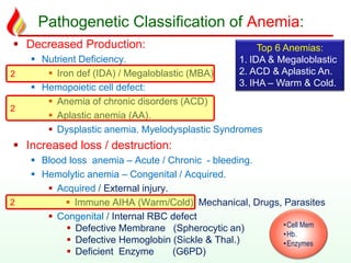
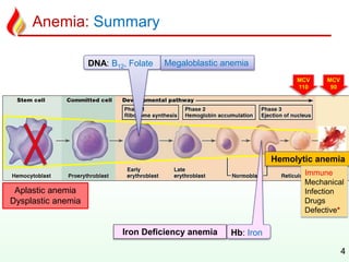
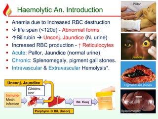
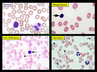
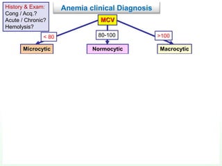
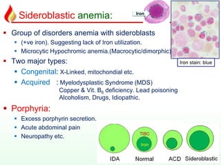
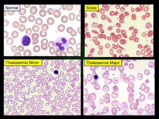
….. foundation of clinical medicine
Shashidhar Venkatesh Murthy
A/Prof & Head of Pathology
College of Medicine & Dentistry
Clinical Pathology:
RBC 1.3: Anemia Summary
CPC : Term2 Week1 - Haem 1/2.
System : Haematology - RBC Disorders.
Topic : 1: Anemia Intro 2: IDA, MBA & ACD 3: Acquired HA 4: Congenital HA. 5: Others.
Pathogenetic Classification of Anemia:
 Decreased Production:
 Nutrient Deficiency.
 Iron def (IDA) / Megaloblastic (MBA)
 Hemopoietic cell defect:
 Anemia of chronic disorders (ACD)
 Aplastic anemia (AA).
 Dysplastic anemia. Myelodysplastic Syndromes
 Increased loss / destruction:
 Blood loss anemia – Acute / Chronic - bleeding.
 Hemolytic anemia – Congenital / Acquired.
 Acquired / External injury.
 Immune AIHA (Warm/Cold) Mechanical, Drugs, Parasites
 Congenital / Internal RBC defect
 Defective Membrane (Spherocytic an)
 Defective Hemoglobin (Sickle & Thal.)
 Deficient Enzyme (G6PD)
2
•Cell Mem
•Hb.
•Enzymes
Top 6 Anemias:
1. IDA & Megaloblastic
2. ACD & Aplastic An.
3. IHA – Warm & Cold.
2
2
Anemia: Summary
4
MCV
90
MCV
110
DNA: B12, Folate
Hb: Iron
Megaloblastic anemia
Iron Deficiency anemia
Aplastic anemia
Dysplastic anemia
Hemolytic anemia
Immune
Mechanical
Infection
Drugs
Defective*
Haemolytic An. Introduction
 Anemia due to Increased RBC destruction
  life span (<120d) - Abnormal forms
 Bilirubin  Unconj. Jaundice (N. urine)
 Increased RBC production - ↑ Reticulocytes
 Acute: Pallor, Jaundice (normal urine)
 Chronic: Splenomegaly, pigment gall stones.
 Intravascular & Extravascular Hemolysis*.
Unconj. Jaundice
Immune
Mech.
Infection
Porphyrin  Bil. Unconj
Globins
Iron
Bil. Conj
Jaundice
Splenomegaly
Pigment Gall stones
Pallor
Iron Deficiency
Megaloblastic
Hemolytic
Normal
MCV
Microcytic Normocytic Macrocytic
Iron studies - Ferritin
Low Normal/high
IDA ACD / Thalassemia
Reticulocyte count
high low ACD /
Aplastic anemia
Hemolytic anemia or
blood loss
Measure B12 + folate
Megaloblastic
Normal Low
Anemia clinical DiagnosisHistory & Exam:
Cong / Acq.?
Acute / Chronic?
Hemolysis?
< 80 80-100 >100
Sideroblastic anemia:
 Group of disorders anemia with sideroblasts
 (+ve iron). Suggesting lack of Iron utilization.
 Microcytic Hypochromic anemia.(Macrocytic/dimorphic)
 Two major types:
 Congenital: X-Linked, mitochondial etc.
 Acquired : Myelodysplastic Syndrome (MDS)
Copper & Vit. B6 deficiency. Lead poisoning
Alcoholism, Drugs, Idiopathic.
 Porphyria:
 Excess porphyrin secretion.
 Acute abdominal pain
 Neuropathy etc.
Iron
Iron stain: blue
Polycythemia: (high Hb)
 Relative or spurious erythrocytosis
 Dehydration: Diarrhea, vomiting, diuretics,
excess alcohol. etc.
 Absolute erythrocytosis (True ):
 Secondary: Tissue Hypoxia: Smoking (CO),
High altitude, Lung disease. Cardiac shunts,
High O2 affinity Hb.
High Erythropoietin – Paraneoplastic
Syndromes, Androgen therapy.
 Primary - Polycythemia Rubra vera:
Myeloproliferative disorder: Neoplastic
proliferation of erythroid cells in bone
marrow – old age, hepatosplenomegaly.
10
 Hb, skin flushing &
Hepatosplenomegaly
Need help? contact me…
1. Office location: DB39-136 (Townsville)
2. Office Tel: 4781 4566
3. Email: venkatesh.shashidhar@jcu.edu.au
4. Emergency?: 0416933704
Need personal coaching?
Email me for an appointment.
You are the stone..
Be true to your work,
your word, & your friend.
-- Henry David Thoreau
Integrity is doing the right thing when no one is watching….!
Thalassemia Minor
Sickle
Thalassemia Major
Normal
Haem15 - Anemia conclusions & Polycythemia

Haem15 - Anemia conclusions & Polycythemia

  • 2.
    ….. foundation ofclinical medicine Shashidhar Venkatesh Murthy A/Prof & Head of Pathology College of Medicine & Dentistry Clinical Pathology: RBC 1.3: Anemia Summary CPC : Term2 Week1 - Haem 1/2. System : Haematology - RBC Disorders. Topic : 1: Anemia Intro 2: IDA, MBA & ACD 3: Acquired HA 4: Congenital HA. 5: Others.
  • 3.
    Pathogenetic Classification ofAnemia:  Decreased Production:  Nutrient Deficiency.  Iron def (IDA) / Megaloblastic (MBA)  Hemopoietic cell defect:  Anemia of chronic disorders (ACD)  Aplastic anemia (AA).  Dysplastic anemia. Myelodysplastic Syndromes  Increased loss / destruction:  Blood loss anemia – Acute / Chronic - bleeding.  Hemolytic anemia – Congenital / Acquired.  Acquired / External injury.  Immune AIHA (Warm/Cold) Mechanical, Drugs, Parasites  Congenital / Internal RBC defect  Defective Membrane (Spherocytic an)  Defective Hemoglobin (Sickle & Thal.)  Deficient Enzyme (G6PD) 2 •Cell Mem •Hb. •Enzymes Top 6 Anemias: 1. IDA & Megaloblastic 2. ACD & Aplastic An. 3. IHA – Warm & Cold. 2 2
  • 4.
    Anemia: Summary 4 MCV 90 MCV 110 DNA: B12,Folate Hb: Iron Megaloblastic anemia Iron Deficiency anemia Aplastic anemia Dysplastic anemia Hemolytic anemia Immune Mechanical Infection Drugs Defective*
  • 5.
    Haemolytic An. Introduction Anemia due to Increased RBC destruction   life span (<120d) - Abnormal forms  Bilirubin  Unconj. Jaundice (N. urine)  Increased RBC production - ↑ Reticulocytes  Acute: Pallor, Jaundice (normal urine)  Chronic: Splenomegaly, pigment gall stones.  Intravascular & Extravascular Hemolysis*. Unconj. Jaundice Immune Mech. Infection Porphyrin  Bil. Unconj Globins Iron Bil. Conj Jaundice Splenomegaly Pigment Gall stones Pallor
  • 6.
  • 7.
    MCV Microcytic Normocytic Macrocytic Ironstudies - Ferritin Low Normal/high IDA ACD / Thalassemia Reticulocyte count high low ACD / Aplastic anemia Hemolytic anemia or blood loss Measure B12 + folate Megaloblastic Normal Low Anemia clinical DiagnosisHistory & Exam: Cong / Acq.? Acute / Chronic? Hemolysis? < 80 80-100 >100
  • 9.
    Sideroblastic anemia:  Groupof disorders anemia with sideroblasts  (+ve iron). Suggesting lack of Iron utilization.  Microcytic Hypochromic anemia.(Macrocytic/dimorphic)  Two major types:  Congenital: X-Linked, mitochondial etc.  Acquired : Myelodysplastic Syndrome (MDS) Copper & Vit. B6 deficiency. Lead poisoning Alcoholism, Drugs, Idiopathic.  Porphyria:  Excess porphyrin secretion.  Acute abdominal pain  Neuropathy etc. Iron Iron stain: blue
  • 10.
    Polycythemia: (high Hb) Relative or spurious erythrocytosis  Dehydration: Diarrhea, vomiting, diuretics, excess alcohol. etc.  Absolute erythrocytosis (True ):  Secondary: Tissue Hypoxia: Smoking (CO), High altitude, Lung disease. Cardiac shunts, High O2 affinity Hb. High Erythropoietin – Paraneoplastic Syndromes, Androgen therapy.  Primary - Polycythemia Rubra vera: Myeloproliferative disorder: Neoplastic proliferation of erythroid cells in bone marrow – old age, hepatosplenomegaly. 10  Hb, skin flushing & Hepatosplenomegaly
  • 11.
    Need help? contactme… 1. Office location: DB39-136 (Townsville) 2. Office Tel: 4781 4566 3. Email: venkatesh.shashidhar@jcu.edu.au 4. Emergency?: 0416933704 Need personal coaching? Email me for an appointment. You are the stone..
  • 12.
    Be true toyour work, your word, & your friend. -- Henry David Thoreau Integrity is doing the right thing when no one is watching….!
  • 13.