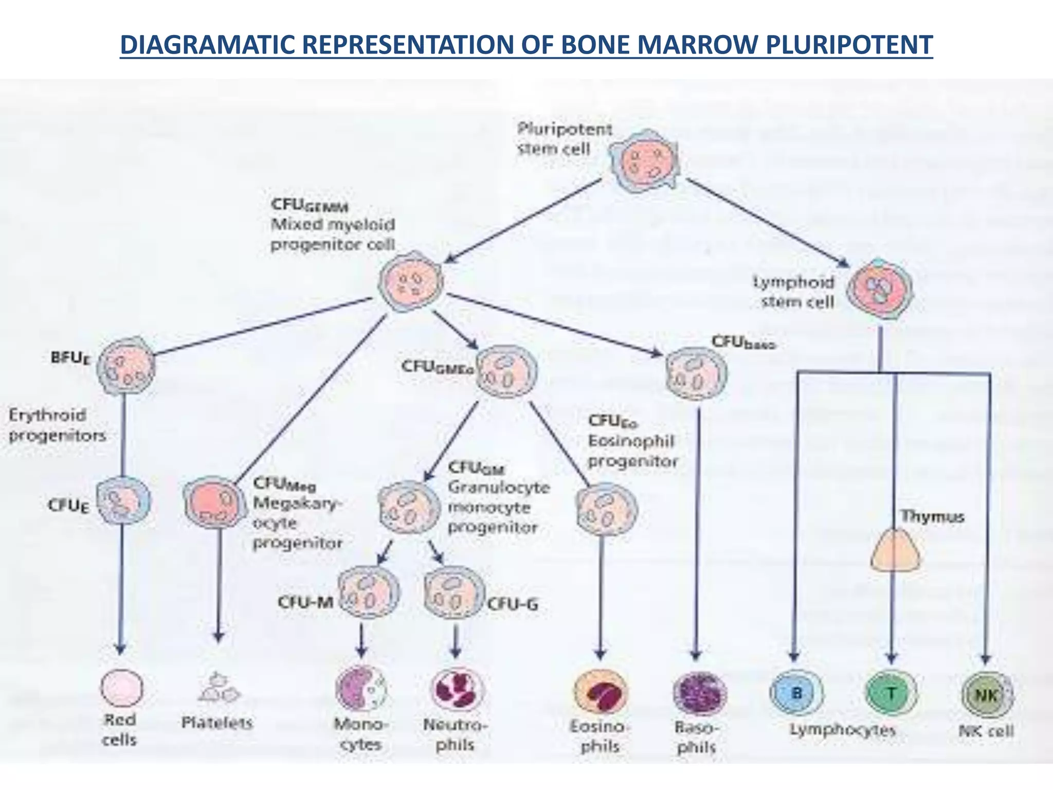
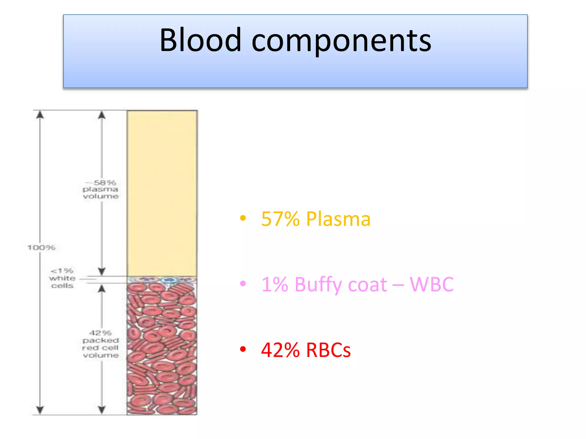
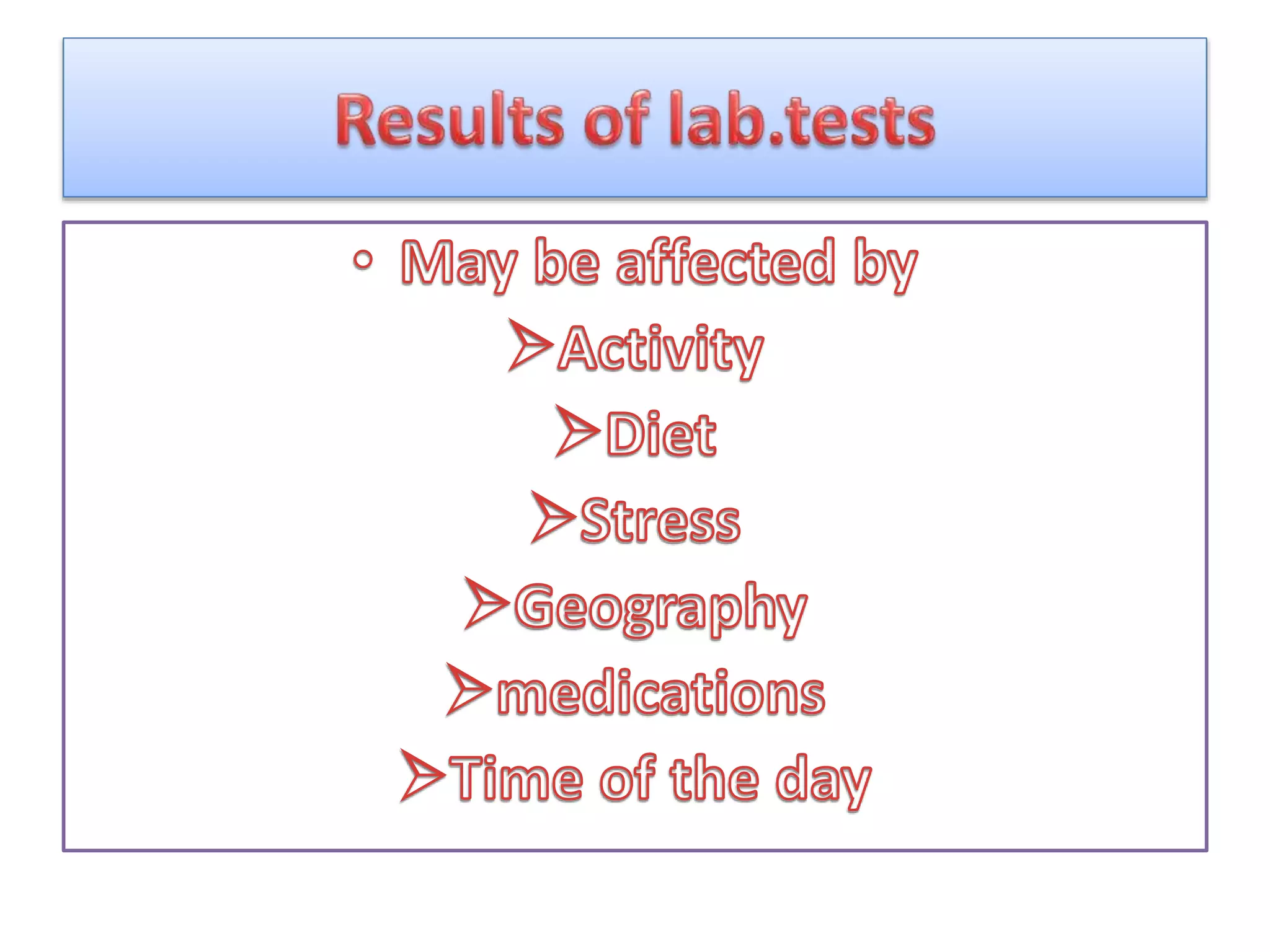
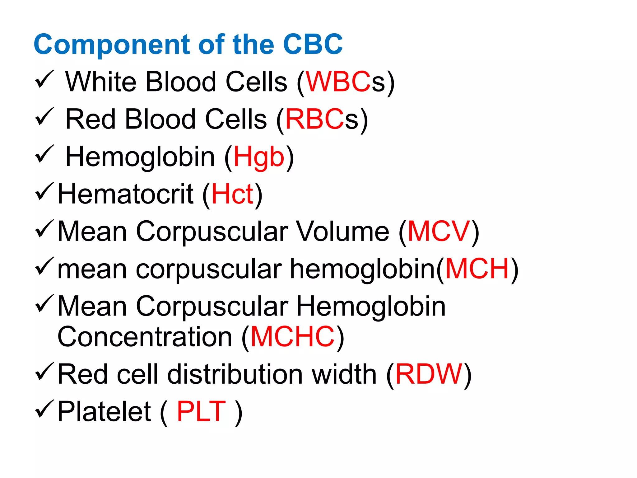
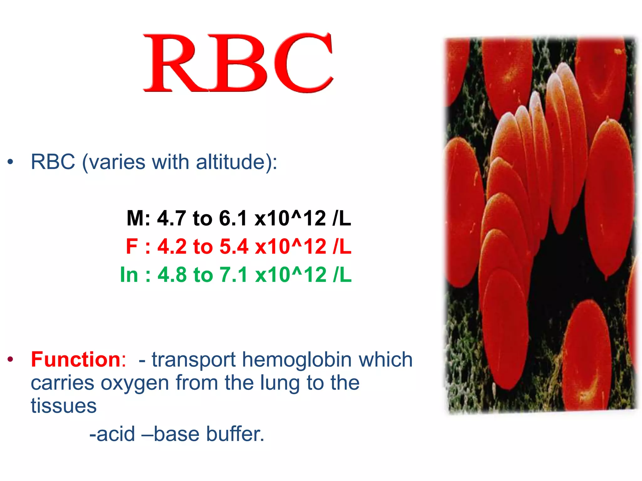
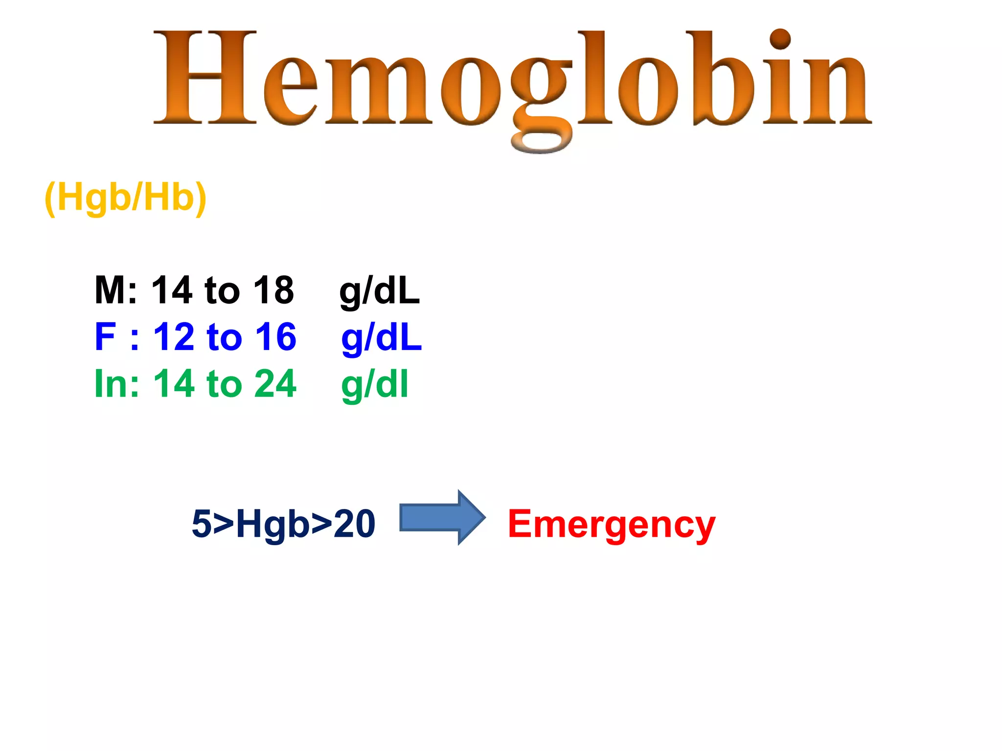
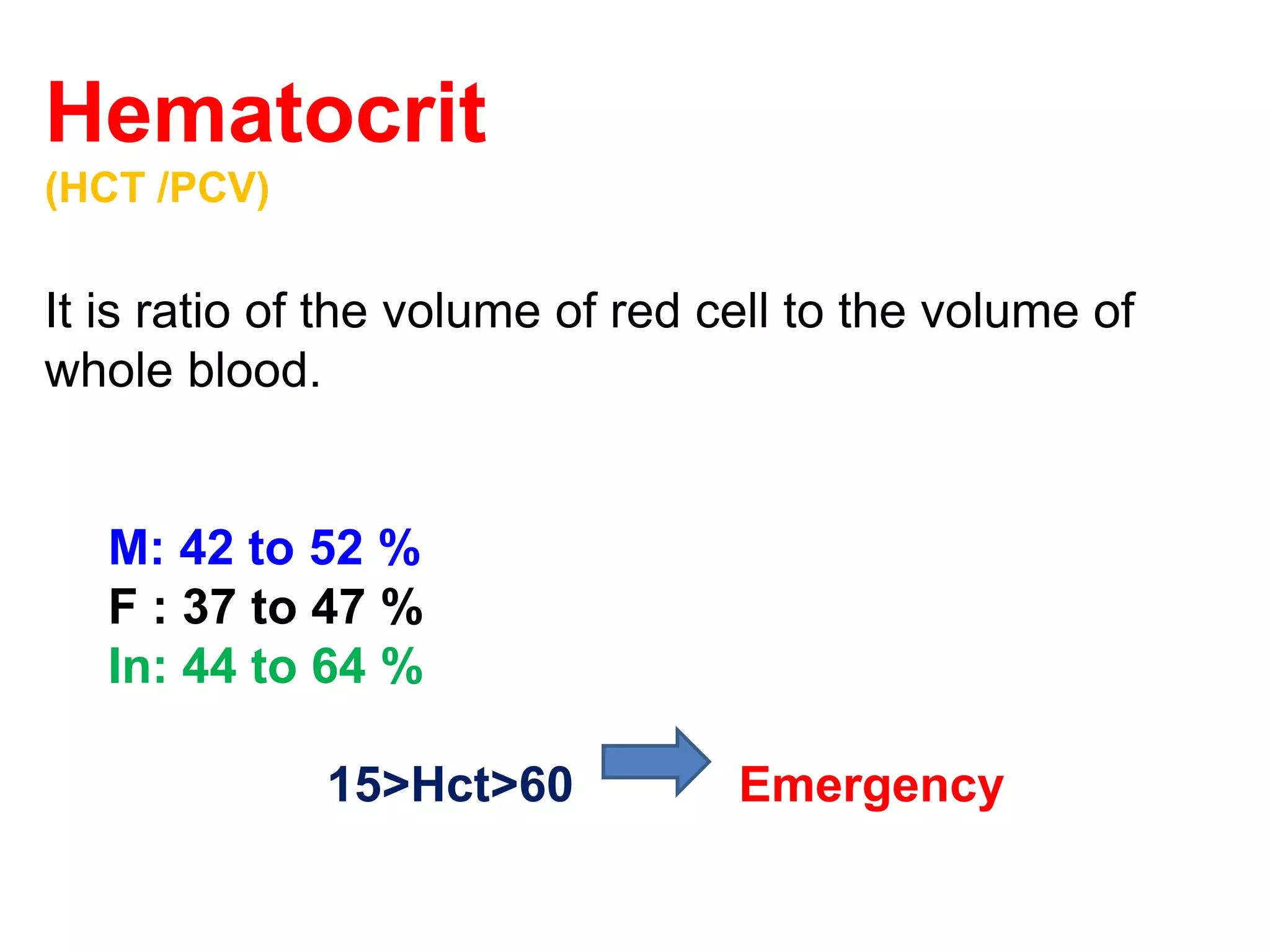
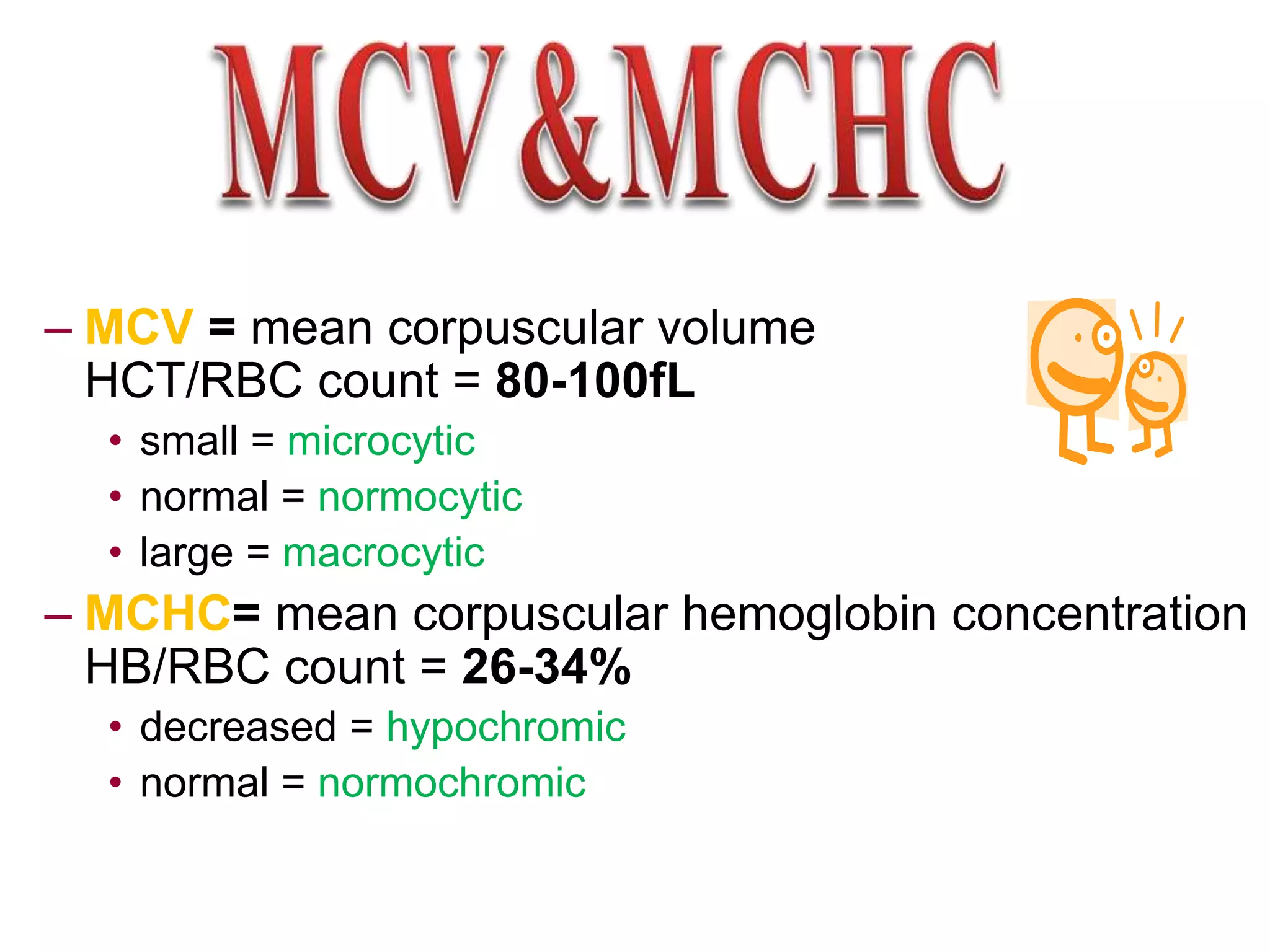
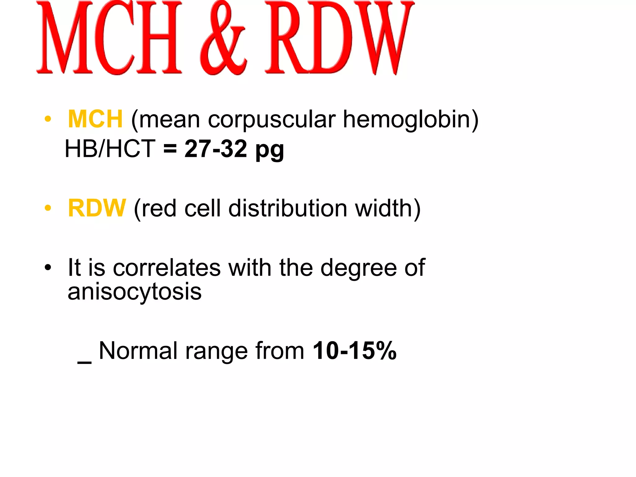
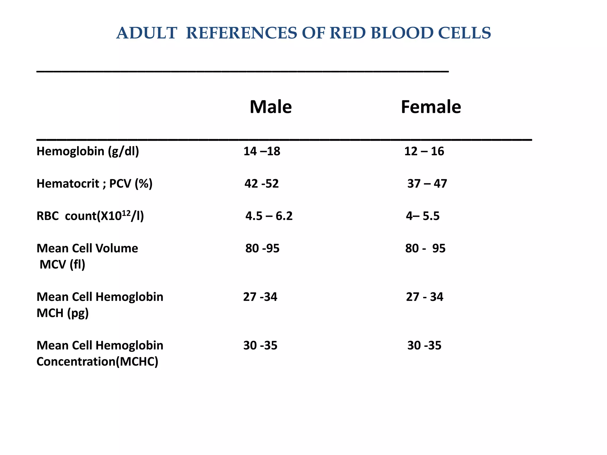
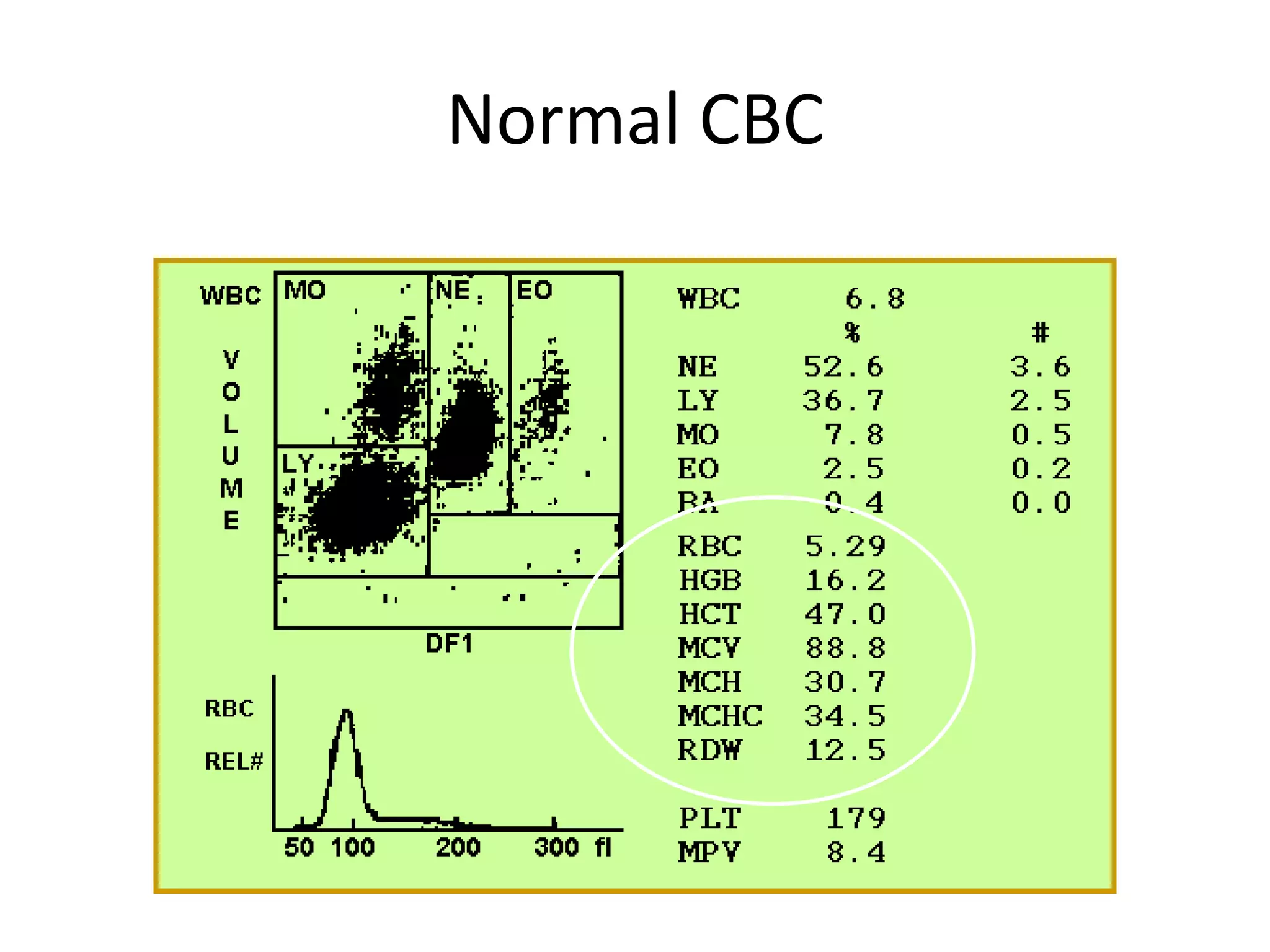
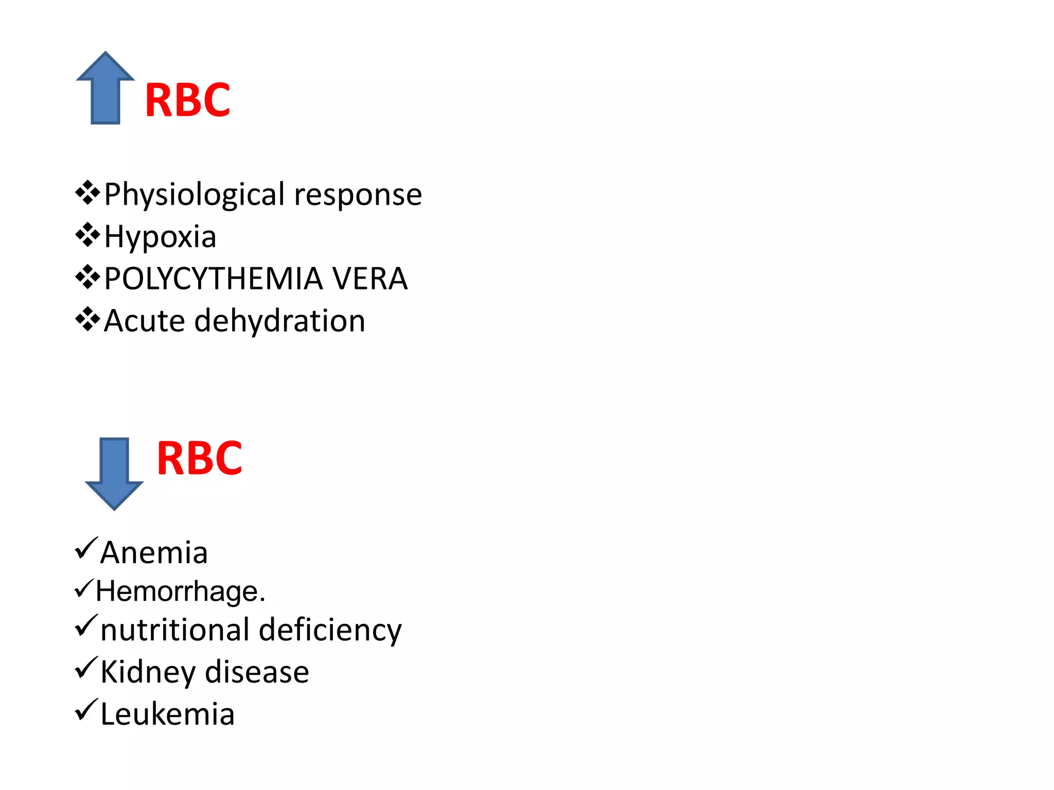
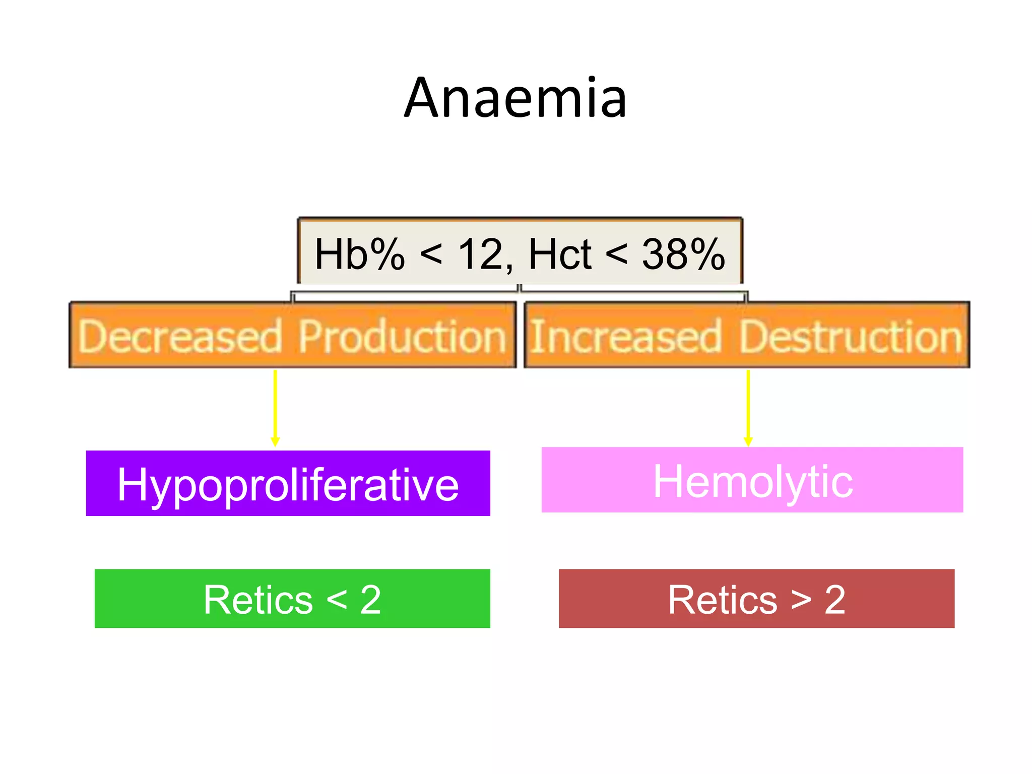
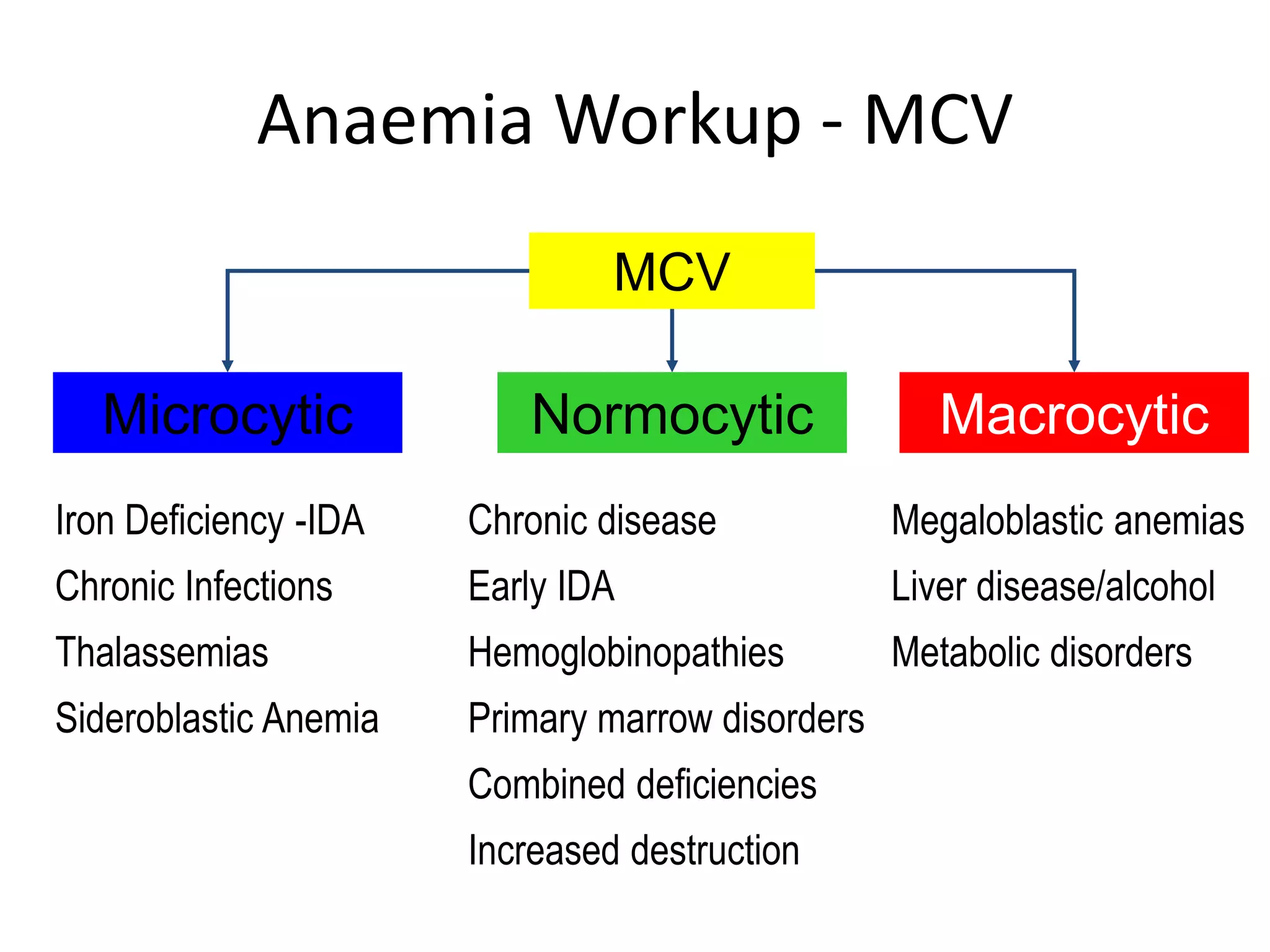
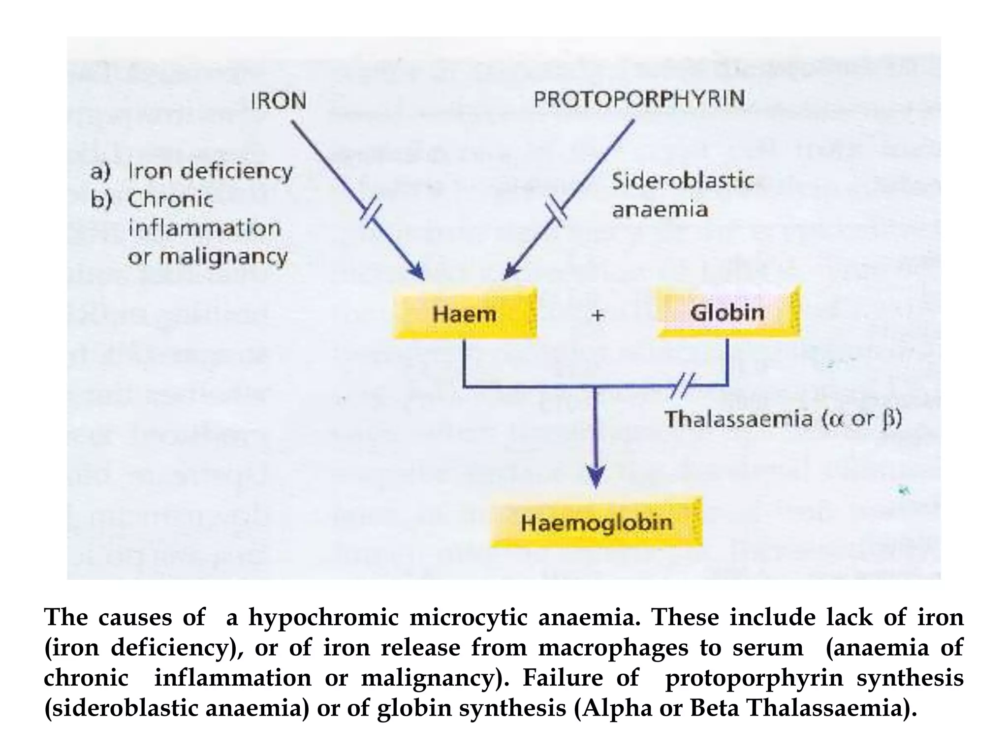
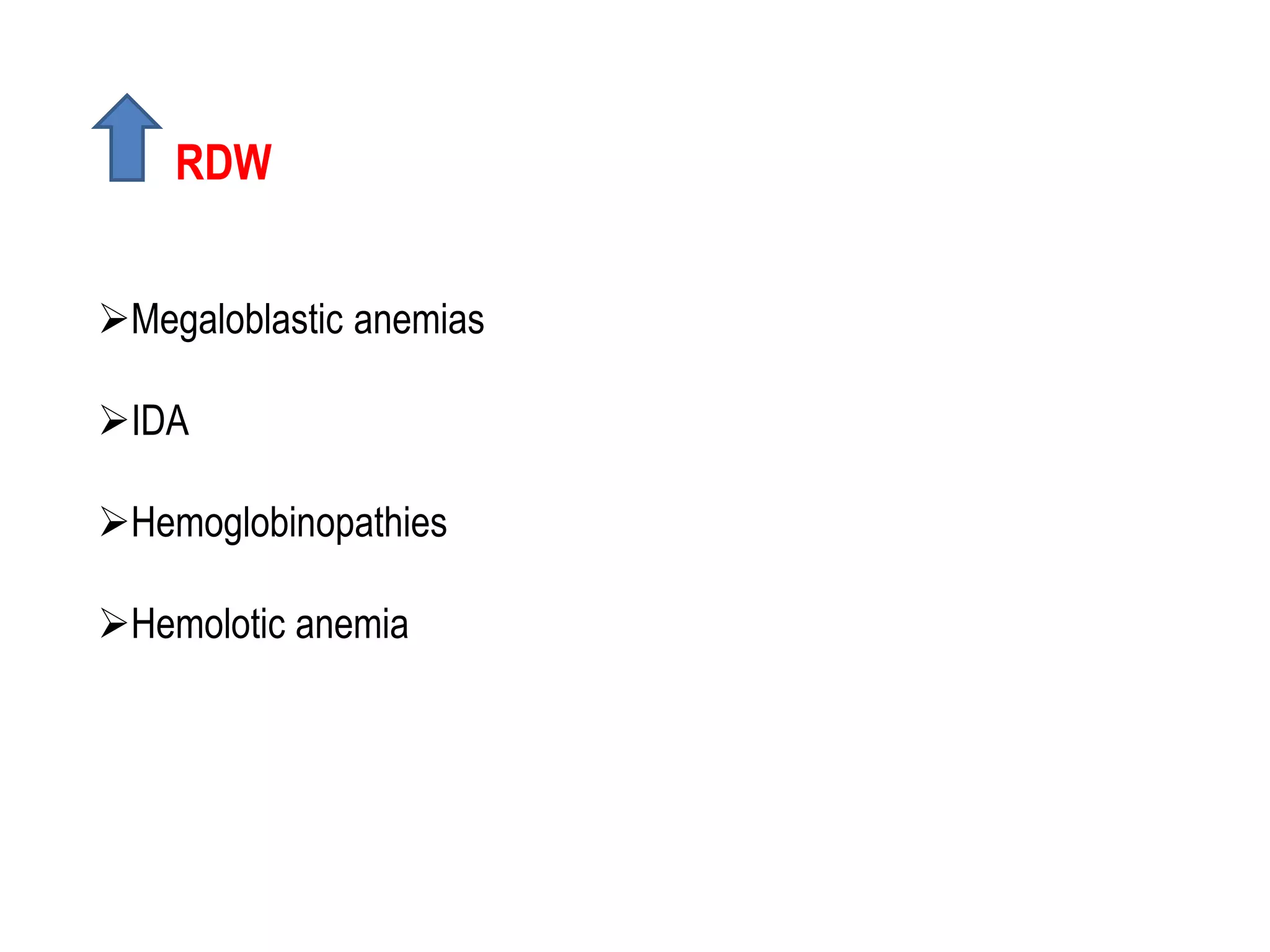
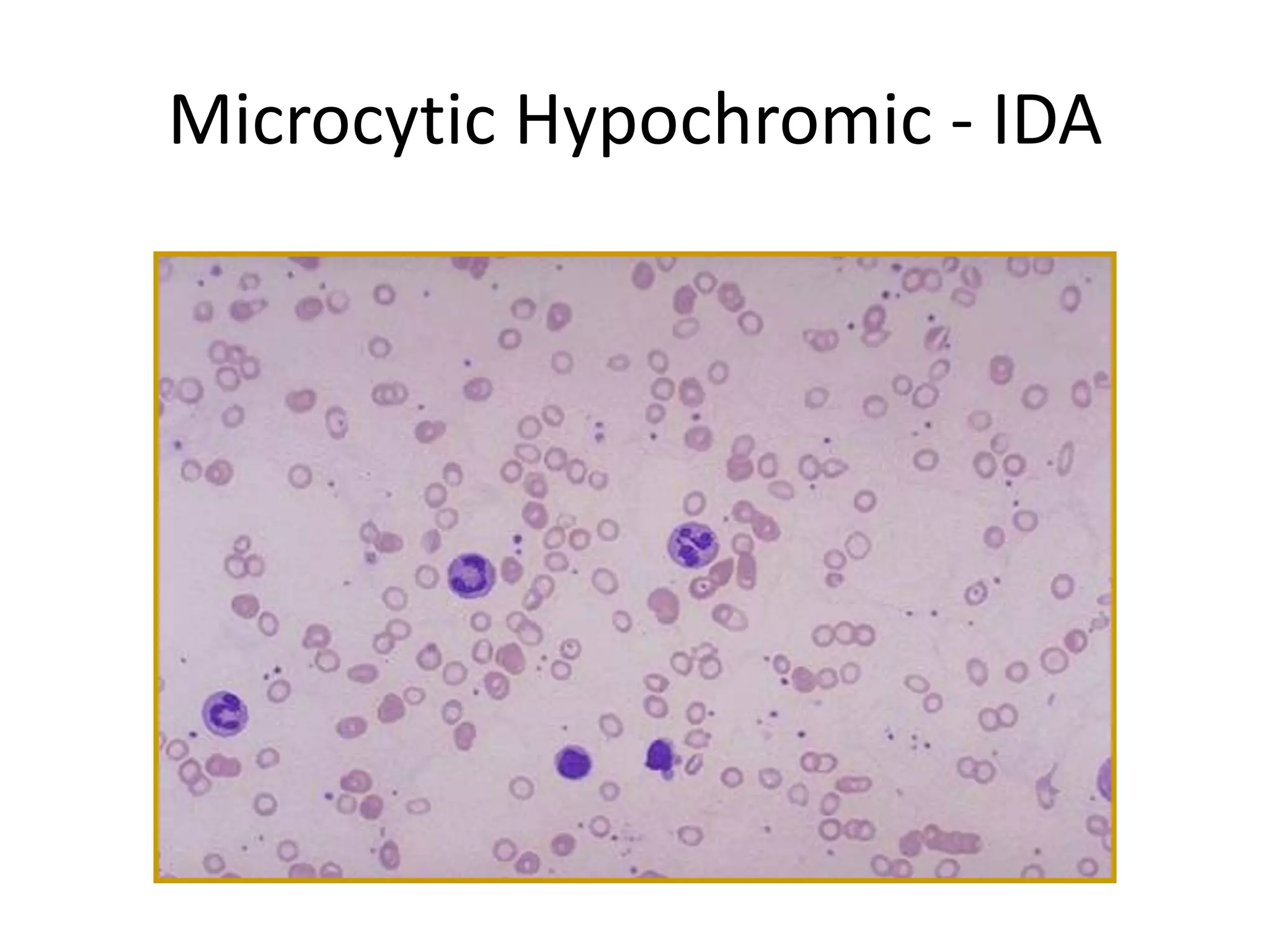
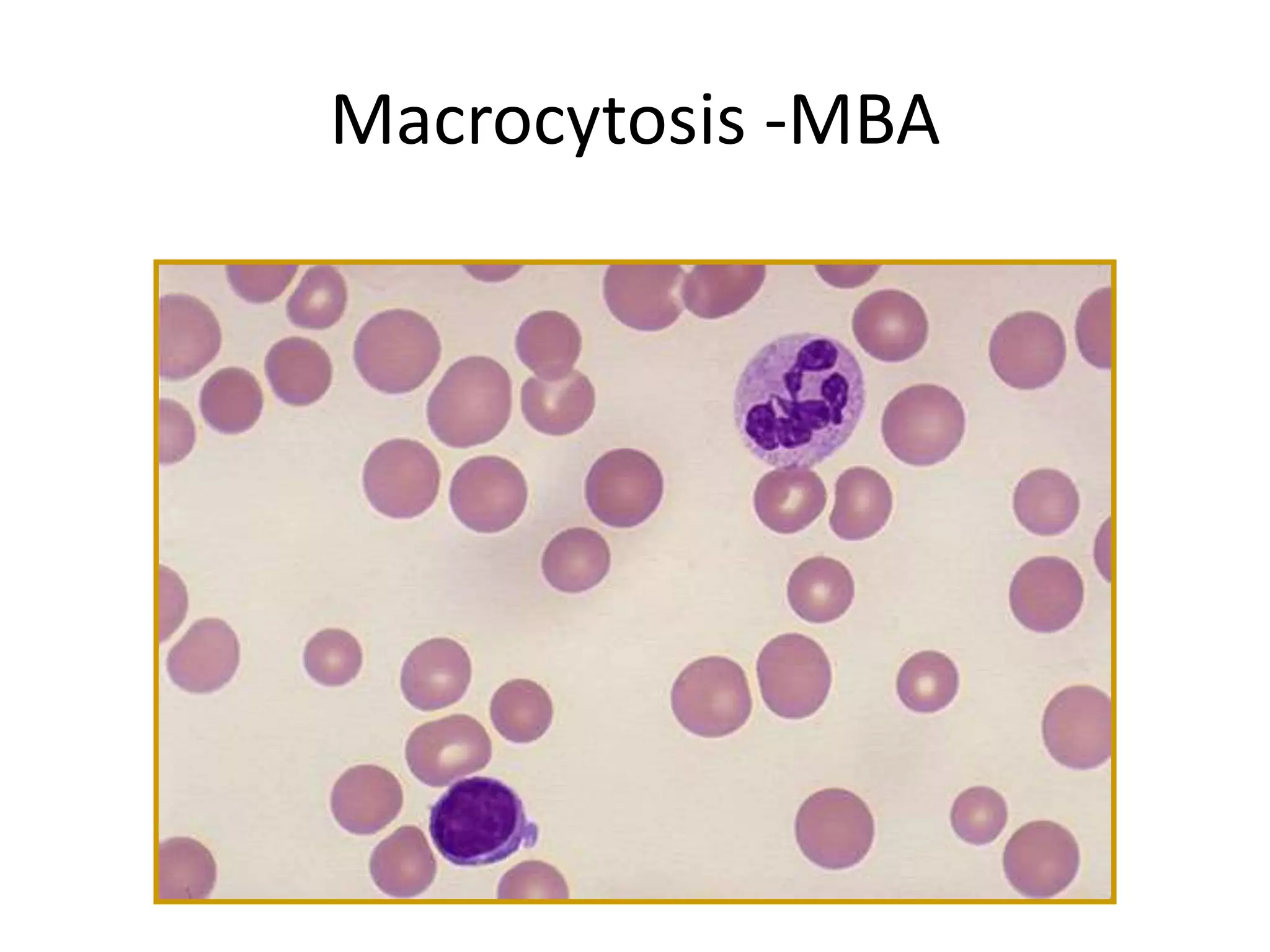
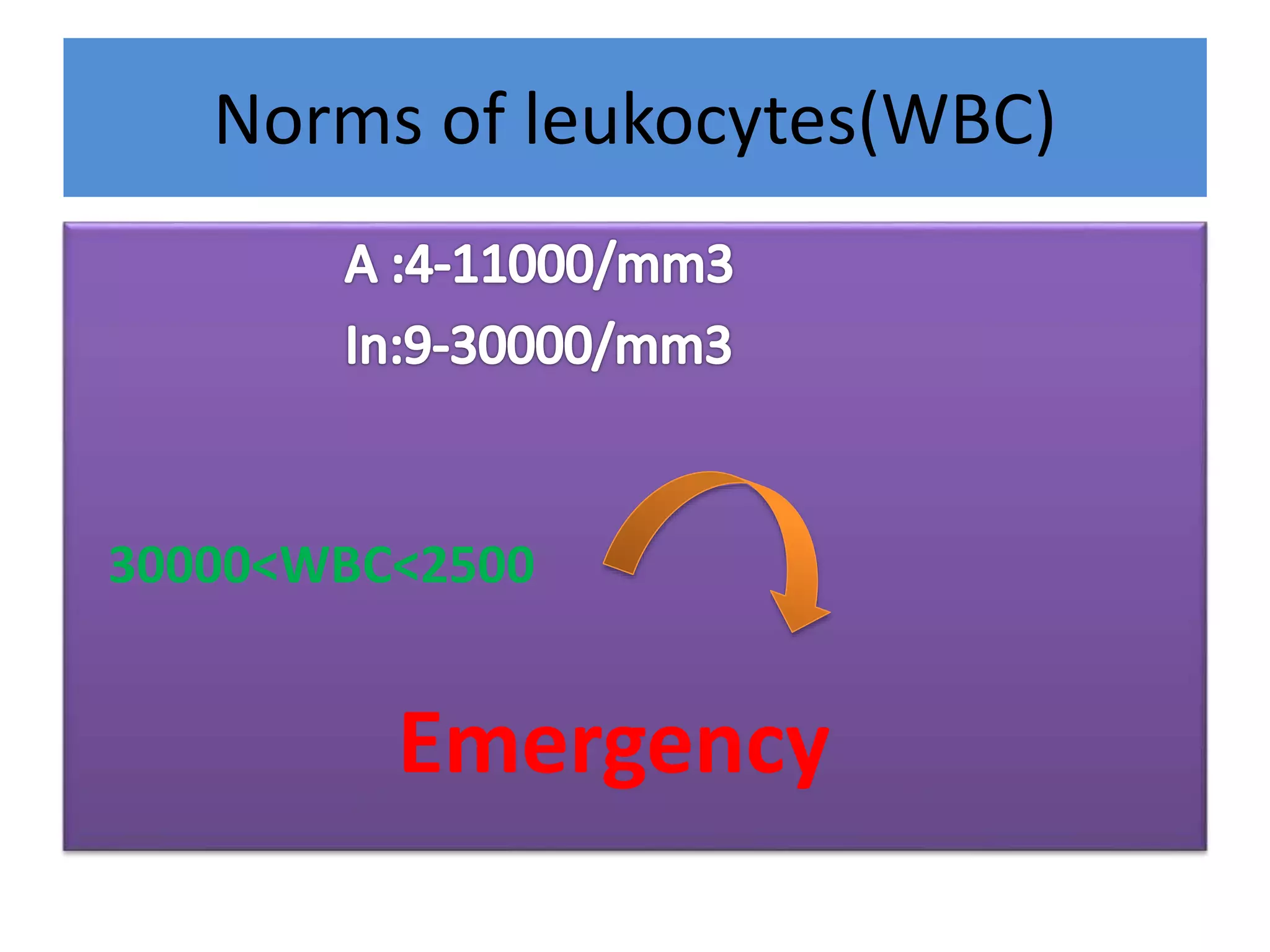
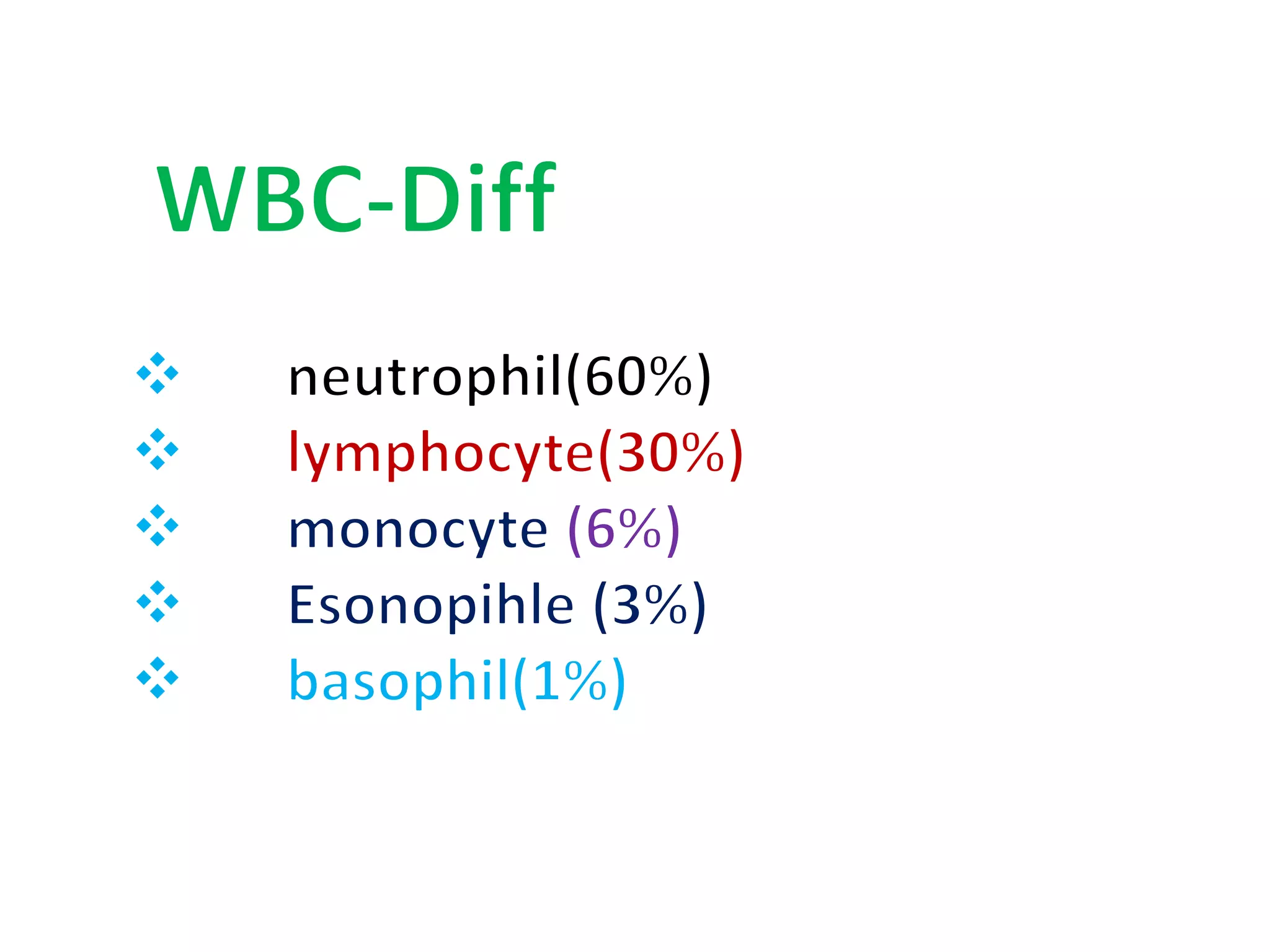
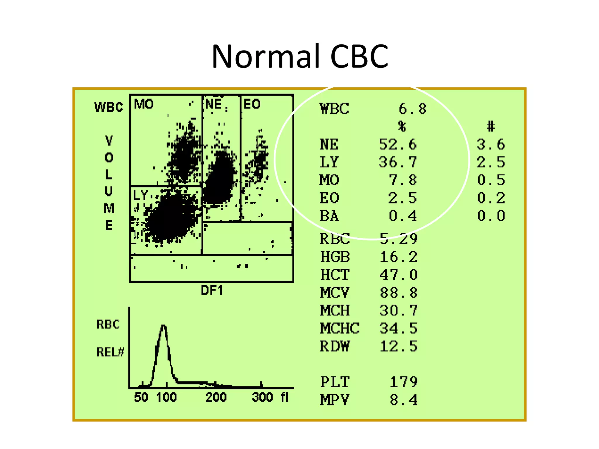
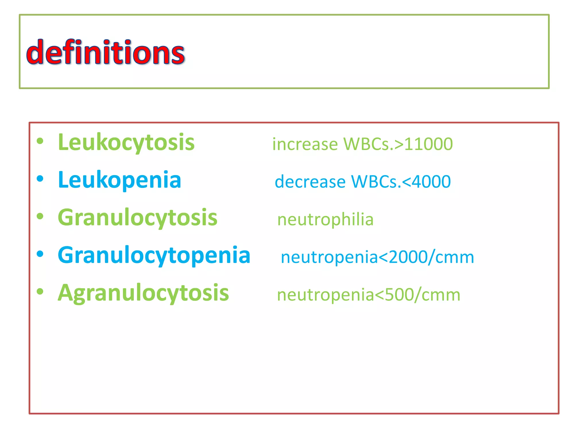
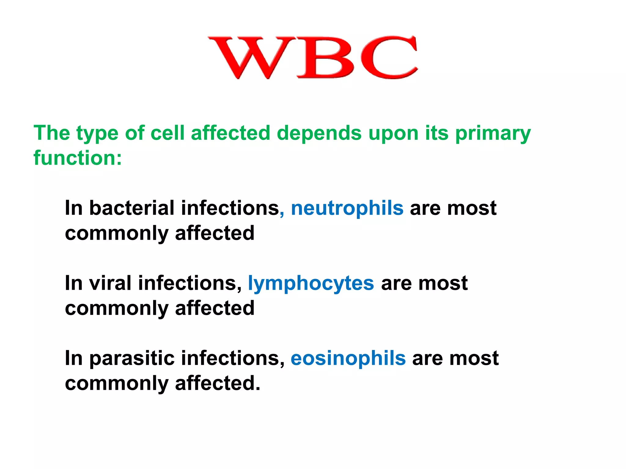
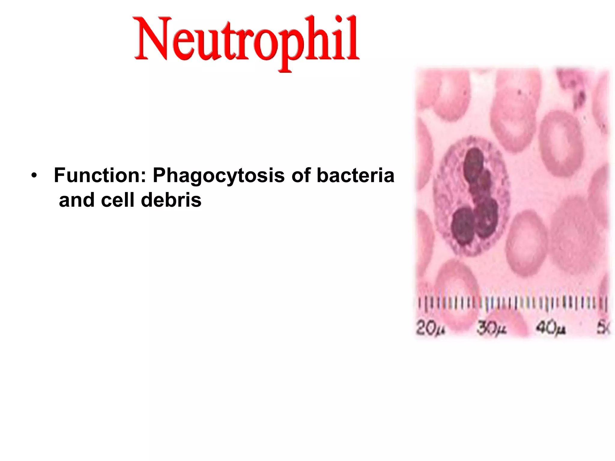
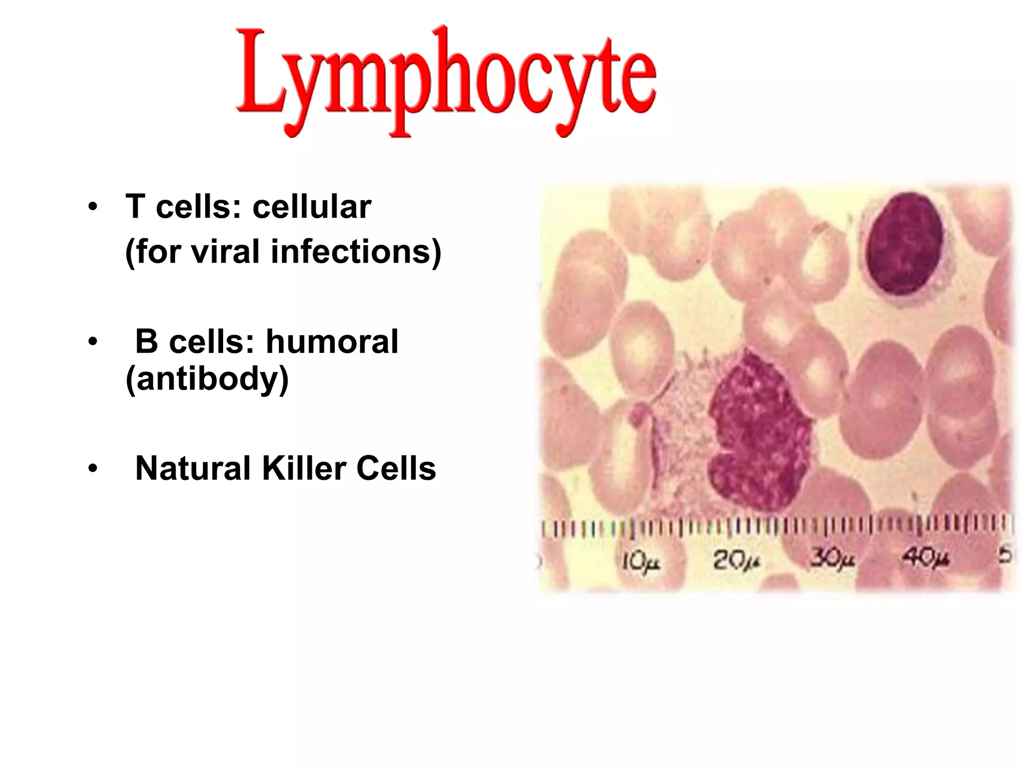
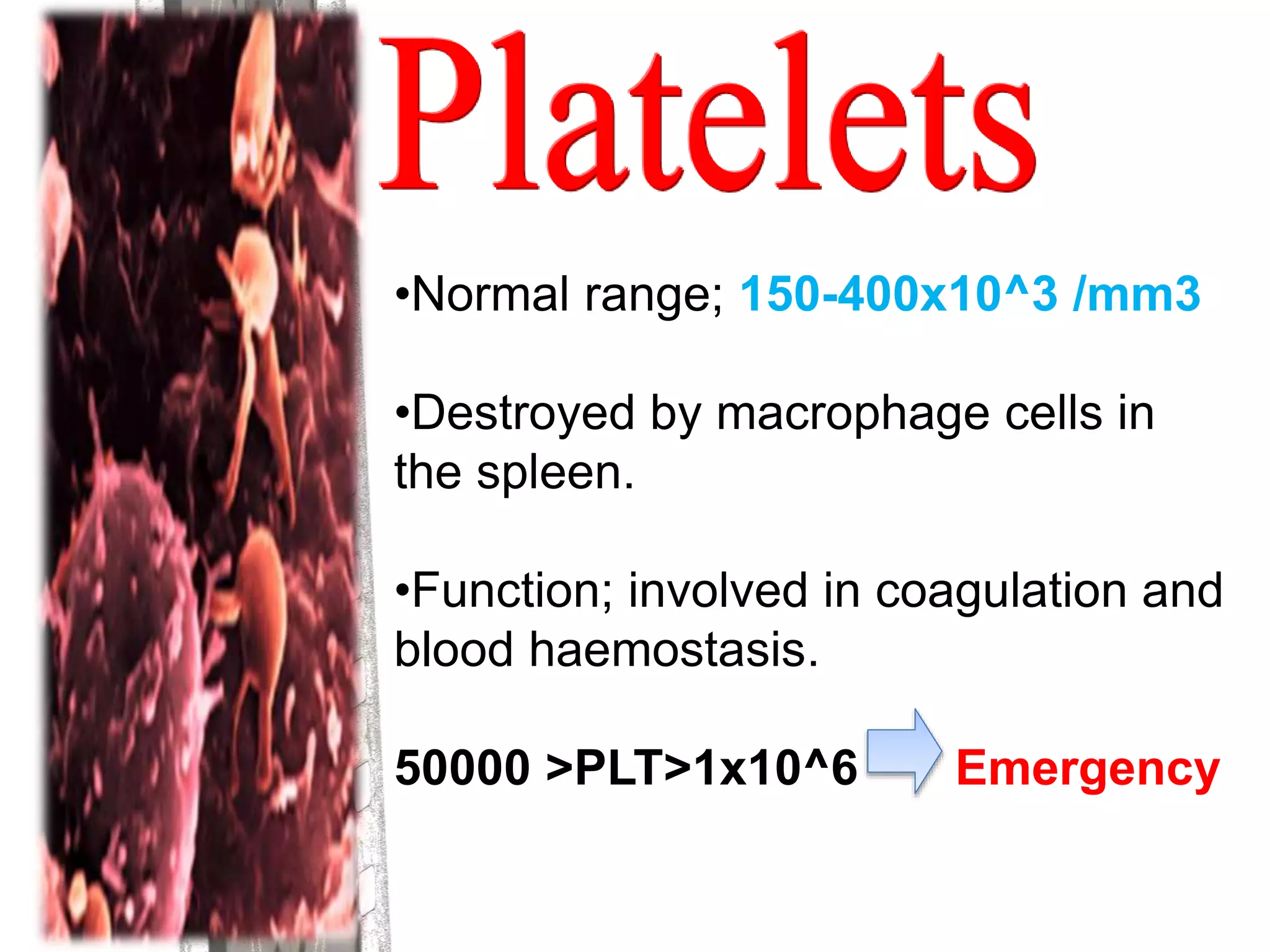
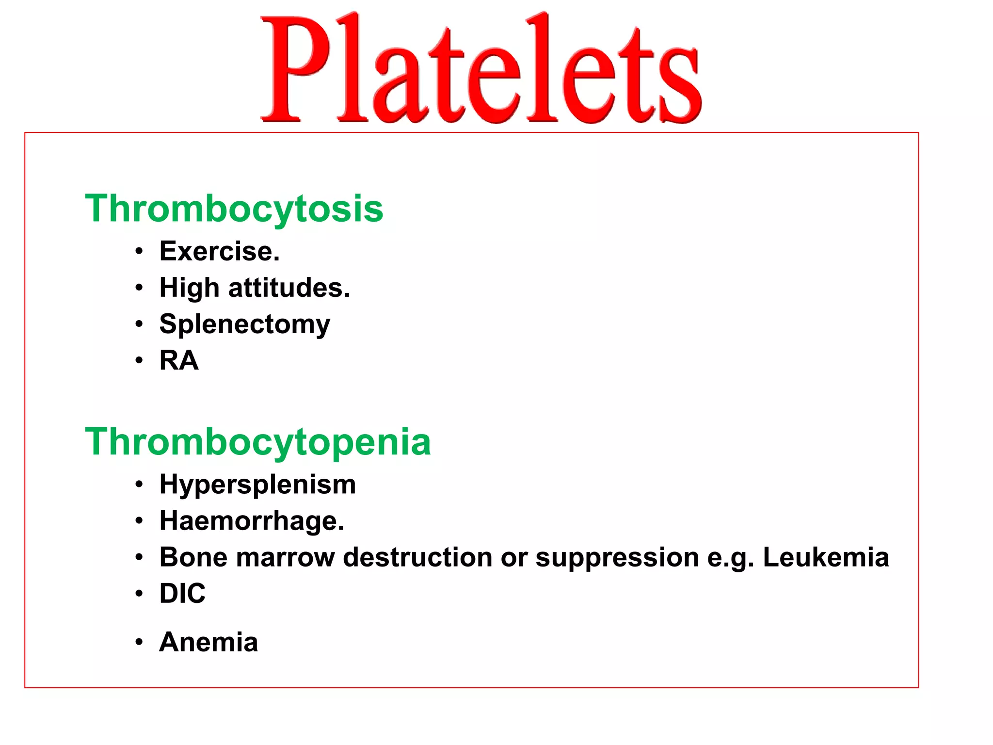
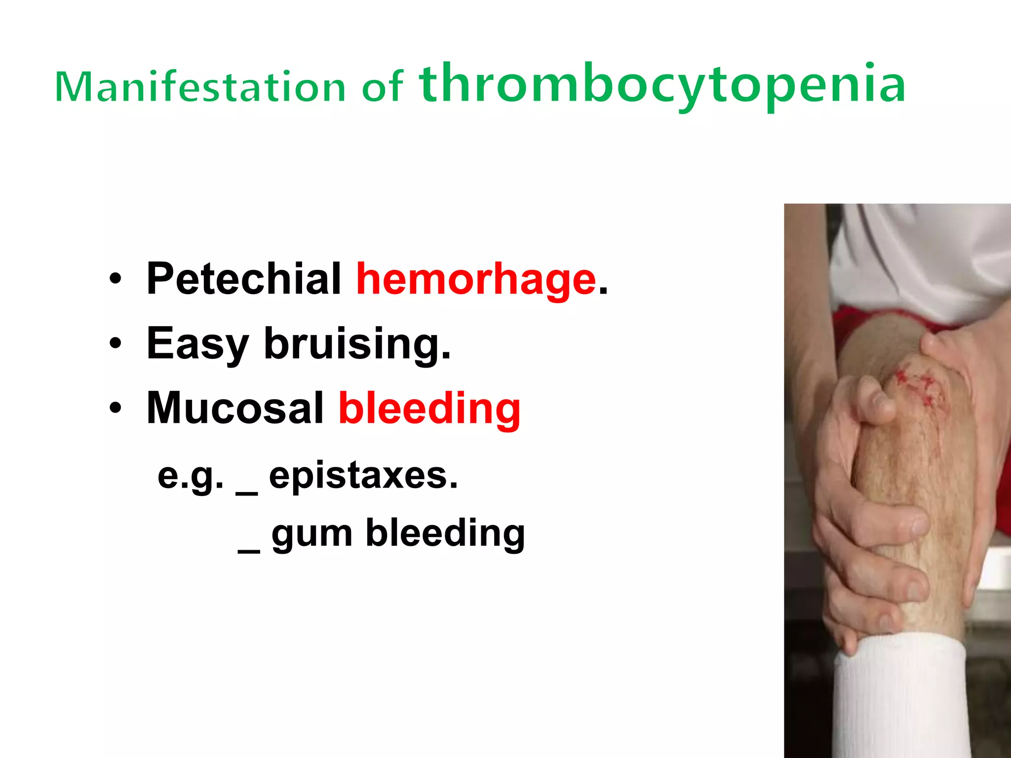
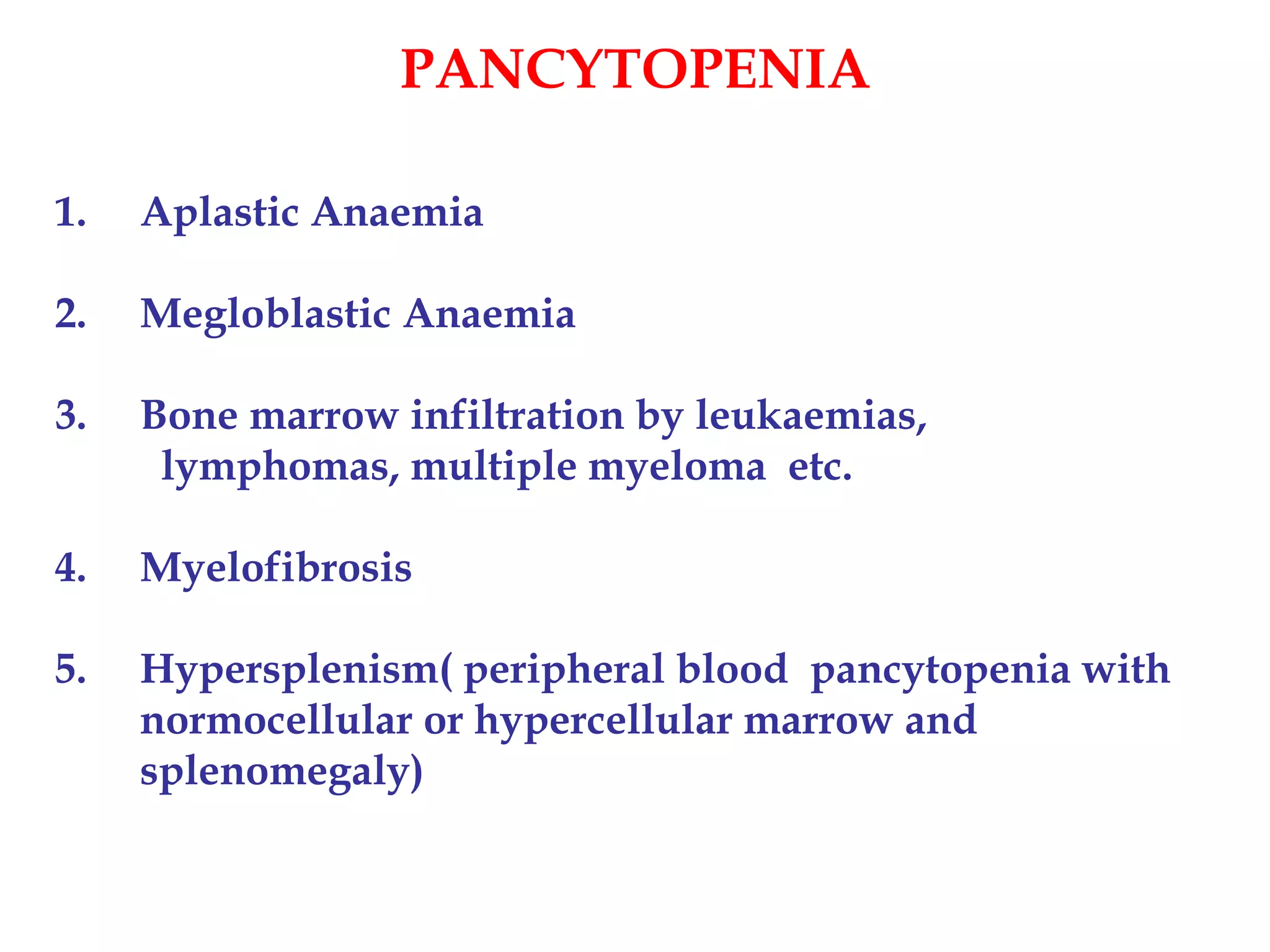
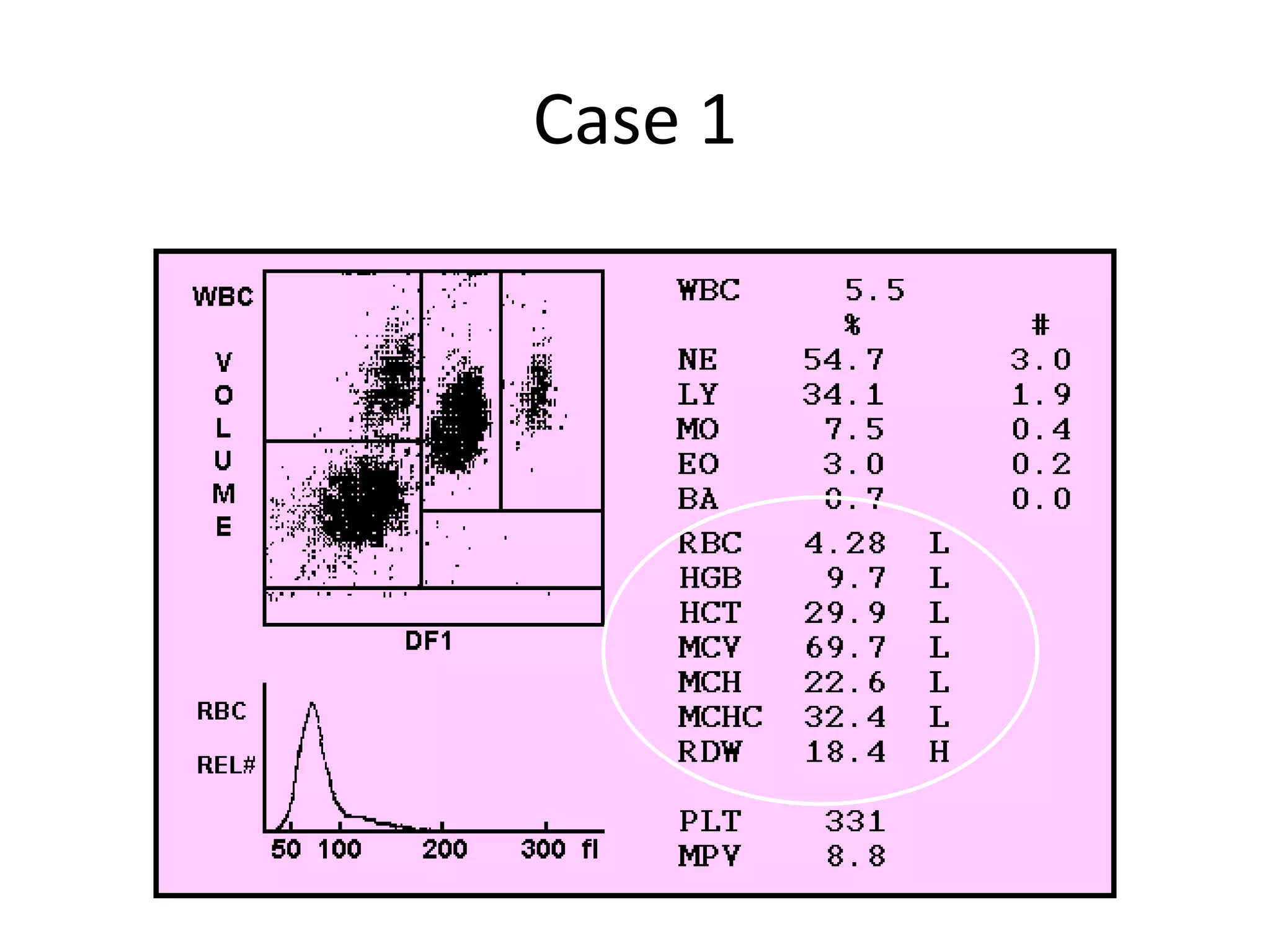
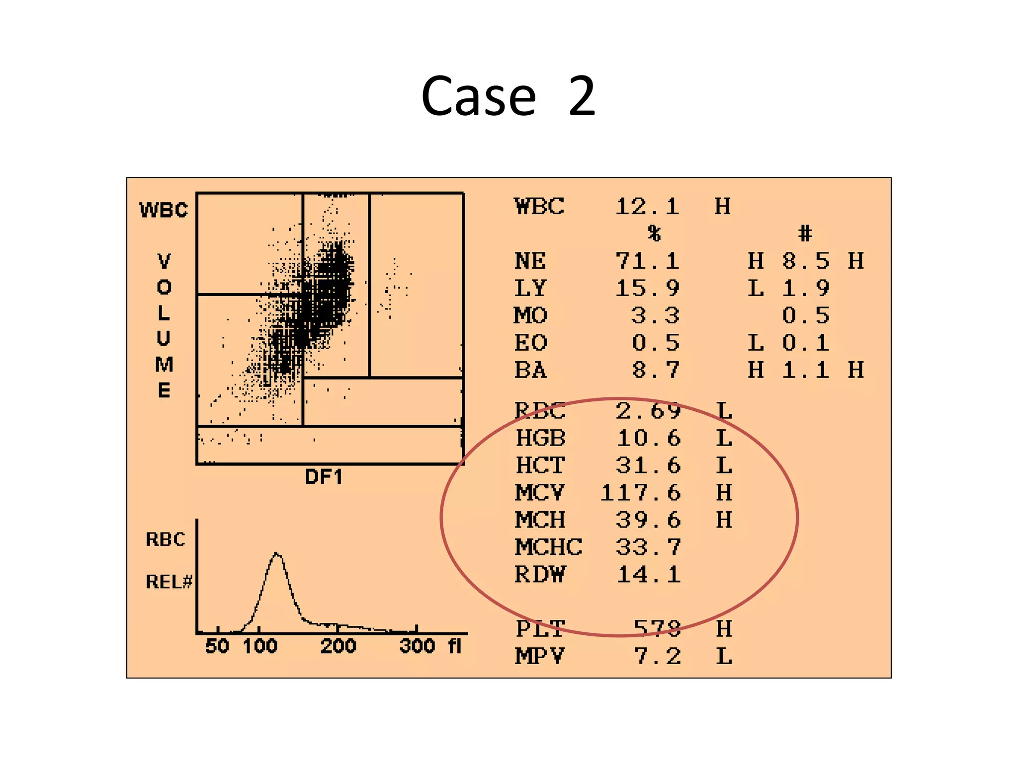
بسم الله الرحمن الرحیم 
In the hgd name ال 
of 
ALLAH
DIAGRAMATIC REPRESENTATION OF BONE MARROW PLURIPOTENT
Blood components 
PCV or Hematocrit 
• 57% Plasma 
• 1% Buffy coat – WBC 
• 42% RBCs
Interpretation of 
CBC- complete blood count
Component of the CBC 
 White Blood Cells (WBCs) 
 Red Blood Cells (RBCs) 
 Hemoglobin (Hgb) 
Hematocrit (Hct) 
Mean Corpuscular Volume (MCV) 
mean corpuscular hemoglobin(MCH) 
Mean Corpuscular Hemoglobin 
Concentration (MCHC) 
Red cell distribution width (RDW) 
Platelet ( PLT )
• RBC (varies with altitude): 
M: 4.7 to 6.1 x10^12 /L 
F : 4.2 to 5.4 x10^12 /L 
In : 4.8 to 7.1 x10^12 /L 
• Function: - transport hemoglobin which 
carries oxygen from the lung to the 
tissues 
-acid –base buffer.
(Hgb/Hb) 
M: 14 to 18 g/dL 
F : 12 to 16 g/dL 
In: 14 to 24 g/dl 
5>Hgb>20 Emergency
Hematocrit 
(HCT /PCV) 
It is ratio of the volume of red cell to the volume of 
whole blood. 
M: 42 to 52 % 
F : 37 to 47 % 
In: 44 to 64 % 
15>Hct>60 Emergency
– MCV = mean corpuscular volume 
HCT/RBC count = 80-100fL 
• small = microcytic 
• normal = normocytic 
• large = macrocytic 
– MCHC= mean corpuscular hemoglobin concentration 
HB/RBC count = 26-34% 
• decreased = hypochromic 
• normal = normochromic
• MCH (mean corpuscular hemoglobin) 
HB/HCT = 27-32 pg 
• RDW (red cell distribution width) 
• It is correlates with the degree of 
anisocytosis 
_ Normal range from 10-15%
ADULT REFERENCES OF RED BLOOD CELLS 
_________________________________________________ 
Male Female 
_________________________________________________ 
Hemoglobin (g/dl) 14 –18 12 – 16 
Hematocrit ; PCV (%) 42 -52 37 – 47 
RBC count(X1012/l) 4.5 – 6.2 4– 5.5 
Mean Cell Volume 80 -95 80 - 95 
MCV (fl) 
Mean Cell Hemoglobin 27 -34 27 - 34 
MCH (pg) 
Mean Cell Hemoglobin 30 -35 30 -35 
Concentration(MCHC)
Normal CBC
RBC 
Physiological response 
Hypoxia 
POLYCYTHEMIA VERA 
Acute dehydration 
RBC 
Anemia 
Hemorrhage. 
nutritional deficiency 
Kidney disease 
Leukemia
Anaemia 
Hb% < 12, Hct < 38% 
Hypoproliferative Hemolytic 
Retics < 2 Retics > 2
Anaemia Workup - MCV 
Microcytic 
MCV 
Normocytic Macrocytic 
Iron Deficiency -IDA 
Chronic Infections 
Thalassemias 
Sideroblastic Anemia 
Chronic disease 
Early IDA 
Hemoglobinopathies 
Primary marrow disorders 
Combined deficiencies 
Increased destruction 
Megaloblastic anemias 
Liver disease/alcohol 
Metabolic disorders
The causes of a hypochromic microcytic anaemia. These include lack of iron 
(iron deficiency), or of iron release from macrophages to serum (anaemia of 
chronic inflammation or malignancy). Failure of protoporphyrin synthesis 
(sideroblastic anaemia) or of globin synthesis (Alpha or Beta Thalassaemia).
RDW 
Megaloblastic anemias 
IDA 
Hemoglobinopathies 
Hemolotic anemia
Microcytic Hypochromic - IDA
Macrocytosis -MBA
Norms of leukocytes(WBC) 
30000<WBC<2500 
Emergency
Normal CBC
• Leukocytosis increase WBCs.>11000 
• Leukopenia decrease WBCs.<4000 
• Granulocytosis neutrophilia 
• Granulocytopenia neutropenia<2000/cmm 
• Agranulocytosis neutropenia<500/cmm
The type of cell affected depends upon its primary 
function: 
In bacterial infections, neutrophils are most 
commonly affected 
In viral infections, lymphocytes are most 
commonly affected 
In parasitic infections, eosinophils are most 
commonly affected.
Abnormal result of WBC 
Leukocytosis 
_ Infectious diseases 
_Inflammatory disease(such as RA &allergy) 
_Leukemia 
_Severe emotional or physical stress 
_Tissue damage (e.g. necrosis,or burns)
Leukopenia: 
 Decreased WBC production from BM 
 Irradiation 
 Exposure to chemical or drugs 
 Autoimmune disease 
 Acute infection
• Function: Phagocytosis of bacteria 
and cell debris
Neutrophilia 
– Conditions associated with : 
1-Bacterial infections (most common cause) 
2-Tissue destruction 
e.g. tissue infarctions, burns. 
3- leukemoid reaction 
4-Leukemia
Neutropenia 
– this may result from: 
1-Decreased bone marrow production 
e.g. BM hypoplasia. 
2-Ineffective bone marrow production 
– E.g. megaloblastic anemias and 
myelodysplastic syndromes. 
3- post acute infection 
_ e.g. typhoid fever, brucellosis.
• Function: Involved in allergy 
, parasitic infections
Eosinophilia 
may be found in 
• Parasitic infections 
• Allergic conditions and hypersensitivity 
reaction
Basophilia. 
Most uncommon 
Seen in systemic hypersensitivity.
• T cells: cellular 
(for viral infections) 
• B cells: humoral 
(antibody) 
• Natural Killer Cells
Lymphocytosis 
_ Viral infection 
e.g. Infectious mononucleosis, CMV or pertussis. 
_ Bacterial infection 
e.g. TB 
Lymphopenia 
_Stress 
_Steroid therapy 
_ Irradiation & immunodeficincy disease
•Normal range; 150-400x10^3 /mm3 
•Destroyed by macrophage cells in 
the spleen. 
•Function; involved in coagulation and 
blood haemostasis. 
50000 >PLT>1x10^6 Emergency
Thrombocytosis 
• Exercise. 
• High attitudes. 
• Splenectomy 
• RA 
Thrombocytopenia 
• Hypersplenism 
• Haemorrhage. 
• Bone marrow destruction or suppression e.g. Leukemia 
• DIC 
• Anemia
• Petechial hemorhage. 
• Easy bruising. 
• Mucosal bleeding 
e.g. _ epistaxes. 
_ gum bleeding
PANCYTOPENIA 
1. Aplastic Anaemia 
2. Megloblastic Anaemia 
3. Bone marrow infiltration by leukaemias, 
lymphomas, multiple myeloma etc. 
4. Myelofibrosis 
5. Hypersplenism( peripheral blood pancytopenia with 
normocellular or hypercellular marrow and 
splenomegaly)
Case 1
Case 2
Any 
Question ?
Tnx 
4 
yr 
attention 
Be 
happy

CBC interpretation

  • 1.
    بسم الله الرحمنالرحیم In the hgd name ال of ALLAH
  • 2.
    DIAGRAMATIC REPRESENTATION OFBONE MARROW PLURIPOTENT
  • 3.
    Blood components PCVor Hematocrit • 57% Plasma • 1% Buffy coat – WBC • 42% RBCs
  • 4.
    Interpretation of CBC-complete blood count
  • 6.
    Component of theCBC  White Blood Cells (WBCs)  Red Blood Cells (RBCs)  Hemoglobin (Hgb) Hematocrit (Hct) Mean Corpuscular Volume (MCV) mean corpuscular hemoglobin(MCH) Mean Corpuscular Hemoglobin Concentration (MCHC) Red cell distribution width (RDW) Platelet ( PLT )
  • 7.
    • RBC (varieswith altitude): M: 4.7 to 6.1 x10^12 /L F : 4.2 to 5.4 x10^12 /L In : 4.8 to 7.1 x10^12 /L • Function: - transport hemoglobin which carries oxygen from the lung to the tissues -acid –base buffer.
  • 8.
    (Hgb/Hb) M: 14to 18 g/dL F : 12 to 16 g/dL In: 14 to 24 g/dl 5>Hgb>20 Emergency
  • 9.
    Hematocrit (HCT /PCV) It is ratio of the volume of red cell to the volume of whole blood. M: 42 to 52 % F : 37 to 47 % In: 44 to 64 % 15>Hct>60 Emergency
  • 10.
    – MCV =mean corpuscular volume HCT/RBC count = 80-100fL • small = microcytic • normal = normocytic • large = macrocytic – MCHC= mean corpuscular hemoglobin concentration HB/RBC count = 26-34% • decreased = hypochromic • normal = normochromic
  • 11.
    • MCH (meancorpuscular hemoglobin) HB/HCT = 27-32 pg • RDW (red cell distribution width) • It is correlates with the degree of anisocytosis _ Normal range from 10-15%
  • 12.
    ADULT REFERENCES OFRED BLOOD CELLS _________________________________________________ Male Female _________________________________________________ Hemoglobin (g/dl) 14 –18 12 – 16 Hematocrit ; PCV (%) 42 -52 37 – 47 RBC count(X1012/l) 4.5 – 6.2 4– 5.5 Mean Cell Volume 80 -95 80 - 95 MCV (fl) Mean Cell Hemoglobin 27 -34 27 - 34 MCH (pg) Mean Cell Hemoglobin 30 -35 30 -35 Concentration(MCHC)
  • 13.
  • 14.
    RBC Physiological response Hypoxia POLYCYTHEMIA VERA Acute dehydration RBC Anemia Hemorrhage. nutritional deficiency Kidney disease Leukemia
  • 15.
    Anaemia Hb% <12, Hct < 38% Hypoproliferative Hemolytic Retics < 2 Retics > 2
  • 16.
    Anaemia Workup -MCV Microcytic MCV Normocytic Macrocytic Iron Deficiency -IDA Chronic Infections Thalassemias Sideroblastic Anemia Chronic disease Early IDA Hemoglobinopathies Primary marrow disorders Combined deficiencies Increased destruction Megaloblastic anemias Liver disease/alcohol Metabolic disorders
  • 17.
    The causes ofa hypochromic microcytic anaemia. These include lack of iron (iron deficiency), or of iron release from macrophages to serum (anaemia of chronic inflammation or malignancy). Failure of protoporphyrin synthesis (sideroblastic anaemia) or of globin synthesis (Alpha or Beta Thalassaemia).
  • 18.
    RDW Megaloblastic anemias IDA Hemoglobinopathies Hemolotic anemia
  • 19.
  • 20.
  • 22.
    Norms of leukocytes(WBC) 30000<WBC<2500 Emergency
  • 24.
  • 25.
    • Leukocytosis increaseWBCs.>11000 • Leukopenia decrease WBCs.<4000 • Granulocytosis neutrophilia • Granulocytopenia neutropenia<2000/cmm • Agranulocytosis neutropenia<500/cmm
  • 26.
    The type ofcell affected depends upon its primary function: In bacterial infections, neutrophils are most commonly affected In viral infections, lymphocytes are most commonly affected In parasitic infections, eosinophils are most commonly affected.
  • 27.
    Abnormal result ofWBC Leukocytosis _ Infectious diseases _Inflammatory disease(such as RA &allergy) _Leukemia _Severe emotional or physical stress _Tissue damage (e.g. necrosis,or burns)
  • 28.
    Leukopenia:  DecreasedWBC production from BM  Irradiation  Exposure to chemical or drugs  Autoimmune disease  Acute infection
  • 29.
    • Function: Phagocytosisof bacteria and cell debris
  • 30.
    Neutrophilia – Conditionsassociated with : 1-Bacterial infections (most common cause) 2-Tissue destruction e.g. tissue infarctions, burns. 3- leukemoid reaction 4-Leukemia
  • 31.
    Neutropenia – thismay result from: 1-Decreased bone marrow production e.g. BM hypoplasia. 2-Ineffective bone marrow production – E.g. megaloblastic anemias and myelodysplastic syndromes. 3- post acute infection _ e.g. typhoid fever, brucellosis.
  • 32.
    • Function: Involvedin allergy , parasitic infections
  • 33.
    Eosinophilia may befound in • Parasitic infections • Allergic conditions and hypersensitivity reaction
  • 34.
    Basophilia. Most uncommon Seen in systemic hypersensitivity.
  • 35.
    • T cells:cellular (for viral infections) • B cells: humoral (antibody) • Natural Killer Cells
  • 36.
    Lymphocytosis _ Viralinfection e.g. Infectious mononucleosis, CMV or pertussis. _ Bacterial infection e.g. TB Lymphopenia _Stress _Steroid therapy _ Irradiation & immunodeficincy disease
  • 37.
    •Normal range; 150-400x10^3/mm3 •Destroyed by macrophage cells in the spleen. •Function; involved in coagulation and blood haemostasis. 50000 >PLT>1x10^6 Emergency
  • 38.
    Thrombocytosis • Exercise. • High attitudes. • Splenectomy • RA Thrombocytopenia • Hypersplenism • Haemorrhage. • Bone marrow destruction or suppression e.g. Leukemia • DIC • Anemia
  • 39.
    • Petechial hemorhage. • Easy bruising. • Mucosal bleeding e.g. _ epistaxes. _ gum bleeding
  • 40.
    PANCYTOPENIA 1. AplasticAnaemia 2. Megloblastic Anaemia 3. Bone marrow infiltration by leukaemias, lymphomas, multiple myeloma etc. 4. Myelofibrosis 5. Hypersplenism( peripheral blood pancytopenia with normocellular or hypercellular marrow and splenomegaly)
  • 41.
  • 42.
  • 43.
  • 44.
    Tnx 4 yr attention Be happy