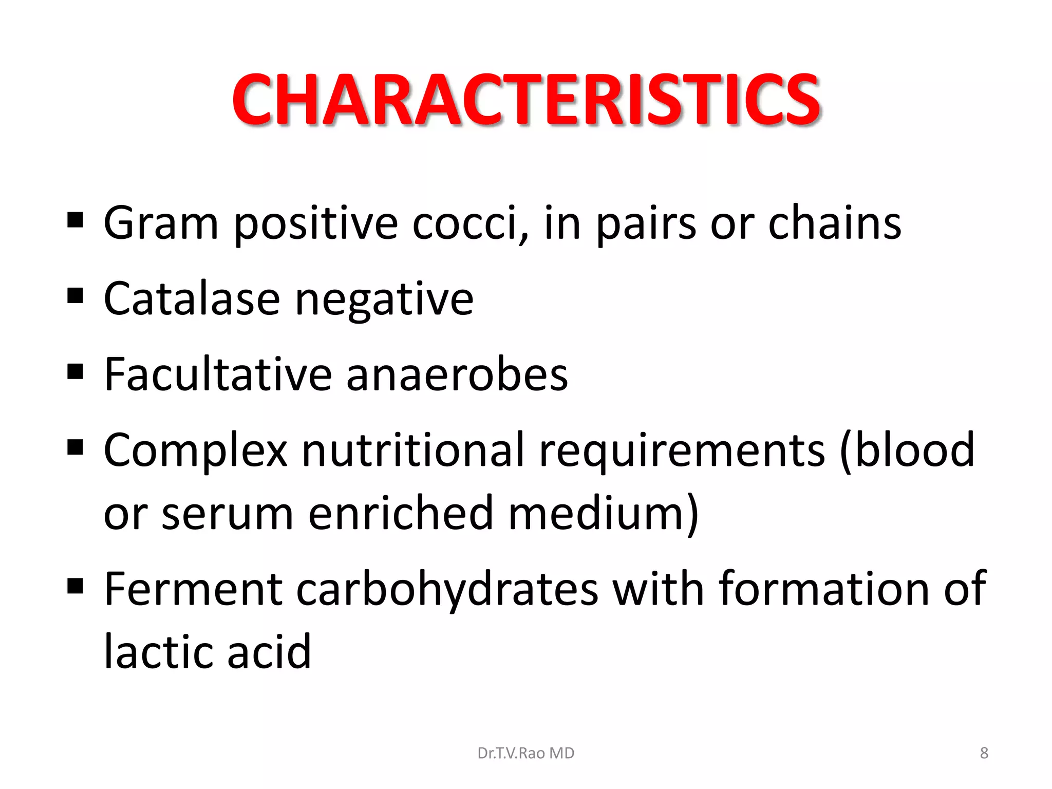
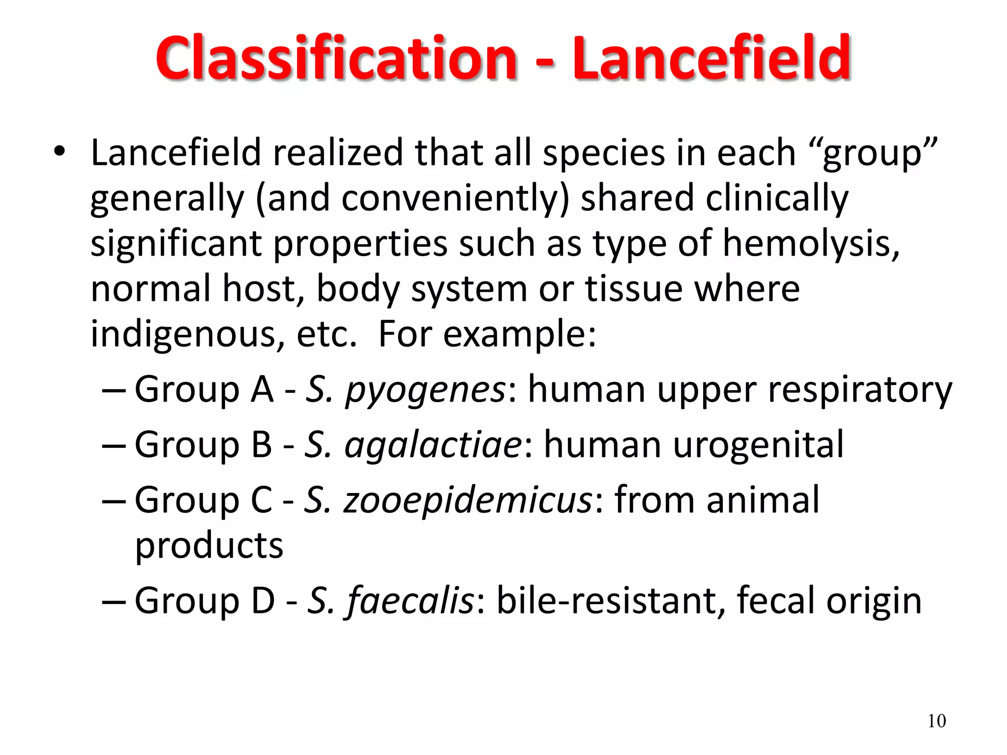
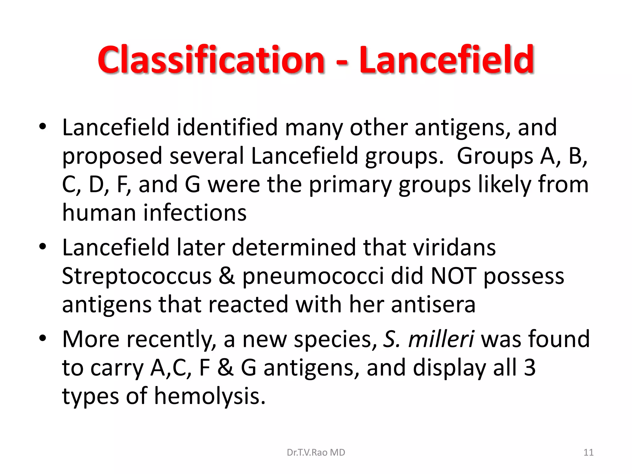
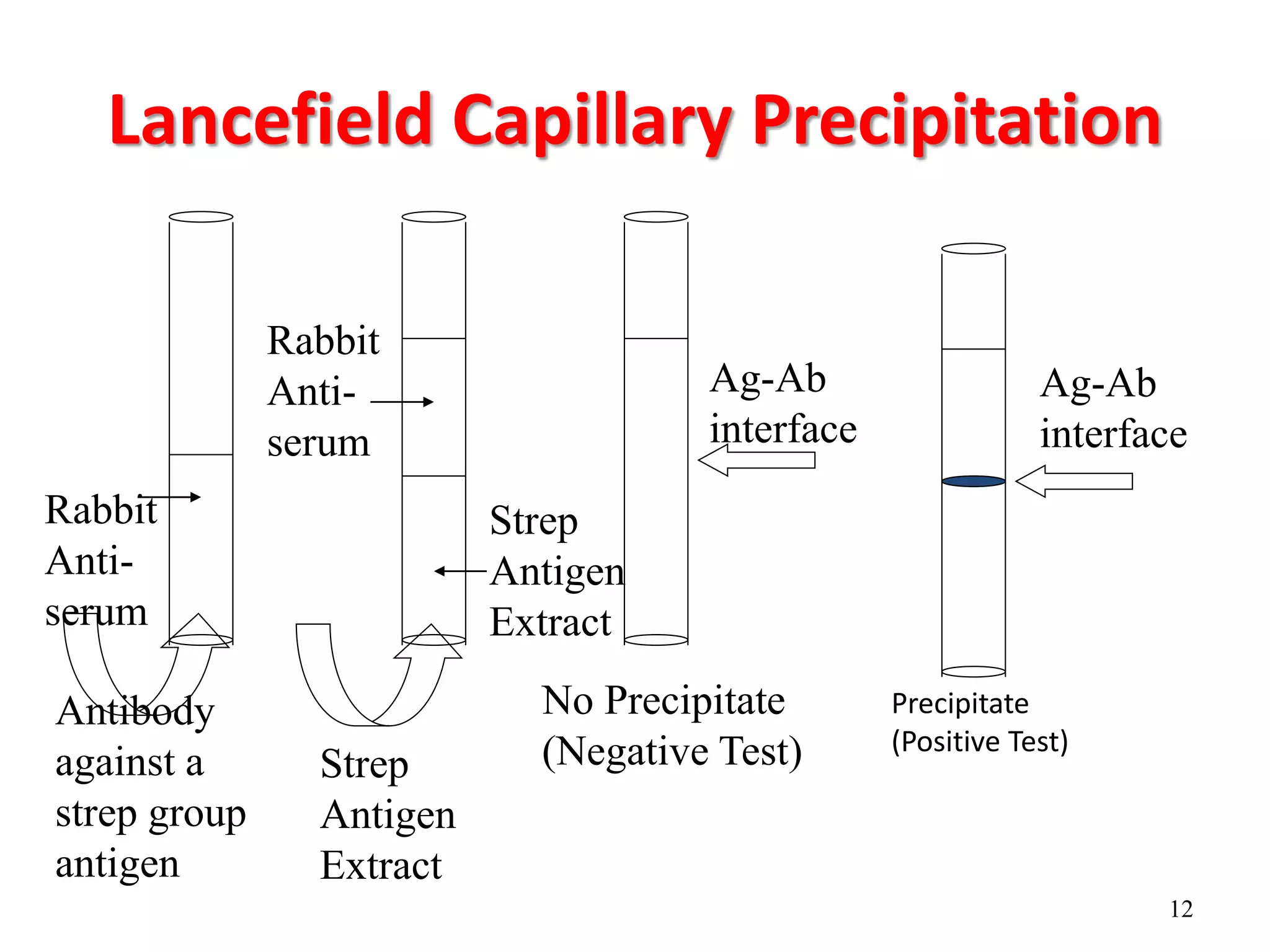
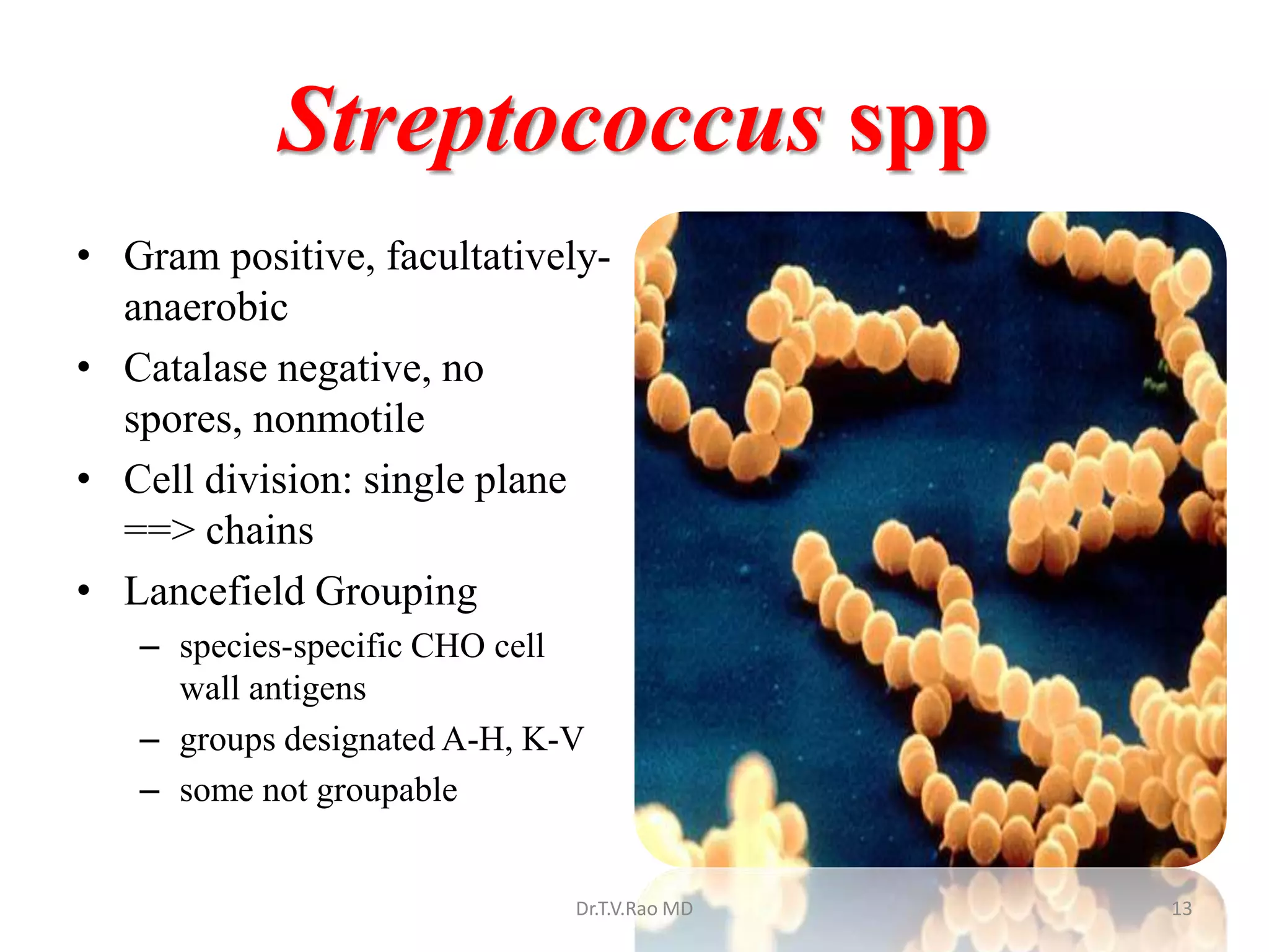
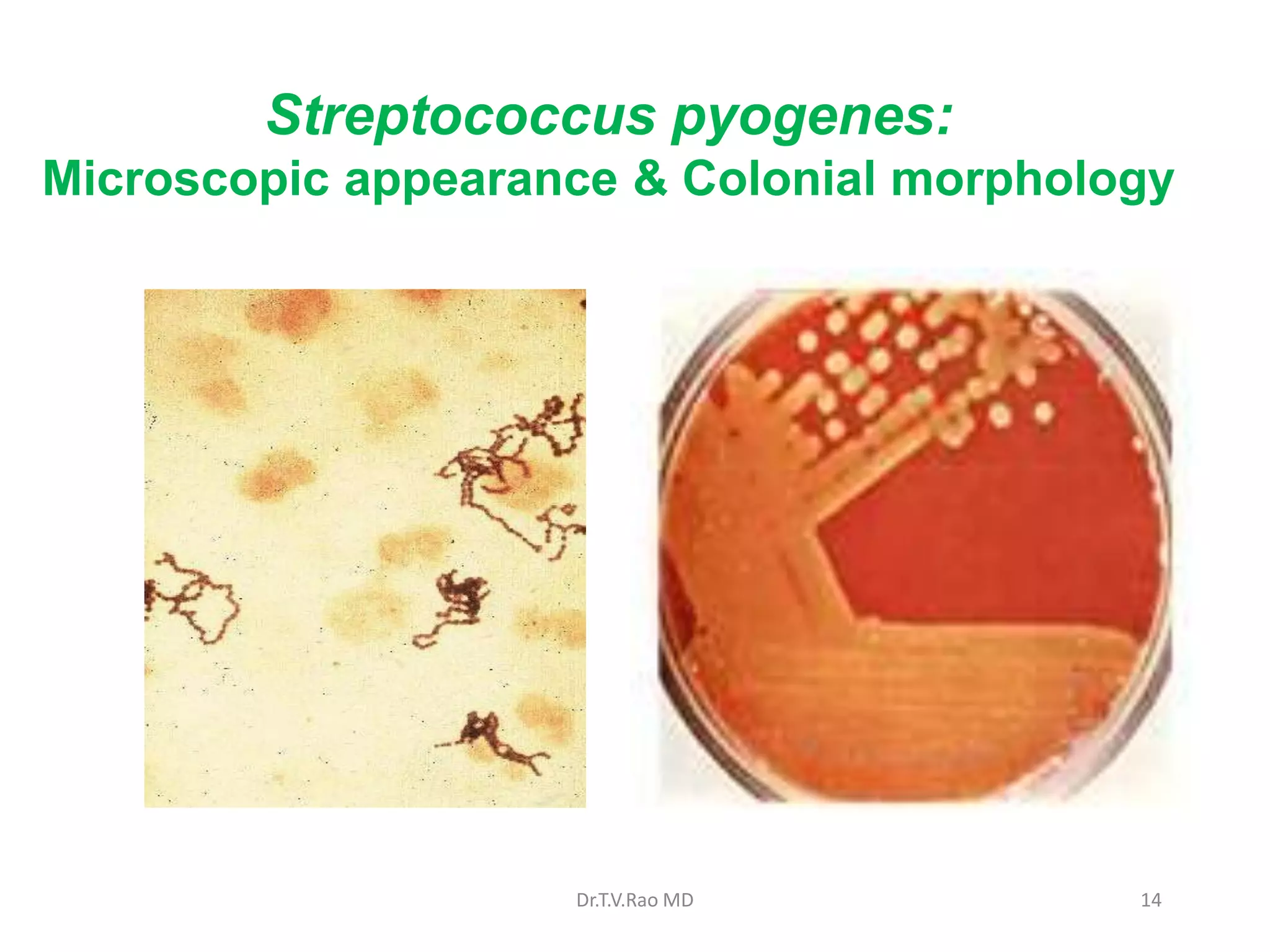
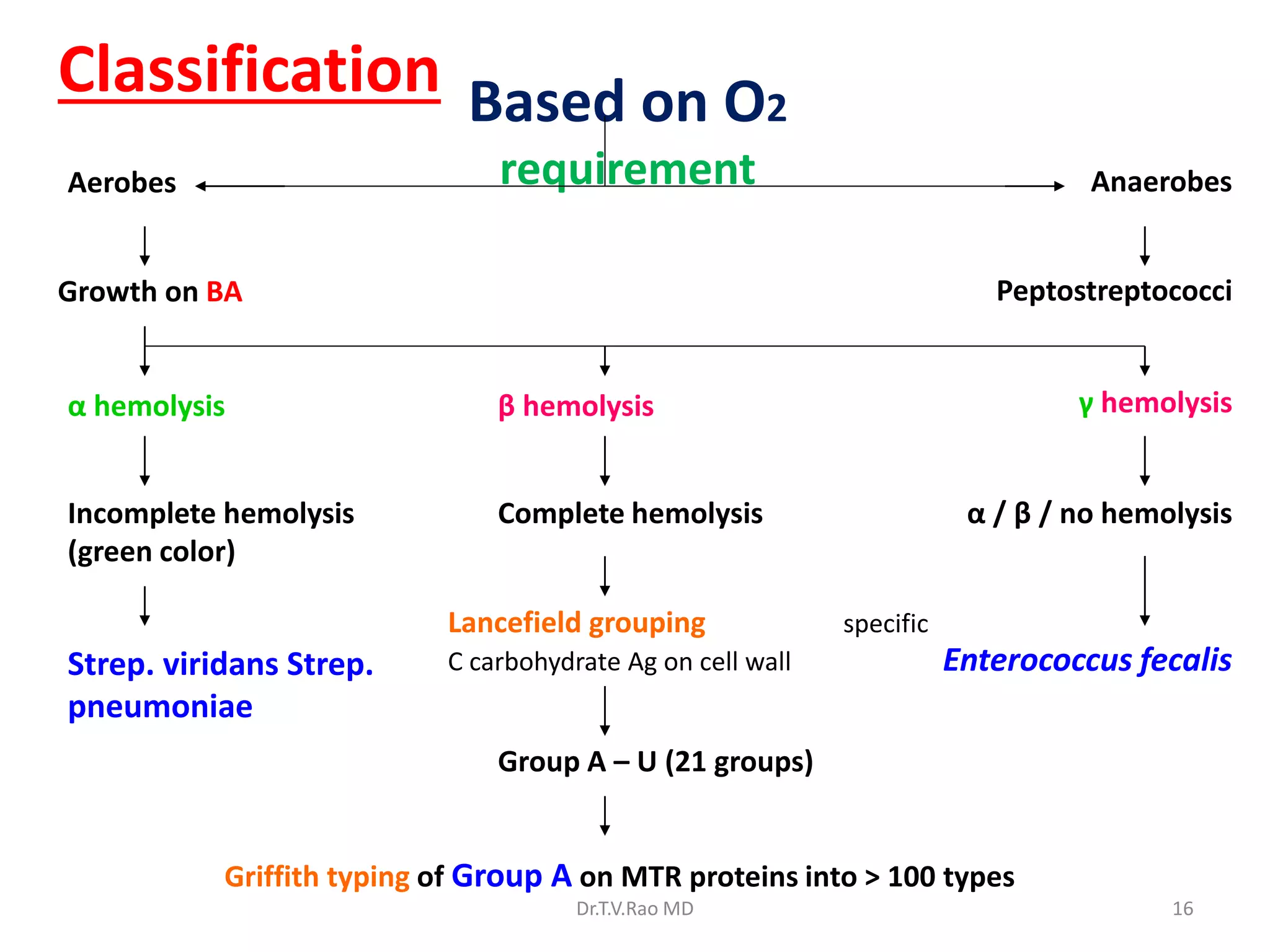
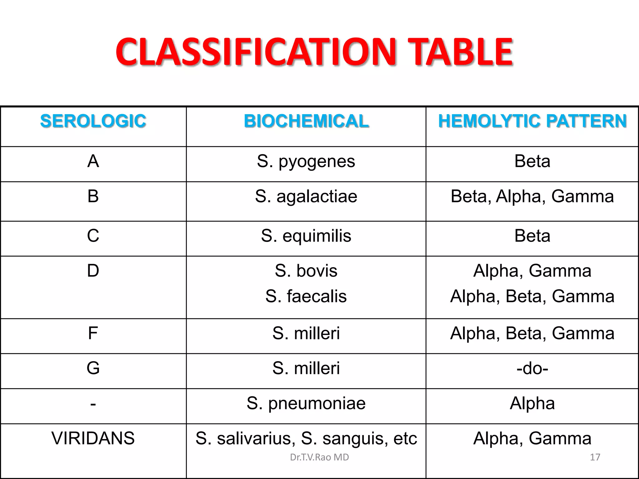
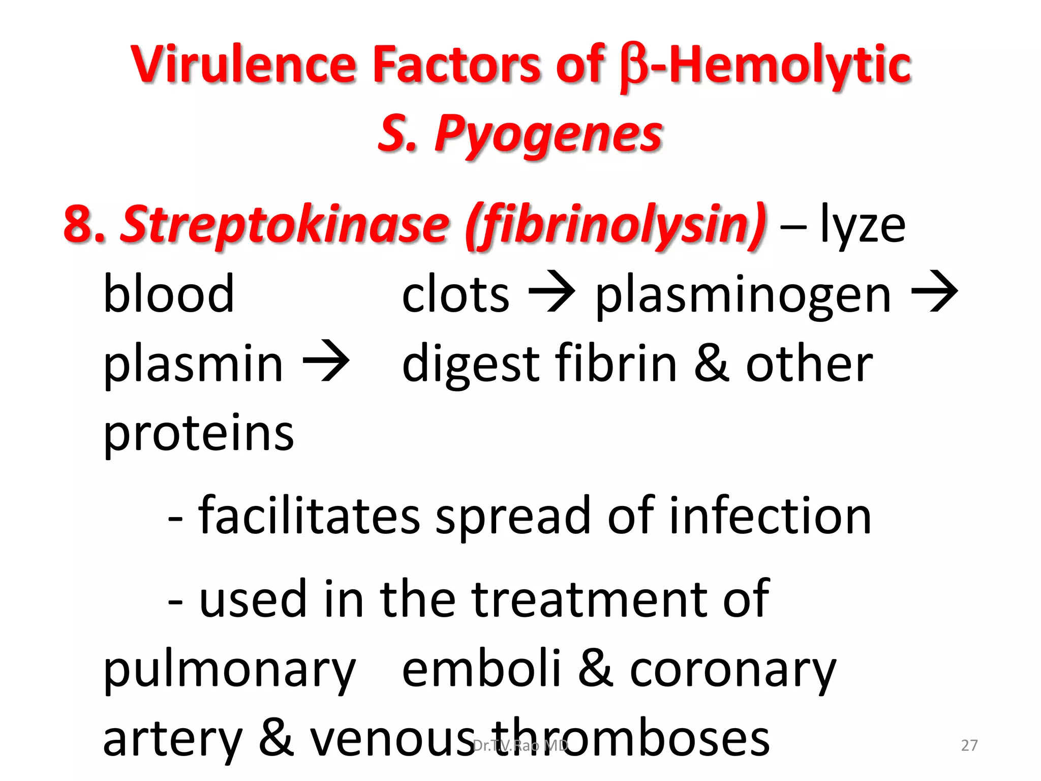
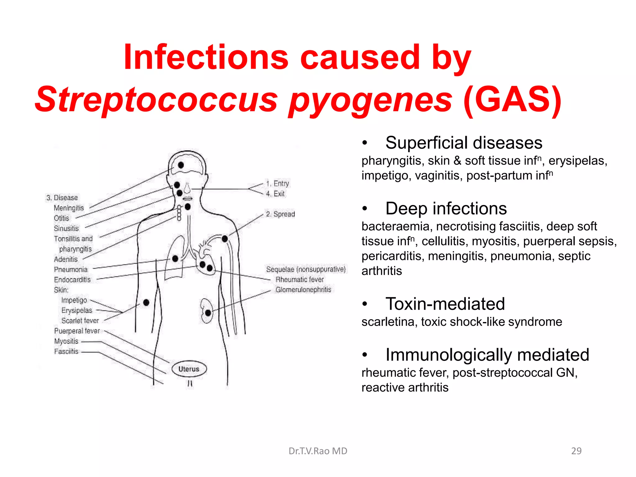
Group A
Streptococcus
Streptococcus pyogenes
Dr.T.V.Rao MD
Dr.T.V.Rao MD 1
2
Preliminary Grouping of Gram Positive Cocci
Gram Positive Coccus
Catalase
Rothia
Staphylococcus
Micrococcus
PYR
+ _
+
“A Disk*
chains _
S.pyogenes
“GAS”
*A disc contains bacitracin
Enterococcus
Streptococcus sp & other
Group genera
+
Other Strep
Group genera
S R
_
See “Staph” PP
Note: SBA hemolysis as
alt to PYR?
18.05.09 Phase I/ Module VII Dr Ekta 3
Overview of the Medically Important Gram Positive Cocci
Family, Genus, species Characteristics Clinical manifestations
Staphylococcaceae Cocci in cluster; catalase-positive
Staphylococcus aureus Coagulase +ve, yellow-pigmented colonies Pyogenic infections,
toxicoses
S. epidermidis Coagulase -ve, whitish colonies, normal
flora
Foreign body infections
Streptococcaceae Cocci in chains and in pairs,
catalase-negative
Streptococcus pyogenes Cocci in chains, Lancefield group A, β -
hemolysis
Tonsillitis, scarlet fever,
skin infections
S. pneumoniae Diplococci, α-hemolysis Pneumonia, otitis media,
sinusitis
S. agalactiae Chain-forming cocci, group antigen B, β-
hemolysis
Meningitis/sepsis in
neonates
S. viridans Cocci in chains, α-hemolysis Endocarditis, dental caries
Enterococcaceae In chains & pairs, α, β, or γ-hemolysis,
group antigen D, catalase -ve
Flora of intestines of
humans and animals
Enterococcus faecalis
Enterococcus faecium
Aesculin-positive, growth in 6.5% NaCl, pH
9.6
Opportunistic infections
Group A Streptococcal infection and
Health Care
Alexander Gordon
(1752-1799)
“... seized such women only as were
visited, or delivered, by a practitioner or
nurse, who had previously attended
patients affected by the disease….a
specific contagion, or infection....
…I could venture to foretell what women
would be affected with the disease, upon
hearing by what midwife they were to be
delivered..”
1795
Dr.T.V.Rao MD 4
Group A streptococcal infection and
health care
Ignaz Philipp Semmelweis
(1818-1865)
All students or doctors who enter the
wards for the purpose of making an
examination must wash their hands
thoroughly in a solution of chlorinated
lime which will be placed in
convenient basins near the entrance
of the wards. This disinfection will be
considered sufficient for this visit.
Between examinations the hands
must be washed in soap and water.
1847
Dr.T.V.Rao MD 5
Group A Streptococcal infection and
health care
Louis Pasteur
(1822-1895)
”It is the nursing and medical staff who
carry the microbe from an infected woman
to a healthy one….
This water, this sponge, this lint with which
you wash or cover a wound, may deposit
germs which have the power of multiplying
rapidly within the tissue....
If I had the honour of being a surgeon....not
only would I use none but perfectly clean
instruments, but I would clean my hands
with the greatest care...”
1879
Dr.T.V.Rao MD 6
Rebecca Lancefield Classifies
Streptococcus
Dr.T.V.Rao MD 7
CHARACTERISTICS
 Gram positive cocci, in pairs or chains
 Catalase negative
 Facultative anaerobes
 Complex nutritional requirements (blood
or serum enriched medium)
 Ferment carbohydrates with formation of
lactic acid
Dr.T.V.Rao MD 8
LANCEFIELD
CLASSIFICATION
• Group A – rhamnose-N-acetylglucosamine
• Group B – rhamnose-glucosamine
polysaccharide
• Group C –rhamnose-N-acetylglucosamine
• Group D – glycerol teichoic acid containing
alanine & glucose
• Group F – glucopyrasonyl-N-
acetylgalactosamine
Dr.T.V.Rao MD 9
10
Classification - Lancefield
• Lancefield realized that all species in each “group”
generally (and conveniently) shared clinically
significant properties such as type of hemolysis,
normal host, body system or tissue where
indigenous, etc. For example:
– Group A - S. pyogenes: human upper respiratory
– Group B - S. agalactiae: human urogenital
– Group C - S. zooepidemicus: from animal
products
– Group D - S. faecalis: bile-resistant, fecal origin
Classification - Lancefield
• Lancefield identified many other antigens, and
proposed several Lancefield groups. Groups A, B,
C, D, F, and G were the primary groups likely from
human infections
• Lancefield later determined that viridans
Streptococcus & pneumococci did NOT possess
antigens that reacted with her antisera
• More recently, a new species, S. milleri was found
to carry A,C, F & G antigens, and display all 3
types of hemolysis.
Dr.T.V.Rao MD 11
12
Lancefield Capillary Precipitation
Antibody
against a
strep group
antigen
Strep
Antigen
Extract
No Precipitate
(Negative Test)
Precipitate
(Positive Test)
Ag-Ab
interface
Ag-Ab
interface
Rabbit
Anti-
serum
Rabbit
Anti-
serum
Strep
Antigen
Extract
Streptococcus spp
• Gram positive, facultatively-
anaerobic
• Catalase negative, no
spores, nonmotile
• Cell division: single plane
==> chains
• Lancefield Grouping
– species-specific CHO cell
wall antigens
– groups designated A-H, K-V
– some not groupable
Dr.T.V.Rao MD 13
Streptococcus pyogenes:
Microscopic appearance & Colonial morphology
Dr.T.V.Rao MD 14
Structure of Streptococci
Dr.T.V.Rao MD 15
Dr.T.V.Rao MD 16
Classification Based on O2
requirement
Aerobes Anaerobes
Peptostreptococci
Growth on BA
α hemolysis β hemolysis γ hemolysis
Incomplete hemolysis
(green color)
Complete hemolysis α / β / no hemolysis
Strep. viridans Strep.
pneumoniae
Enterococcus fecalis
Lancefield grouping specific
C carbohydrate Ag on cell wall
Group A – U (21 groups)
Griffith typing of Group A on MTR proteins into > 100 types
CLASSIFICATION TABLE
SEROLOGIC BIOCHEMICAL HEMOLYTIC PATTERN
A S. pyogenes Beta
B S. agalactiae Beta, Alpha, Gamma
C S. equimilis Beta
D S. bovis
S. faecalis
Alpha, Gamma
Alpha, Beta, Gamma
F S. milleri Alpha, Beta, Gamma
G S. milleri -do-
- S. pneumoniae Alpha
VIRIDANS S. salivarius, S. sanguis, etc Alpha, Gamma
Dr.T.V.Rao MD 17
PRESUMPTIVE IDENTIFICATION OF
STREPTOCOCCI
Organism Susceptibility
A P
Hydrolysis
hippurate esculin
Growth
Bile NaCl
Lysis
bile
S. pyogenes S R - - - - -
S. agalactiae R R + - - + -
Grp D
S. faecalis
S. bovis
R R
R R
- +
- -
+ +
+ -
-
-
Viridans R R
(var)
- - - - -
Pneumococcus R S - - - - +
Dr.T.V.Rao MD 18
Group A Streptococcus
(S. pyogenes)
 Structure:
1. Capsule – hyaluronic acid
2. Cell wall
a. protein antigens M,T,R
M protein  major virulence factor
T & R protein  no role in the
virulence
b. group specific carbohydrates – rhamnose-N-acetylglucosamine
3. Pili  consists partly of M protein & covered with
lipoteichoic acid  for attachment
Dr.T.V.Rao MD 19
Streptococcus pyogenes
• Streptococcus pyogenes is one of the most
frequent pathogens of humans. It is estimated
that between 5-15% of normal individuals harbor
the bacterium, usually in the respiratory tract,
without signs of disease. As normal flora, S.
pyogenes can infect when defenses are
compromised or when the organisms are able to
penetrate the constitutive defenses. When the
bacteria are introduced or transmitted to
vulnerable tissues, a variety of types of
suppurative infections can occur
Dr.T.V.Rao MD 20
VIRULENCE FACTORS
1. Capsule – non-immunogenic
2. M protein – hair-like projections on the cell
wall
- major virulence factor
- promotes adherence
- antiphagocytic
- anticomplement
- type specific
Dr.T.V.Rao MD 21
22
Virulence Factors of b-Hemolytic
S. Pyogenes
Produces surface antigens:
– C-carbohydrates – protect against lysozyme
– Fimbriae – adherence
– M-protein – contributes to resistance to
phagocytosis
– Hyaluronic acid capsule – provokes no
immune response
–C5a protease hinders complement and
neutrophil response
Dr.T.V.Rao MD
Virulence Factors
• Streptolysin O: thiol-activated toxin (Groups A,C,G)
– damages membranes: RBCs, myocardial cells, PMNs
– role in intracellular survival
• Erythrogenic toxins: rash of scarlet fever
– pyrogenicity, lethal shock
– 105-fold increased sensitivity to endotoxin
• Pyrogenic exotoxin A
– contributes to streptococcal TSLS
– stimulates cytokine production by T cells
– endothelial cell damage, hypotensive shock, ischemia-based
necrosis
Dr.T.V.Rao MD 23
Dr.T.V.Rao MD 24
Streptococcus pyogenes – virulence factors
Antigenic – produce ASLO
Streptolysin S (SLS)
Exotoxins
Oxygen stable , non-antigenic
Damage cardiac cells
Streptolysin O (SLO) Oxygen labile
Streptococcal Pyrogenic Exotoxin (SPEs)
Manifestation of scarlet fever
Exoenzymes Streptokinase (fibrinolysin) / Streptodornase
(DNAase) / Hyalarunidase
Virulence Factors of b-Hemolytic
S. Pyogenes
Extracellular enzymes
1 Streptokinase –
digests fibrin clots
2 Hyaluronidase –
breaks down
connective tissue
3 DNase –
hydrolyzes DNA
25
Dr.T.V.Rao MD
Virulence Factors of b-Hemolytic
S. Pyogenes
4. Lipoteichoic acid – for adherence
5. Erythrogenic toxin – pyrogenic exotoxins
A,B,C
- responsible for the rash of Scarlet fever
6. Streptolysin O – lyses WBC, platelets, RBC
- immunogenic
7. Streptolysin S – non-immunogenic
- responsible for the hemolytic zones
around colonies
Dr.T.V.Rao MD 26
Virulence Factors of b-Hemolytic
S. Pyogenes
8. Streptokinase (fibrinolysin) – lyze
blood clots  plasminogen 
plasmin  digest fibrin & other
proteins
- facilitates spread of infection
- used in the treatment of
pulmonary emboli & coronary
artery & venous thromboses
Dr.T.V.Rao MD 27
Virulence Factors of b-Hemolytic
S. Pyogenes
9. DNAse (streptodornase) – depolymerizes
cell-free DNA in purulent materials
10. Hyaluronidase – spreading factor
- splits hyaluronic acid
 streptodornase & streptokinase  used in
enzymatic debridement  liquefy
exudates & facilitate removal of pus &
necrotic tissue  antibiotics gain better
access
Dr.T.V.Rao MD 28
Infections caused by
Streptococcus pyogenes (GAS)
• Superficial diseases
pharyngitis, skin & soft tissue infn, erysipelas,
impetigo, vaginitis, post-partum infn
• Deep infections
bacteraemia, necrotising fasciitis, deep soft
tissue infn, cellulitis, myositis, puerperal sepsis,
pericarditis, meningitis, pneumonia, septic
arthritis
• Toxin-mediated
scarletina, toxic shock-like syndrome
• Immunologically mediated
rheumatic fever, post-streptococcal GN,
reactive arthritis
Dr.T.V.Rao MD 29
Group A streptococcal infection
Overall disease burden
Each year
• 1.8 million new cases of
serious infection
• at least 500,000 deaths
• 110 million cases of soft tissue
infection
• 610 million cases of
pharyngitis
At least 18 million people suffer
the consequences of serious
GAS diseases
Dr.T.V.Rao MD 30
CLINICAL SYNDROMES
A. Suppurative Infections
1. Skin Infections
a. impetigo – superficial blisters
covered with pus or honey–colored
crust
b. erysipelas – acute superficial
cellulitis of the skin with lymphatic
involvement
Dr.T.V.Rao MD 31
Pharyngitis the Prominent and
common Infection
Dr.T.V.Rao MD 32
Pharyngitis and tonsillitis
33
Dr.T.V.Rao MD
Infection of Tonsils
Dr.T.V.Rao MD 34
URI continues to be common
presentation
Dr.T.V.Rao MD 35
Skin lesions
Dr.T.V.Rao MD 36
CLINICAL SYNDROMES
. Scarlet fever – a complication of
pharyngitis if the causative agent is
capable of producing erythrogenic toxin
 initial symptoms of pharyngitis, diffuse
erythematous rash with sparing of the
palms & soles
Circumoral pallor
“strawberry tongue”
Dr.T.V.Rao MD 37
CLINICAL SYNDROMES
Pneumonia –
rapidly progressive
& severe
 most commonly a
sequela to viral
infections like
influenza or
measles
Dr.T.V.Rao MD 38
Erysipelas
Dr.T.V.Rao MD 39
Streptococcus pyogenes
Necrotizing Fasciitis
• Invasive, nonTSLS
disease
– necrotizing fasciitis
(“flesh-eating
bacteria”)
– rapid development of
shock, multiple organ
system failure
– high fatality rate
Dr.T.V.Rao MD 40
Rheumatic Heart disease is leading
cause of Morbidity
Dr.T.V.Rao MD 41
Streptococcus pyogenes
Sequellae to strep throat
• Heart valve damage
(rheumatic heart disease)
– < 3% of people with strep
throat, weeks after sore
throat
– migrating arthritis; heart
valve damage (50%), some
fatal
– recurrences common, lifelong
penicillin therapy
– shared antigen: M protein,
cardiac myosin
– attack by T cells, Ab:
inflammation, valve damage
Dr.T.V.Rao MD 42
Post streptococcal diseases
• Rheumatic Fever-
autoimmune disease
involving heart valves,
joints, nervous system.
Follows a strep throat
• Acute glomerulonephritis
or Bright’s Disease-
inflammatory disease of
renal glomeruli and
structures involved in
blood filter of kidney. Due
to deposition of Ag/Ab
complexes
Dr.T.V.Rao MD 43
CLINICAL SYNDROMES
B. Non-suppurative sequelae
1. Rheumatic fever – associated with M
types causing URI & skin infections
 fever, malaise, migratory nonsuppurative
polyarthritis, evidence of inflammation of
the heart
 carditis  leads to thickened & deformed
valves & to small perivascular granulomas in
the myocardium (Aschoff bodies)
Dr.T.V.Rao MD 44
Rheumatic Fever
• Most common cause of
permanent heart valve
damage in children
• Exact cause not yet
known but there
appears to be some
antibody cross reactivity
between the cell wall of
S. pyogenes and heart
muscle
Dr.T.V.Rao MD 45
Lesions in the Heart
Dr.T.V.Rao MD 46
Rheumatic Fever
• Diagnosis is based on
symptoms and is
difficult
• Occurs most frequently
between ages of 6 and
15
• US it is about 0.05% of
pop having strep
infections
• 100x more frequent in
tropical countries Dr.T.V.Rao MD 47
• Glomerulonephritis
– symptoms 10 days after 1˚ infection: edema
– decreased urination, hematuria, hypertension
– Ag:Ab complexes accumulate, C’ activated
– provoke inflammatory response, interferes with
normal kidney function
– young children: self-limiting
– teenagers/adults: rare permanent kidney damage,
chronic glomerulonephritis
Streptococcus pyogenes
Sequellae to strep throat or Skin Infections
Dr.T.V.Rao MD 48
Glomerulonephritis
2. Acute Glomerulonephritis – associated with M
types producing URI & skin infections
 particularly associated with types 12, 4, 2 &
49 which are nephritogenic
 initiated by ag-ab complexes on the
glomerular basement membrane
 hematuria, proteinuria, edema &
hypertension
Dr.T.V.Rao MD 49
Glomerulonephritis
• Diagnosis based on
history of Strep throat
and clinical findings.
• Symptoms include
fever, malaise, edema,
hypertension and blood
or protein in urine
• Occurs in 0.5% of those
having strep throat.
Dr.T.V.Rao MD 50
DIAGNOSIS
1. Microscopy
2. Culture – Bacitracin Test (Taxo-A)
3. Antigen detection tests – Enzyme-linked
immunosorbent assay (ELISA) or
agglutination tests
4. Antibody detection
 ASO titer – for respiratory disease
 antiDNAse & antihyaluronidase – for skin
infections
Dr.T.V.Rao MD 51
Diagnosis and treatment of Strep Throat
• Tell tale symptoms are
slight fever associated
with sore throat and
visual of pus in back of
throat
• Quick diagnostic tests
(Molecular) available
but must be confirmed
by throat swab and
growth on blood agar
(beta hemolysis)
Dr.T.V.Rao MD 52
Dr.T.V.Rao MD 53
Lab diagnosis – Strep. pyogenes
• Specimens: throat swab, pus,
blood
• Microscopy :Gram stain - GPC in
chains
• Culture: BA - beta hemolytic
colonies
• Identification tests -
– Catalase Negative
– Bacitracin sensitive
– Penicillin sensitive
– ASO titre / DNAase B test
B
B
DIAGNOSIS
1. Microscopy
2. Culture – Bacitracin Test (Taxo-A)
3. Antigen detection tests – Enzyme-linked
immunosorbent assay (ELISA) or
agglutination tests
4. Antibody detection
 ASO titer – for respiratory disease
 antiDNAse & antihyaluronidase – for skin
infections
Dr.T.V.Rao MD 54
Streptococci grown Blood agar
Dr.T.V.Rao MD 55
TREATMENT
1. Penicillin G – drug of choice
2. Erythromycin
 Antistreptococcal chemoprophylaxis in
persons who have suffered an acute attack
of rheumatic fever  Penicillin G 1.2 M units
IM every 3-4 weeks or daily oral penicillin or
oral sulfonamide
Dr.T.V.Rao MD 56
• Programme created by Dr.T.V.Rao MD
for Medical and Paramedical Students in
the Developing World
• Email
• doctortvrao@gmail.com
Dr.T.V.Rao MD 57

Group A Streptococcus.pptx

  • 1.
  • 2.
    2 Preliminary Grouping ofGram Positive Cocci Gram Positive Coccus Catalase Rothia Staphylococcus Micrococcus PYR + _ + “A Disk* chains _ S.pyogenes “GAS” *A disc contains bacitracin Enterococcus Streptococcus sp & other Group genera + Other Strep Group genera S R _ See “Staph” PP Note: SBA hemolysis as alt to PYR?
  • 3.
    18.05.09 Phase I/Module VII Dr Ekta 3 Overview of the Medically Important Gram Positive Cocci Family, Genus, species Characteristics Clinical manifestations Staphylococcaceae Cocci in cluster; catalase-positive Staphylococcus aureus Coagulase +ve, yellow-pigmented colonies Pyogenic infections, toxicoses S. epidermidis Coagulase -ve, whitish colonies, normal flora Foreign body infections Streptococcaceae Cocci in chains and in pairs, catalase-negative Streptococcus pyogenes Cocci in chains, Lancefield group A, β - hemolysis Tonsillitis, scarlet fever, skin infections S. pneumoniae Diplococci, α-hemolysis Pneumonia, otitis media, sinusitis S. agalactiae Chain-forming cocci, group antigen B, β- hemolysis Meningitis/sepsis in neonates S. viridans Cocci in chains, α-hemolysis Endocarditis, dental caries Enterococcaceae In chains & pairs, α, β, or γ-hemolysis, group antigen D, catalase -ve Flora of intestines of humans and animals Enterococcus faecalis Enterococcus faecium Aesculin-positive, growth in 6.5% NaCl, pH 9.6 Opportunistic infections
  • 4.
    Group A Streptococcalinfection and Health Care Alexander Gordon (1752-1799) “... seized such women only as were visited, or delivered, by a practitioner or nurse, who had previously attended patients affected by the disease….a specific contagion, or infection.... …I could venture to foretell what women would be affected with the disease, upon hearing by what midwife they were to be delivered..” 1795 Dr.T.V.Rao MD 4
  • 5.
    Group A streptococcalinfection and health care Ignaz Philipp Semmelweis (1818-1865) All students or doctors who enter the wards for the purpose of making an examination must wash their hands thoroughly in a solution of chlorinated lime which will be placed in convenient basins near the entrance of the wards. This disinfection will be considered sufficient for this visit. Between examinations the hands must be washed in soap and water. 1847 Dr.T.V.Rao MD 5
  • 6.
    Group A Streptococcalinfection and health care Louis Pasteur (1822-1895) ”It is the nursing and medical staff who carry the microbe from an infected woman to a healthy one…. This water, this sponge, this lint with which you wash or cover a wound, may deposit germs which have the power of multiplying rapidly within the tissue.... If I had the honour of being a surgeon....not only would I use none but perfectly clean instruments, but I would clean my hands with the greatest care...” 1879 Dr.T.V.Rao MD 6
  • 7.
  • 8.
    CHARACTERISTICS  Gram positivecocci, in pairs or chains  Catalase negative  Facultative anaerobes  Complex nutritional requirements (blood or serum enriched medium)  Ferment carbohydrates with formation of lactic acid Dr.T.V.Rao MD 8
  • 9.
    LANCEFIELD CLASSIFICATION • Group A– rhamnose-N-acetylglucosamine • Group B – rhamnose-glucosamine polysaccharide • Group C –rhamnose-N-acetylglucosamine • Group D – glycerol teichoic acid containing alanine & glucose • Group F – glucopyrasonyl-N- acetylgalactosamine Dr.T.V.Rao MD 9
  • 10.
    10 Classification - Lancefield •Lancefield realized that all species in each “group” generally (and conveniently) shared clinically significant properties such as type of hemolysis, normal host, body system or tissue where indigenous, etc. For example: – Group A - S. pyogenes: human upper respiratory – Group B - S. agalactiae: human urogenital – Group C - S. zooepidemicus: from animal products – Group D - S. faecalis: bile-resistant, fecal origin
  • 11.
    Classification - Lancefield •Lancefield identified many other antigens, and proposed several Lancefield groups. Groups A, B, C, D, F, and G were the primary groups likely from human infections • Lancefield later determined that viridans Streptococcus & pneumococci did NOT possess antigens that reacted with her antisera • More recently, a new species, S. milleri was found to carry A,C, F & G antigens, and display all 3 types of hemolysis. Dr.T.V.Rao MD 11
  • 12.
    12 Lancefield Capillary Precipitation Antibody againsta strep group antigen Strep Antigen Extract No Precipitate (Negative Test) Precipitate (Positive Test) Ag-Ab interface Ag-Ab interface Rabbit Anti- serum Rabbit Anti- serum Strep Antigen Extract
  • 13.
    Streptococcus spp • Grampositive, facultatively- anaerobic • Catalase negative, no spores, nonmotile • Cell division: single plane ==> chains • Lancefield Grouping – species-specific CHO cell wall antigens – groups designated A-H, K-V – some not groupable Dr.T.V.Rao MD 13
  • 14.
    Streptococcus pyogenes: Microscopic appearance& Colonial morphology Dr.T.V.Rao MD 14
  • 15.
  • 16.
    Dr.T.V.Rao MD 16 ClassificationBased on O2 requirement Aerobes Anaerobes Peptostreptococci Growth on BA α hemolysis β hemolysis γ hemolysis Incomplete hemolysis (green color) Complete hemolysis α / β / no hemolysis Strep. viridans Strep. pneumoniae Enterococcus fecalis Lancefield grouping specific C carbohydrate Ag on cell wall Group A – U (21 groups) Griffith typing of Group A on MTR proteins into > 100 types
  • 17.
    CLASSIFICATION TABLE SEROLOGIC BIOCHEMICALHEMOLYTIC PATTERN A S. pyogenes Beta B S. agalactiae Beta, Alpha, Gamma C S. equimilis Beta D S. bovis S. faecalis Alpha, Gamma Alpha, Beta, Gamma F S. milleri Alpha, Beta, Gamma G S. milleri -do- - S. pneumoniae Alpha VIRIDANS S. salivarius, S. sanguis, etc Alpha, Gamma Dr.T.V.Rao MD 17
  • 18.
    PRESUMPTIVE IDENTIFICATION OF STREPTOCOCCI OrganismSusceptibility A P Hydrolysis hippurate esculin Growth Bile NaCl Lysis bile S. pyogenes S R - - - - - S. agalactiae R R + - - + - Grp D S. faecalis S. bovis R R R R - + - - + + + - - - Viridans R R (var) - - - - - Pneumococcus R S - - - - + Dr.T.V.Rao MD 18
  • 19.
    Group A Streptococcus (S.pyogenes)  Structure: 1. Capsule – hyaluronic acid 2. Cell wall a. protein antigens M,T,R M protein  major virulence factor T & R protein  no role in the virulence b. group specific carbohydrates – rhamnose-N-acetylglucosamine 3. Pili  consists partly of M protein & covered with lipoteichoic acid  for attachment Dr.T.V.Rao MD 19
  • 20.
    Streptococcus pyogenes • Streptococcuspyogenes is one of the most frequent pathogens of humans. It is estimated that between 5-15% of normal individuals harbor the bacterium, usually in the respiratory tract, without signs of disease. As normal flora, S. pyogenes can infect when defenses are compromised or when the organisms are able to penetrate the constitutive defenses. When the bacteria are introduced or transmitted to vulnerable tissues, a variety of types of suppurative infections can occur Dr.T.V.Rao MD 20
  • 21.
    VIRULENCE FACTORS 1. Capsule– non-immunogenic 2. M protein – hair-like projections on the cell wall - major virulence factor - promotes adherence - antiphagocytic - anticomplement - type specific Dr.T.V.Rao MD 21
  • 22.
    22 Virulence Factors ofb-Hemolytic S. Pyogenes Produces surface antigens: – C-carbohydrates – protect against lysozyme – Fimbriae – adherence – M-protein – contributes to resistance to phagocytosis – Hyaluronic acid capsule – provokes no immune response –C5a protease hinders complement and neutrophil response Dr.T.V.Rao MD
  • 23.
    Virulence Factors • StreptolysinO: thiol-activated toxin (Groups A,C,G) – damages membranes: RBCs, myocardial cells, PMNs – role in intracellular survival • Erythrogenic toxins: rash of scarlet fever – pyrogenicity, lethal shock – 105-fold increased sensitivity to endotoxin • Pyrogenic exotoxin A – contributes to streptococcal TSLS – stimulates cytokine production by T cells – endothelial cell damage, hypotensive shock, ischemia-based necrosis Dr.T.V.Rao MD 23
  • 24.
    Dr.T.V.Rao MD 24 Streptococcuspyogenes – virulence factors Antigenic – produce ASLO Streptolysin S (SLS) Exotoxins Oxygen stable , non-antigenic Damage cardiac cells Streptolysin O (SLO) Oxygen labile Streptococcal Pyrogenic Exotoxin (SPEs) Manifestation of scarlet fever Exoenzymes Streptokinase (fibrinolysin) / Streptodornase (DNAase) / Hyalarunidase
  • 25.
    Virulence Factors ofb-Hemolytic S. Pyogenes Extracellular enzymes 1 Streptokinase – digests fibrin clots 2 Hyaluronidase – breaks down connective tissue 3 DNase – hydrolyzes DNA 25 Dr.T.V.Rao MD
  • 26.
    Virulence Factors ofb-Hemolytic S. Pyogenes 4. Lipoteichoic acid – for adherence 5. Erythrogenic toxin – pyrogenic exotoxins A,B,C - responsible for the rash of Scarlet fever 6. Streptolysin O – lyses WBC, platelets, RBC - immunogenic 7. Streptolysin S – non-immunogenic - responsible for the hemolytic zones around colonies Dr.T.V.Rao MD 26
  • 27.
    Virulence Factors ofb-Hemolytic S. Pyogenes 8. Streptokinase (fibrinolysin) – lyze blood clots  plasminogen  plasmin  digest fibrin & other proteins - facilitates spread of infection - used in the treatment of pulmonary emboli & coronary artery & venous thromboses Dr.T.V.Rao MD 27
  • 28.
    Virulence Factors ofb-Hemolytic S. Pyogenes 9. DNAse (streptodornase) – depolymerizes cell-free DNA in purulent materials 10. Hyaluronidase – spreading factor - splits hyaluronic acid  streptodornase & streptokinase  used in enzymatic debridement  liquefy exudates & facilitate removal of pus & necrotic tissue  antibiotics gain better access Dr.T.V.Rao MD 28
  • 29.
    Infections caused by Streptococcuspyogenes (GAS) • Superficial diseases pharyngitis, skin & soft tissue infn, erysipelas, impetigo, vaginitis, post-partum infn • Deep infections bacteraemia, necrotising fasciitis, deep soft tissue infn, cellulitis, myositis, puerperal sepsis, pericarditis, meningitis, pneumonia, septic arthritis • Toxin-mediated scarletina, toxic shock-like syndrome • Immunologically mediated rheumatic fever, post-streptococcal GN, reactive arthritis Dr.T.V.Rao MD 29
  • 30.
    Group A streptococcalinfection Overall disease burden Each year • 1.8 million new cases of serious infection • at least 500,000 deaths • 110 million cases of soft tissue infection • 610 million cases of pharyngitis At least 18 million people suffer the consequences of serious GAS diseases Dr.T.V.Rao MD 30
  • 31.
    CLINICAL SYNDROMES A. SuppurativeInfections 1. Skin Infections a. impetigo – superficial blisters covered with pus or honey–colored crust b. erysipelas – acute superficial cellulitis of the skin with lymphatic involvement Dr.T.V.Rao MD 31
  • 32.
    Pharyngitis the Prominentand common Infection Dr.T.V.Rao MD 32
  • 33.
  • 34.
  • 35.
    URI continues tobe common presentation Dr.T.V.Rao MD 35
  • 36.
  • 37.
    CLINICAL SYNDROMES . Scarletfever – a complication of pharyngitis if the causative agent is capable of producing erythrogenic toxin  initial symptoms of pharyngitis, diffuse erythematous rash with sparing of the palms & soles Circumoral pallor “strawberry tongue” Dr.T.V.Rao MD 37
  • 38.
    CLINICAL SYNDROMES Pneumonia – rapidlyprogressive & severe  most commonly a sequela to viral infections like influenza or measles Dr.T.V.Rao MD 38
  • 39.
  • 40.
    Streptococcus pyogenes Necrotizing Fasciitis •Invasive, nonTSLS disease – necrotizing fasciitis (“flesh-eating bacteria”) – rapid development of shock, multiple organ system failure – high fatality rate Dr.T.V.Rao MD 40
  • 41.
    Rheumatic Heart diseaseis leading cause of Morbidity Dr.T.V.Rao MD 41
  • 42.
    Streptococcus pyogenes Sequellae tostrep throat • Heart valve damage (rheumatic heart disease) – < 3% of people with strep throat, weeks after sore throat – migrating arthritis; heart valve damage (50%), some fatal – recurrences common, lifelong penicillin therapy – shared antigen: M protein, cardiac myosin – attack by T cells, Ab: inflammation, valve damage Dr.T.V.Rao MD 42
  • 43.
    Post streptococcal diseases •Rheumatic Fever- autoimmune disease involving heart valves, joints, nervous system. Follows a strep throat • Acute glomerulonephritis or Bright’s Disease- inflammatory disease of renal glomeruli and structures involved in blood filter of kidney. Due to deposition of Ag/Ab complexes Dr.T.V.Rao MD 43
  • 44.
    CLINICAL SYNDROMES B. Non-suppurativesequelae 1. Rheumatic fever – associated with M types causing URI & skin infections  fever, malaise, migratory nonsuppurative polyarthritis, evidence of inflammation of the heart  carditis  leads to thickened & deformed valves & to small perivascular granulomas in the myocardium (Aschoff bodies) Dr.T.V.Rao MD 44
  • 45.
    Rheumatic Fever • Mostcommon cause of permanent heart valve damage in children • Exact cause not yet known but there appears to be some antibody cross reactivity between the cell wall of S. pyogenes and heart muscle Dr.T.V.Rao MD 45
  • 46.
    Lesions in theHeart Dr.T.V.Rao MD 46
  • 47.
    Rheumatic Fever • Diagnosisis based on symptoms and is difficult • Occurs most frequently between ages of 6 and 15 • US it is about 0.05% of pop having strep infections • 100x more frequent in tropical countries Dr.T.V.Rao MD 47
  • 48.
    • Glomerulonephritis – symptoms10 days after 1˚ infection: edema – decreased urination, hematuria, hypertension – Ag:Ab complexes accumulate, C’ activated – provoke inflammatory response, interferes with normal kidney function – young children: self-limiting – teenagers/adults: rare permanent kidney damage, chronic glomerulonephritis Streptococcus pyogenes Sequellae to strep throat or Skin Infections Dr.T.V.Rao MD 48
  • 49.
    Glomerulonephritis 2. Acute Glomerulonephritis– associated with M types producing URI & skin infections  particularly associated with types 12, 4, 2 & 49 which are nephritogenic  initiated by ag-ab complexes on the glomerular basement membrane  hematuria, proteinuria, edema & hypertension Dr.T.V.Rao MD 49
  • 50.
    Glomerulonephritis • Diagnosis basedon history of Strep throat and clinical findings. • Symptoms include fever, malaise, edema, hypertension and blood or protein in urine • Occurs in 0.5% of those having strep throat. Dr.T.V.Rao MD 50
  • 51.
    DIAGNOSIS 1. Microscopy 2. Culture– Bacitracin Test (Taxo-A) 3. Antigen detection tests – Enzyme-linked immunosorbent assay (ELISA) or agglutination tests 4. Antibody detection  ASO titer – for respiratory disease  antiDNAse & antihyaluronidase – for skin infections Dr.T.V.Rao MD 51
  • 52.
    Diagnosis and treatmentof Strep Throat • Tell tale symptoms are slight fever associated with sore throat and visual of pus in back of throat • Quick diagnostic tests (Molecular) available but must be confirmed by throat swab and growth on blood agar (beta hemolysis) Dr.T.V.Rao MD 52
  • 53.
    Dr.T.V.Rao MD 53 Labdiagnosis – Strep. pyogenes • Specimens: throat swab, pus, blood • Microscopy :Gram stain - GPC in chains • Culture: BA - beta hemolytic colonies • Identification tests - – Catalase Negative – Bacitracin sensitive – Penicillin sensitive – ASO titre / DNAase B test B B
  • 54.
    DIAGNOSIS 1. Microscopy 2. Culture– Bacitracin Test (Taxo-A) 3. Antigen detection tests – Enzyme-linked immunosorbent assay (ELISA) or agglutination tests 4. Antibody detection  ASO titer – for respiratory disease  antiDNAse & antihyaluronidase – for skin infections Dr.T.V.Rao MD 54
  • 55.
    Streptococci grown Bloodagar Dr.T.V.Rao MD 55
  • 56.
    TREATMENT 1. Penicillin G– drug of choice 2. Erythromycin  Antistreptococcal chemoprophylaxis in persons who have suffered an acute attack of rheumatic fever  Penicillin G 1.2 M units IM every 3-4 weeks or daily oral penicillin or oral sulfonamide Dr.T.V.Rao MD 56
  • 57.
    • Programme createdby Dr.T.V.Rao MD for Medical and Paramedical Students in the Developing World • Email • doctortvrao@gmail.com Dr.T.V.Rao MD 57