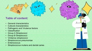
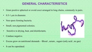
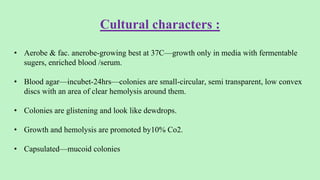
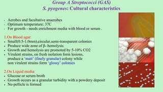
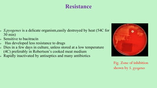
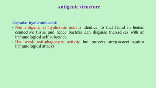
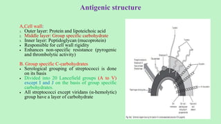
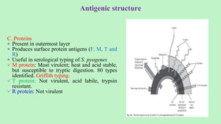
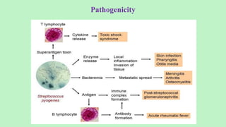
2. Table of content:
• General characteristics
• Cultural characteristics
• Pathogenic and Virulance factors
• Classification
• Group A Streptococci
• Group B Streptococci
• Viridance streptococci
• Streptococcus pneumoniae
• Enterococcus
• Streptococcus mutans and dental caries
3. • Gram positive spherical or ovoid cocci arranged in long chains, commonly in pairs.
• 0.5-1 µm in diameter.
• Non spore forming bacteria.
• Small, non pigmented colonies.
• Sensitive to drying, heat, and disinfectants.
• Catalase negative.
• Excess grow on nutritional demands – Blood , serum , sugars (only acid , no gas)
• It can be capsulated.
GENERAL CHARACTERISTICS
4. Cultural characters :
• Aerobe & fac. anerobe-growing best at 37C—growth only in media with fermentable
sugers, enriched blood /serum.
• Blood agar—incubet-24hrs—colonies are small-circular, semi transparent, low convex
discs with an area of clear hemolysis around them.
• Colonies are glistening and look like dewdrops.
• Growth and hemolysis are promoted by10% Co2.
• Capsulated—mucoid colonies
7. Streptococcus
Oxygen requirement
Aerobes & facultative anaerobes
Hemolysis
Alpha HS
Beta HS Gama HS/NHS
Obligate anaerobes
e.g. Peptostreptococci
GREEN
Partial H’lysis
Small zone
Viridans Group
Pneumococci
Clear, Complete, 2 – 4mm
Streptolysin O
O2 labile, Seen in deep subsurface colonies,
Antigenic in nature
Streptolysin – S
O2 stable, surface h’lysis, non-antigenic
20 Lancefiled groups , based on carbohydrate “C”
antigen , A-V without I & J
Group – A ( S. pyogenes )
Group B ( S Agalactiae)
Enterocococcus
Non- enterococcus group
9. Serological Lancefield Classification:
• Streptococci classified into many groups from A to V except I &J
• One or more species per group
• Classification based on C- carbohydrate antigen of cell wall
– Groupable streptococci
• A, B and D (more frequent)
• C, G and F (Less frequent)
– Non-groupable streptococci
• S. pneumoniae (pneumonia)
• viridans streptococci
– e.g. S. mutans
– Causing dental carries
Streptococci
Group A
S. pyogenes
Group B
S. agalactiae
Group C
S. equisimitis
Group D
Enterococcus
Lanciefield classification
Other groups
(E-U)
10. Group A Streptococci (GAS)
S. pyogenes: Cultural characteristics
• Aerobes and facultative anaerobes
• Optimum temperature: 37C
• For growth - needs enrichment media with blood or serum .
1.On Blood agar:
Small(0.5-1.0mm),circular,semi-transparent colonies
Produce wide zone of β- hemolysis
Growth and hemolysis are promoted by 5-10% CO2
Virulent strains, on fresh isolation form lesions,
produce a ‘matt’ (finely granular) colony while
non virulent strains form ‘glossy’ colonies
2.On Liquid media:
Glucose or serum broth
Growth occurs as a granular turbidity with a powdery deposit
No pellicle is formed
11. Resistance
S.pyogenes is a delicate organism,easily destroyed by heat (54C for
30 min)
Sensitive to bacitracin
Has developed less resistance to drugs
Dies in a few days in culture, unless stored at a low temperature
(4C) preferably in Robertson’s cooked meat medium
Rapidly inactivated by antiseptics and many antibiotics
Fig. Zone of inhibition
shown by S. pyogenes
12. Antigenic structure
Capsular hyaluronic acid:
• Non antigenic as hyaluronic acid is identical to that found in human
connective tissue and hence bacteria can disguise themselves with an
immunological self substance
• Has weak anti-phagocytic activity but protects streptococci against
immunological attacks
13. Antigenic structure
A,Cell wall:
1. Outer layer: Protein and lipoteichoic acid
2. Middle layer: Group specific carbohydrate
3. Inner layer: Peptidoglycan (mucoprotein)
Responsible for cell wall rigidity
Enhances non-specific resistance (pyrogenic
and thrombolytic activity)
B. Group specific C-carbohydrates
Serological grouping of streptococci is done
on its basis
Divided into 20 Lancefield groups (A to V)
except I and J on the basis of group specific
carbohydrates.
All streptococci except viridans (α-hemolytic)
group have a layer of carbohydrate
14. C. Proteins
Present in outermost layer
Produces surface protein antigens (F, M, T and
R)
Useful in serological typing of S. pyogenes
M protein: Most virulent; heat and acid stable,
but susceptible to tryptic digestion. 80 types
identified. Griffith typing.
T protein: Not virulent, acid labile, trypsin
resistant.
R protein: Not virulent
Antigenic structure
15. Antigenic structure
F-protein
Recognizes host fibronectin, a matrix protein that is present in eukaryotic cells. Hence
helps in attachment together with lipoteichoic acid and M protein
M protein
Most antigenic
Covered with lipoteichoic acid that enable the organism to attach to epithelial cell
M protein is heat and acid stable but susceptible to tryptic digestion
On the basis of antigenic difference in M protein, S. pyogenes can be divided into about 100
types
The most distal part of M protein shows extensive variability among strains hence
individual may suffer from recurrent S. pyogenes infections with strains expressing different
versions of M protein.
Acts as a virulence factor by inhibiting phagocytosis
T-Protein
• Common to many M- types
• Not associated with virulence and is not a protective antigen
• It is strongly antigenic
R protein
• Non-type-specific and is associated with M- proteins of types 2,3 28 and 48 known as M-
associated protein (MAP)
• Not associated to virulence and not a protective antigen
• Strongly antigenic
16. Antigenic Structure
D. Pili (Fimbriae)
Hair like and project from capsule
Consist partly of M-protein
Covered with lipoteichoic acid
Important in the attachment of streptococci to epithelial cells
17. Hemolysins (Streptolysins)
Produce complete disruption of RBC
Contribute to tissue invasion and destruction
There are two types of Streptolysins
Streptolysin O: sensitive to oxygen
Streptolysin S: insensitive to oxygen
18. Erythrogenic toxin
• Also known as pyrogenic exotoxin/ Dick/ Scarlatinal toxin
• Primary effect of the toxin is production of fever hence also called
Streptococcal pyrogenic exotoxin (SPE)
• Known as erythrogenic because its intradermal injection into
susceptible individuals produced an erythematous reaction (Dick
test)
• DICK TEST- Used to identify susceptibility to scarlet fever
• Mediate production of rash e.g. (scarlet fever)
• SUPERANTIGENS hence massive release of cytokines occur that
leads to variety of clinical signs including inflammation, shock and
organ failure
Rash in scarlet fever
19. Streptokinase (Fibrinolysin)
Two types of streptokinase (A and B)
Antigenic protein
Fibrinolysin facilitates spread of infection by breaking down the fibrin barrier
around the lesions also known as spreading factor
Promotes the lysis of human blood clot by converting plasminogen to plasmin
It is given intravenously for the treatment of early myocardial infarction and other
thromboembolic disorders
20. Deoxyribonucleases (Streptodornase, DNAase)
• Degrades DNA
• Four antigenically distinct DNAases: A,B,C,D; B most antigenic
• Capable of liquefying DNA accumulated in thick pus derived from nuclei of necrotic
cells, hence the exudate is thin in streptococcal infections
• Important therapeutically in liquefying localised collections of thick exudates
(Empyema)
• Demonstration of anti-DNAase B antibody in the diagnosis of S. pyogenes infections
when ASO titres is low
21. Hyaluronidase
• Breaks down hyaluronic acid of connective tissue and favors spread of infection
• Antigenic and specific antibodies are formed
• Degrades capsule
• Others are proteinase, phosphatase, amylase, esterases, NADase, C5a
peptidase, lipase, Serum opacity factor (SOP) etc.
22. Pathogenicity
Produces pyrogenic infection with a tendency
to spread locally, along lymphatics and
through blood stream
Disease caused can be:
1. Suppurative diseases:
a. Respiratory infections
b. Skin and soft tissue infections
c. Genital infections
2. Non suppurative sequelae:
a. Acute rheumatic fever
b. Acute glomerulonephritis
24. Group A Streptococcal Diseases
Suppurative:
1 Pharyngitis (“strep throat”)
• inflammation of the pharynx
2 Pyoderma/Impetigo
• confined, pus-producing lesion that usually occurs on the face,
arms, or legs
3 cellulitis and Necrotizing fasciitis
• toxin production destroys tissues and eventually muscle and fat
tissue
4 Toxic shock syndrome
• S. pyogenes is also known as ‘FLESH EATING BACTERIA’ -
extensive necrosis of subcutaneous and muscular tissue and
adjacent fascia – causes Toxic shock like syndrome
25. Non suppurative:
1 Scarlet fever –
• Rash that begins on the chest and spreads across the body.
• Complication of S. pyogenes pharyngitis due to specific M protein
types
• Characterized by aschoff nodules (sub cutaneous nodule)
• Aschoff bodies are nodules found in the hearts of individuals with
rheumatic fever. They result from inflammation in the heart
muscle and are characteristic of rheumatic heart disease
2 acute glomerulonephritis-
• Caused by specific nephritogenic strains of group A streptococcus
• Characterized by acute inflammation of renal glomeruli with
edema, hypertension, hematuria and proteinuria
• Sequela of both pharyngeal and pyodermal streptococcal infection
26. Treatment, prevention and control
DRUGS USED:
• For streptococcal pharyngitis: Oral penicillin V or amoxicillin
• Oral cephalosporin or macrolides can be used for penicillin sensitive patients
• For severe, systemic infection: Combined use of intravenous penicillin with protein
synthesis inhibiting antibiotics (clindamycin) is recommended
• Streptococcal pyogenes have developed resistance over tetracyclines and
sulfonamides, newer macrolides
• Antimicrobial drugs has no effect on glomerulonephritis and rheumatic fever
27. Prophylaxis
• Rheumatic fever requires long term antibiotic prophylaxis to prevent recurrence of
disease
• Penicillin is used in patients who have developed early signs of rheumatic fever
• For acute glomerulonephritis no need of antibiotic therapy and prophylactic therapy (no
re-infection)
• For patients with serious soft tissue infection, drainage and aggressive surgical
debridement must be initiated
28. Group B beta hemolytic Streptococci
Streptococcus agalactiae
• Infect newborns - Neonatal infection
• Source – Infection acquired through maternal vagina during birth
• Group B Streptococci (GBS) – puerperal sepsis, pneumonia
• Presents as meningitis, pneumonia or septicemia
• Most common cause of neonatal meningitis
• Diagnostic markers – Hippurate hydrolysis, CAMP test
29. CAMP test
• Christie, Atkins and Munch-Peterson
• When S. agalactiae is inoculated perpendicular to a streak of S. aureus grown on
blood agar an accentuated zone of hemolysis occurs
S. aureus
(Spingomyelinase C)
Group A
Streptococcus
Group B
Streptococcus
(CAMP Factor)
Enhanced Zone of
Hemolysis
30. Group C beta hemolytic Streptococci
Streptococci equisimilis
• Upper respiratory infections
• Endocarditis, osteomyelitis, brain abscess
• Treat with penicillin and gentamicin
• Source of streptokinase used for thrombolytic therapy in patients
31. Classified into 2 groups:
1. Non enterococcal group: containing S. suis, S. bovis and S. equinus.
2.The enterococcus group: which have been reclassified as a separate
genus called Enterococcus, containing- E. faecalis, E. faecium and
E. durans.
Group D Streptococci:
32. • E. faecalis and E. faecium
– Previously classified as group D streptococci but reclassified as a separate genus
– Normal microbiota of the human colon
• Opportunistic disease:
– Urinary Tact Infection
– Endocarditis
– Common cause of nosocomial infections
• Treatment: Difficult to treat due to resistance
– Ampicillin and ceftriaxone
Enterococcus
33. Alpha-Hemolytic Streptococci:
The Viridans Group
• Alpha-hemolytic (“viridans = green”)
• No Lancefield group
– Lack group-specific carbohydrates
• Normal microbiota
– mouth, pharynx, GI tract, GU tract
• Opportunistic Disease:
– One of the causes of dental caries and dental plaques; produces dextran; leads to
biofilm formation
– Can cause meningitis and endocarditis
Types:
1. S. mitis
2. S. mutans
3. S. salivarius
4. S. sanguis
34. Alpha-Hemolytic Streptococci:
Streptococcus pneumoniae
• Gram-positive diplococcus
• Alpha-hemolytic
• Normally colonizes the mouths and pharynx
• Can cause disease if travels to the lungs
• Disease is highest in children and the elderly
36. 36
Disease caused:
• Middle ear – otitis media
• Para nasal sinuses – sinusitis
• Respiratory tract – pneumonia, bronchitis, empyema
• Meningitis is secondary to otitis media, penumonia, sinusitis and conjunctivitis
Treatment:
o Penicillin
o Amoxycillin
o ceftriaxone/ceftazidime
o Vancomycin
For prophylaxis:
Pneumococcal conjugate vaccine is available
37. Lab Diagnosis of Streptococci:
Sample:Throat swab, pus / swab, Joint fluid (arthritis), Blood (septicemia) etc,
Gram Stain- Gram Positive in chains
Culture on Blood agar- hemolytic colonies, catalase negative
38.
39.
40.
41.
42. Bacitracin sensitivity
• Principle:
– for presumptive identification of group A
– distinguish between S. pyogenes from other beta hemolytic streptococci
– Strep. Pyogenes sensitive to Bacitracin giving zone of inhibition around disk
43. Our
Adviser
Mr. Alexander Lee
Lorem ipsum dolor sit
amet, consectetur
adipiscing elit, sed do
eiusmod tempor
incididunt ut labore et
dolore magna aliqua.
44. Agenda Page
Agenda 01
Lorem ipsum dolor sit
amet, consectetur
adipiscing elit.
Agenda 02
Lorem ipsum dolor sit
amet, consectetur
adipiscing elit.
Agenda 03
Lorem ipsum dolor sit
amet, consectetur
adipiscing elit.
Agenda 04
Lorem ipsum dolor sit
amet, consectetur
adipiscing elit.
45. Reminders for
this Class
Lorem ipsum dolor sit amet,
consectetur adipiscing elit.
Lorem ipsum
dolor sit amet,
consectetur
adipiscing .
Title Here Title Here Title Here
Lorem ipsum
dolor sit amet,
consectetur
adipiscing.
Lorem ipsum
dolor sit amet,
consectetur
adipiscing.
46. Lesson 01:
Classifying Viruses
Lorem ipsum dolor sit amet, consectetur
adipiscing elit, sed do eiusmod tempor
incididunt ut labore et dolore magna aliqua.
47. What is a Virus?
A virus is an infectious organism made of genetic
material surrounded by a protein coat. Viruses are
commonly classified using two key characteristics:
Type of Genetic Material and Shape.
48. Genetic
Material
Viruses can be classified
based on the type of
genetic material they
contain, either DNA
(deoxyribonucleic acid) or
RNA (ribonucliec acid).
DNA (deoxyribonucleic acid)
Double Stranded
RNA (ribonucliec acid)
Single Stranded
50. Virus
Shape
Viruses can be classified
based on their general shape.
Common virus shapes
include spherical, helical,
polyhedral or complex.
Spherical
The genetic material in a
spherical virus is protected
inside a sphere-shaped
outer envelope.
Complex
The genetic material in a
complex virus sits inside a
complex structure made up of a
head, body and legs (tail fibers).
Polyhedral
The genetic material in a
polyhedral virus is protected
inside an outer envelope with
many flat-sided faces.
Helical
The genetic material in a
helical virus is twisted
into a coil-like shape,
similar to a metal spring.
51. Lesson 02:
Bacteria Reproduction
Lorem ipsum dolor sit amet, consectetur
adipiscing elit, sed do eiusmod tempor
incididunt ut labore et dolore magna aliqua.
53. Binary Fission
Most bacteria reproduce through
binary fission. This process involves
a 'parent' bacteria cell dividing into
two identical 'daughter' cells.
Binary fission allows bacteria to rapidly
increase in number, which is important for
their survival. It is also a simple and efficient
way to reproduce that allows bacteria to adapt
quickly to changes in their environment.
Stage 01
DNA begins to replicate in the parent
cell in preparation for segregation.
Stage 02
Cells start to elongate. The
cell wall, cytoplasm and
DNA begin to separate.
Stage 03
Cell walls fully separate to form
two identical daughter cells.
54. Add topic here
Lorem ipsum dolor sit amet,
consectetur adipiscing elit, sed do
eiusmod tempor incididunt ut
labore et dolore magna aliqua.
Lorem ipsum dolor sit amet,
consectetur adipiscing elit, sed
do eiusmod tempor incididunt ut
labore et dolore magna aliqua.
Title here Title here
56. Education is the
passport to the future,
for tomorrow belongs
to those who prepare
for it today.
Malcolm X
57. Lesson 03:
Activity Time!
Lorem ipsum dolor sit amet, consectetur
adipiscing elit, sed do eiusmod tempor
incididunt ut labore et dolore magna aliqua.
58. Research Activity
Task: Complete each of the questions by conducting
research online. Make sure to keep a list of your
references in the space provided.
Question 1:
What is the basic
structure of a virus
and how does it
allow the virus to
infect cells?
Question 2:
How do viruses
replicate and
reproduce
themselves once
they have infected
a cell?
Question 3:
What are some
common illnesses
caused by viruses,
and how do they
affect the
human body?
Question 4:
How do viruses 'hide'
from our
immune system?
59. Introduce yourself!
Name 01
Lorem ipsum dolor sit amet,
consectetur adipiscing elit.
Name 02
Lorem ipsum dolor sit amet,
consectetur adipiscing elit.
Name 03
Lorem ipsum dolor sit amet,
consectetur adipiscing elit.
61. Resource
Page
Use these design resources in your
Canva Presentation.
This presentation template uses the
following free fonts:
Titles: Fredoka One Bold
Headers: Roboto Bold
Body Copy: Roboto Regular
You can find these fonts online too.
Happy designing!
Don't forget to delete this page before
presenting.
62. Credits
This presentation template is free for everyone to
use thanks to the following:
SlidesCarnival for the presentation template
Pexels and Pixabay for the photos
Happy designing!