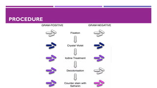
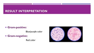
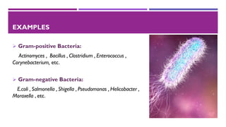
GRAM STAINING
TECHNIQUE
By Mehak Saeed
MSc. Biotechnology
University Of Karachi
INTRODUCTION
Gram staining
The Gram’s staining is an important technique in microbiology, used to differentiate
between Gram-positive organisms and Gram-negative organisms.
HISTORY
➢ In 1884, Danish scientist “Hans Christian Gram” developed the technique while
working with “Carl Friedlander” in the morgue of the city hospital in Berlin.
REAGENTS USED IN GRAM STAINING
➢ Primary stain:
CrystalViolet
➢ Mordant:
Iodine
➢ Decolorizer:
Alcohol and Acetone(95%)
➢ Counter stain:
Safranin
PRINCIPLE
➢ When the bacteria is stained with crystal violet and fixed by mordant, some bacteria
retain the primary stain while some are decolorized by alcohol.
➢ Gram-positive bacteria have thick layer of peptidoglycan and low lipid content. So,
ethanol cannot remove the crystal violet-iodine complex bound to cell wall and bacteria
appears blue or purple in color.
➢ Gram-negative bacteria have thin layer of peptidoglycan and high lipid content.The
decolorizer ethanol, dissolves the lipids in the cell wall and allows crystal violet-iodine
complex to leached out of the cell. So, bacteria appear red in color when stained with
safranin.
APPLICATIONS OF GRAM STAINING
➢ To differentiate between gram-positive and gram-negative bacteria.
➢ To determine the chemical composition of cell wall of bacteria.
➢ To select suitable culture media based on gram stain findings.
➢ To rapid presumptive diagnosis of diseases of such as bacterial meningitis.
PROCEDURE
RESULT INTERPRETATION
 Gram-positive:
Blue/purple color
 Gram-negative:
Red color
EXAMPLES
➢ Gram-positive Bacteria:
Actinomyces , Bacillus , Clostridium , Enterococcus ,
Corynebacterium, etc.
➢ Gram-negative Bacteria:
E.coli , Salmonella , Shigella , Pseudomonas , Helicobacter ,
Moraxella , etc.
Thank You

Gram staining technique

  • 1.
    GRAM STAINING TECHNIQUE By MehakSaeed MSc. Biotechnology University Of Karachi
  • 2.
    INTRODUCTION Gram staining The Gram’sstaining is an important technique in microbiology, used to differentiate between Gram-positive organisms and Gram-negative organisms.
  • 3.
    HISTORY ➢ In 1884,Danish scientist “Hans Christian Gram” developed the technique while working with “Carl Friedlander” in the morgue of the city hospital in Berlin.
  • 4.
    REAGENTS USED INGRAM STAINING ➢ Primary stain: CrystalViolet ➢ Mordant: Iodine ➢ Decolorizer: Alcohol and Acetone(95%) ➢ Counter stain: Safranin
  • 5.
    PRINCIPLE ➢ When thebacteria is stained with crystal violet and fixed by mordant, some bacteria retain the primary stain while some are decolorized by alcohol. ➢ Gram-positive bacteria have thick layer of peptidoglycan and low lipid content. So, ethanol cannot remove the crystal violet-iodine complex bound to cell wall and bacteria appears blue or purple in color. ➢ Gram-negative bacteria have thin layer of peptidoglycan and high lipid content.The decolorizer ethanol, dissolves the lipids in the cell wall and allows crystal violet-iodine complex to leached out of the cell. So, bacteria appear red in color when stained with safranin.
  • 6.
    APPLICATIONS OF GRAMSTAINING ➢ To differentiate between gram-positive and gram-negative bacteria. ➢ To determine the chemical composition of cell wall of bacteria. ➢ To select suitable culture media based on gram stain findings. ➢ To rapid presumptive diagnosis of diseases of such as bacterial meningitis.
  • 7.
  • 8.
    RESULT INTERPRETATION  Gram-positive: Blue/purplecolor  Gram-negative: Red color
  • 9.
    EXAMPLES ➢ Gram-positive Bacteria: Actinomyces, Bacillus , Clostridium , Enterococcus , Corynebacterium, etc. ➢ Gram-negative Bacteria: E.coli , Salmonella , Shigella , Pseudomonas , Helicobacter , Moraxella , etc.
  • 10.