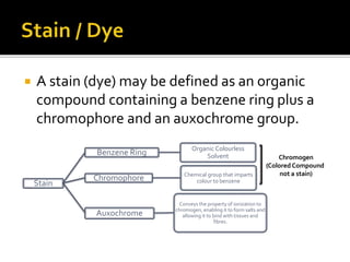
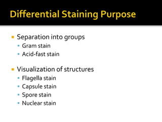
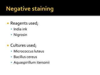
Satin, Staining of bacteria, Basics about Staining types
 To study Microbiological structures and to
divide them into different groups, staining is
used, in light microscopy.
 Numerous staining techniques are available
for visualization, differentiation, and
separation of bacteria in terms of
morphological characteristics and cellular
structures.
 A stain (dye) may be defined as an organic
compound containing a benzene ring plus a
chromophore and an auxochrome group.
Stain
Benzene Ring Organic Colourless
Solvent
Chromophore Chemical group that imparts
colour to benzene
Auxochrome
Conveys the property of ionization to
chromogen, enabling it to form salts and
allowing it to bind with tissues and
fibres.
Chromogen
(Colored Compound
not a stain)
 The ability of stain to bind with
macromolecules such as protein and nucleic
acid depends upon;
 Electrical charge on Chromogen
 Cellular components to be stained
 Acidic Stains are anionic, which means that,
on ionization of the stain, the Chromogen
portion exhibits a negative charge and
therefore has a strong affinity for the positive
constituents of the cell such as Proteins.
 Picric acid is an example of acidic stain.
 Basic stains are cationic, because on
ionization the chromogen portion exhibits a
positive charge and therefore has a strong
affinity for the negative constituents of the
cell such as Nucleic acids.
 Methylene blue is a basic stain.
 Basic stains are commonly used for bacterial
staining.
 There are two types of staining techniques;
 Simple Staining
▪ Use of Single stain
 Differential staining
▪ Use of two contrasting stains
 For visualization of morphological shape
(cocci, bacilli, and spirilli)
 arrangement (chains, clusters, pairs, and
tetrads)
 Basic stains with a positively charged
chromogen are preferred because bacterial
nucleic acids and certain cell wall
components carry a negative charge.
 Commonly used Dyes are;
 Methylene blue
 crystal violet
 carbol fuchsin
 Separation into groups
 Gram stain
 Acid-fast stain
 Visualization of structures
 Flagella stain
 Capsule stain
 Spore stain
 Nuclear stain
 Differential staining requires at least four
chemical reagents, applied sequentially to a
heat-fixed smear;
 primary stain
▪ impart its color to all cells
 decolorizing agent
▪ may or may not remove the primary stain
 Counter stain
▪ has a contrasting color to that of the primary stain
 Gram stain
▪ divides bacterial cells into two major groups, gram-positive
and gram-negative
 Negative staining requires the use of an acidic stain
The acidic stain, with its negatively charged
chromogen, will not penetrate the cells, because of
the negative charge on the surface of bacteria.
 the unstained cells are easily visible against the
colored background.
 heat fixation is not required
 Used for spiralla and fungi that are hard to stain
 Reagents used;
 India ink
 Nigrosin
 Cultures used;
 Micrococcus luteus
 Bacillus cereus
 Aquaspirillum itersonii

Staining? Importance of staining, What is Stain? Types of stain on the basis of charge, Types of staining, Simple staining, Negative staining,Differential Staining

  • 1.
    Satin, Staining ofbacteria, Basics about Staining types
  • 2.
     To studyMicrobiological structures and to divide them into different groups, staining is used, in light microscopy.  Numerous staining techniques are available for visualization, differentiation, and separation of bacteria in terms of morphological characteristics and cellular structures.
  • 3.
     A stain(dye) may be defined as an organic compound containing a benzene ring plus a chromophore and an auxochrome group. Stain Benzene Ring Organic Colourless Solvent Chromophore Chemical group that imparts colour to benzene Auxochrome Conveys the property of ionization to chromogen, enabling it to form salts and allowing it to bind with tissues and fibres. Chromogen (Colored Compound not a stain)
  • 4.
     The abilityof stain to bind with macromolecules such as protein and nucleic acid depends upon;  Electrical charge on Chromogen  Cellular components to be stained
  • 5.
     Acidic Stainsare anionic, which means that, on ionization of the stain, the Chromogen portion exhibits a negative charge and therefore has a strong affinity for the positive constituents of the cell such as Proteins.  Picric acid is an example of acidic stain.
  • 6.
     Basic stainsare cationic, because on ionization the chromogen portion exhibits a positive charge and therefore has a strong affinity for the negative constituents of the cell such as Nucleic acids.  Methylene blue is a basic stain.  Basic stains are commonly used for bacterial staining.
  • 7.
     There aretwo types of staining techniques;  Simple Staining ▪ Use of Single stain  Differential staining ▪ Use of two contrasting stains
  • 8.
     For visualizationof morphological shape (cocci, bacilli, and spirilli)  arrangement (chains, clusters, pairs, and tetrads)
  • 9.
     Basic stainswith a positively charged chromogen are preferred because bacterial nucleic acids and certain cell wall components carry a negative charge.  Commonly used Dyes are;  Methylene blue  crystal violet  carbol fuchsin
  • 10.
     Separation intogroups  Gram stain  Acid-fast stain  Visualization of structures  Flagella stain  Capsule stain  Spore stain  Nuclear stain
  • 11.
     Differential stainingrequires at least four chemical reagents, applied sequentially to a heat-fixed smear;  primary stain ▪ impart its color to all cells  decolorizing agent ▪ may or may not remove the primary stain  Counter stain ▪ has a contrasting color to that of the primary stain  Gram stain ▪ divides bacterial cells into two major groups, gram-positive and gram-negative
  • 12.
     Negative stainingrequires the use of an acidic stain The acidic stain, with its negatively charged chromogen, will not penetrate the cells, because of the negative charge on the surface of bacteria.  the unstained cells are easily visible against the colored background.  heat fixation is not required  Used for spiralla and fungi that are hard to stain
  • 13.
     Reagents used; India ink  Nigrosin  Cultures used;  Micrococcus luteus  Bacillus cereus  Aquaspirillum itersonii