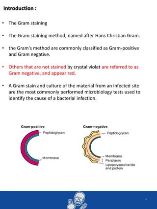
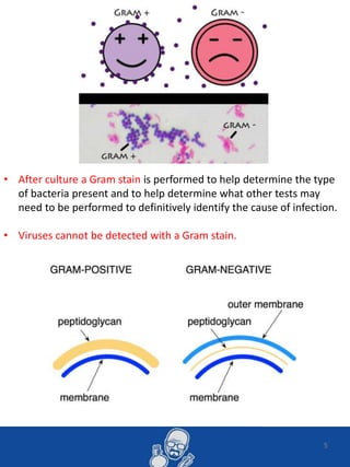
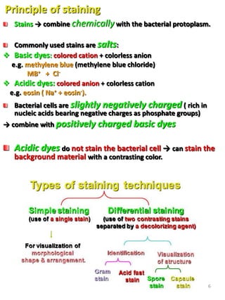
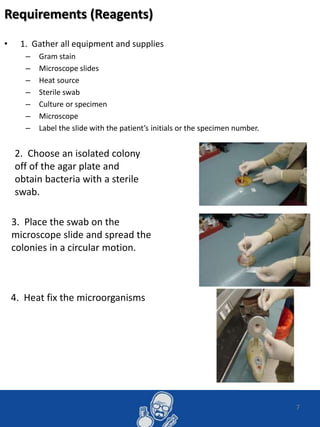
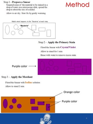
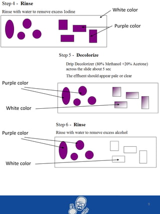
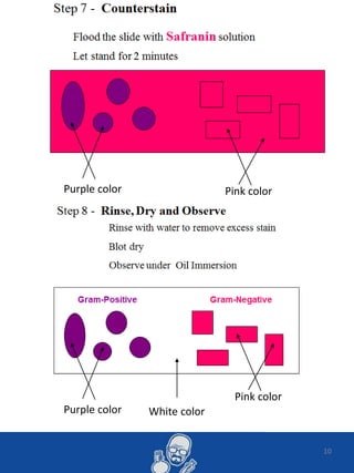
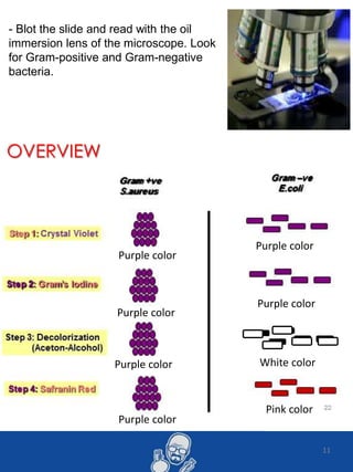
College of Medicine
Al-Imam Mohammad Bin Saud
Islamic University
First year
2013 – 2014
1
By : Asem Shadid – 435032467
Gram Staining
Method
• Asem Mustafa Shadid
• First year
• 435032467
N ame :
2
Microbiology lab report :
1- introduction (Gram Staining)
2-Requirements (Reagents)
3-Method
4-Observations
• Contents : -
3
Introduction :
4
• The Gram staining
• The Gram staining method, named after Hans Christian Gram.
• the Gram's method are commonly classified as Gram-positive
and Gram negative.
• Others that are not stained by crystal violet are referred to as
Gram negative, and appear red.
• A Gram stain and culture of the material from an infected site
are the most commonly performed microbiology tests used to
identify the cause of a bacterial infection.
5
• After culture a Gram stain is performed to help determine the type
of bacteria present and to help determine what other tests may
need to be performed to definitively identify the cause of infection.
• Viruses cannot be detected with a Gram stain.
6
Principle of staining
Stains → combine chemically with the bacterial protoplasm.
Commonly used stains are salts:
 Basic dyes: colored cation + colorless anion
e.g. methylene blue (methylene blue chloride)
MB+ + Cl-
 Acidic dyes: colored anion + colorless cation
e.g. eosin ( Na+ + eosin-).
Bacterial cells are slightly negatively charged ( rich in
nucleic acids bearing negative charges as phosphate groups)
→ combine with positively charged basic dyes
Acidic dyes do not stain the bacterial cell → can stain the
background material with a contrasting color.
7
Requirements (Reagents)
• 1. Gather all equipment and supplies
– Gram stain
– Microscope slides
– Heat source
– Sterile swab
– Culture or specimen
– Microscope
– Label the slide with the patient’s initials or the specimen number.
2. Choose an isolated colony
off of the agar plate and
obtain bacteria with a sterile
swab.
3. Place the swab on the
microscope slide and spread the
colonies in a circular motion.
4. Heat fix the microorganisms
8
Purple color
Orange color
Purple color
Method
9
Purple color
White color
Purple color
White color
Purple color
White color
10
Purple color Pink color
Pink color
Purple color White color
11
- Blot the slide and read with the oil
immersion lens of the microscope. Look
for Gram-positive and Gram-negative
bacteria.
Pink color
White color
Purple color
Purple color
Purple color
Purple color
Purple color
Purple color
OVERVIEW
12
Designed By : Asem Shadid – 435032467 13

Gram stain lab report - by asem shadid

  • 1.
    College of Medicine Al-ImamMohammad Bin Saud Islamic University First year 2013 – 2014 1 By : Asem Shadid – 435032467 Gram Staining Method
  • 2.
    • Asem MustafaShadid • First year • 435032467 N ame : 2
  • 3.
    Microbiology lab report: 1- introduction (Gram Staining) 2-Requirements (Reagents) 3-Method 4-Observations • Contents : - 3
  • 4.
    Introduction : 4 • TheGram staining • The Gram staining method, named after Hans Christian Gram. • the Gram's method are commonly classified as Gram-positive and Gram negative. • Others that are not stained by crystal violet are referred to as Gram negative, and appear red. • A Gram stain and culture of the material from an infected site are the most commonly performed microbiology tests used to identify the cause of a bacterial infection.
  • 5.
    5 • After culturea Gram stain is performed to help determine the type of bacteria present and to help determine what other tests may need to be performed to definitively identify the cause of infection. • Viruses cannot be detected with a Gram stain.
  • 6.
    6 Principle of staining Stains→ combine chemically with the bacterial protoplasm. Commonly used stains are salts:  Basic dyes: colored cation + colorless anion e.g. methylene blue (methylene blue chloride) MB+ + Cl-  Acidic dyes: colored anion + colorless cation e.g. eosin ( Na+ + eosin-). Bacterial cells are slightly negatively charged ( rich in nucleic acids bearing negative charges as phosphate groups) → combine with positively charged basic dyes Acidic dyes do not stain the bacterial cell → can stain the background material with a contrasting color.
  • 7.
    7 Requirements (Reagents) • 1.Gather all equipment and supplies – Gram stain – Microscope slides – Heat source – Sterile swab – Culture or specimen – Microscope – Label the slide with the patient’s initials or the specimen number. 2. Choose an isolated colony off of the agar plate and obtain bacteria with a sterile swab. 3. Place the swab on the microscope slide and spread the colonies in a circular motion. 4. Heat fix the microorganisms
  • 8.
  • 9.
    9 Purple color White color Purplecolor White color Purple color White color
  • 10.
    10 Purple color Pinkcolor Pink color Purple color White color
  • 11.
    11 - Blot theslide and read with the oil immersion lens of the microscope. Look for Gram-positive and Gram-negative bacteria. Pink color White color Purple color Purple color Purple color Purple color Purple color Purple color OVERVIEW
  • 12.
  • 13.
    Designed By :Asem Shadid – 435032467 13

Editor's Notes

  • #6 www.diabeteswellness.net http://www.cdc.gov/diabetes/index.htm, http://ndep.nih.gov/ , http://www.niddk.nih.gov/ r www.diabetes.org. BRIDGES www.idf.org/bridges Diabetes Africa Foot Initiative (DAFI) www.idf.org/diabetes-africa-foot-initiative Diabetes Conversations www.idf.org/node/23553 Diabetes Education www.idf.org/education Diabetes Education Modules www.idf.org/diabetes-education-modules Diabetes Score Card www.idf.org/global-diabetes-scorecard Diabetes Voice www.idf.org/diabetesvoice D-NET www.idf.org/d-net IDF Centres of Education www.idf.org/idf-centres-education
  • #10 http://www.idf.org/types-diabetes   http://en.wikipedia.org/wiki/Diabetes_mellitus#Type_1   http://www.diabetes.co.uk/index.html