SlideShare a Scribd company logo
1 of 6
Report for “Unknown” Identification Project
Jessica Olivares
Francisco Alarcon-Chaidez
12/02/2015
Negative and Positive Identification
Of Unknown Bacterial Strains Report
Introduction/Purpose
The objective of this lab was to classify the unknown bacterial strains provided utilizing a number
of biochemical procedures. In this lab each student was assigned two different strands of bacteria,
one strand being a Gram negative bacteria and the other a Gram positive bacteria. Classifying and
knowing the bacteria we had was done through a series of biochemical test. This lab project took
2 weeks of lab experimentation. Depending on each test the unknown strain transitioned to another
test whether it was a positive result or a negative result we would go from there to another test and
soon to finally identifying both of our bacteria’s. By following the procedures and methods of
inoculation the results I obtained showed that my unknown strains were positive bacteria
enterococcus faecalis, and negative bacteria Salmonella typhimurium.
Materials
Types of media used included broth tube of mixed culture unknown bacteria strain #92. Used
selective and differential media, for our unknown bacteria, such as slants, broth, and semisolid
agar plates to identify negative and positive bacteria. To describe the morphology such as the
growth, color, texture, and shape of our unknown strain we inoculated our bacteria into a
differential media grown on a TSA plate. We used selective media MacConkey Agar and
Mannitol Salt Agar for isolating and inhibit growth of the gram negative bacteria and gram
positive bacteria. MacConkey agar is used for the isolation of gram-negative bacteria and
variates lactose fermenting from lactose non-fermenting gram-negative bacteria we also used the
gram staining technique to view the selected bacteria under the microscope. Catalase test was
used to identify catalase negative bacteria the results were facultative in the production of
bubbles and bacteria that test negative do not require oxygen as a terminal electron acceptor
which anaerobes only ferment. Likewise following this we used the oxidase test as well.
Following this we also used Gram positive test to identify unknown such as the identification of
Hemolysis, Bile Esculin, DNase and the salt broth test. Gram-negative bacteria have a very thin
peptidoglycan layer that is in between an inner cell membrane and a bacterial outer membrane
fallowing the gram positive test we also conducted many of the gram negative test used to
identify the correct gram negative sample.
Methods
The lab project took 2 weeks of observation and identification of our unknown strains which began
from Tuesday 11/10/2015-Thursday 11/19/2015 and consisted of four days of working with the
bacteria sample. On the first day each student picked up an unknown mixed broth tube culture that
was selected to work for two weeks, my unknown bacteria was number 92. This sample was then
inoculated by streaking for the isolation on a TSA agar plate. We then put the inoculated plate
media into the incubator for the next lab period.
On the second lab day we took our TSA plate and inoculated the TSA plate into two mini Nutrient
Agar plates to store for next the following week, the same day we collected our results from our
TSA agar plate and we streaked our growth of bacteria from our TSA plates to be used for the
gram staining technique in order to isolate both of the organisms. We then had to inoculate the two
species to identify if our bacteria’s were Gram negative or Gram positive because from there we
would follow charts that were specific for Gram negative or Gram positive bacteria. Gram staining
is a method of determining bacterial species into two groups, it distinguishes bacteria by the
chemical and physical properties of their cell walls which detects the peptidoglycan, Gram positive
results in a purple/blue color while a Gram negative results in a pink/red color. Gram staining takes
roughly around 5-8 minutes if done correctly. After gram staining we also conducted the catalase
test which used a small sample of our unknown bacteria this test for a catalase enzyme I added a
drop of hydrogen peroxide and positive bubbling occurs to differentiate between staphylococcus
which is catalase positive and streptococcus which is catalase negative, an oxidase test was also
conducted the same day. After initial gram staining was done TSA plate was used to also streak
MAC Agar plates and MSA plates to test for positive and negative bacteria. We then disposed the
TSA plate and incubated the mini NA plates for the next lab period.
On day three we used are MAC and MSA bacterial growth results to gather the test needed to
identify both the species. We used the identification report sheet to figure out what test we needed
to conduct for the negative and positive bacteria. We again inoculated the media appropriately
using the mini NA plates that were made from the last lab period. After everything had been
properly followed and each test was conducted accordingly we then incubated all the test we
needed and disposed the 2 NA plates because we no longer needed for the experiment.
On the fourth and last day of this lab project we gathered all of our results from the incubator and
recorded them into our data sheets for proper identification. We then examined all of our
biochemical test and filled out our report sheet. After, we cleaned up our entire work station and
disposed all our media and we finished by using our identification chart to match our results with
the identity of our microbe. Finally we checked with our professor to see if our results were correct.
Results and Conclusions
From my results I obtained my unknown sample #92 Enterococcus faecalis as my positive
bacteria and Salmonella typhimurium as my negative bacteria. Below I have provide a data sheet
that illustrates my observations which also shows the conducted test and results of both of my
unknown species.
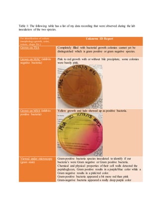
Table 1: The following table has a list of my data recording that were observed during the lab
inoculation of the two species.
For identification of culture
(morphology-growth, color,
texture, shape.Etc.)
Unknown ID Report
Grown on TSA Completely filled with bacterial growth colonies cannot yet be
distinguished which is gram positive or gram negative species.
Grown on MAC (inhibits
negative bacteria)
Pink to red growth with or without bile precipitate, some colonies
were barely pink.
Grown on MSA (inhibits
positive bacteria)
Yellow growth and halo showed up as positive bacteria.
Viewed under microscope
(gram stain)
Gram-positive bacteria species inoculated to identify if our
bacteria’s were Gram negative or Gram positive bacteria.
Chemical and physical properties of their cell walls detected the
peptidoglycan, Gram positive results in a purple/blue color while a
Gram negative results in a pink/red color.
Gram-positive bacteria appeared a bit more red than pink
Gram-negative bacteria appeared a really deep purple color
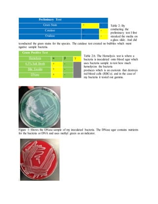
Table 2: By
conducting the
preliminary test I first
streaked the media on
a glass slide. And did
tconducted the gram stains for the species. The catalase test created no bubbles which ment
ngative sample bacteria.
Table 2A: The Hemolysis test is where a
bacteria is inoculated onto blood agar which
uses bacteria sample to test how much
hemolysins the bacteria
produces which is an exotoxin that destroys
red blood cells (RBCs); and in the case of
my bacteria it tested out gamma.
Figure 1: Shows the DNase sample of my inoculated bacteria. The DNase agar contains nutrients
for the bacteria or DNA and uses methyl green as an indicator.
:
Preliminary Test
Gram Stain + -
Catalase + -
Oxidase + -
Gram Positive Test
Hemolysis α β γ
6.5% Salt Broth + -
Bile Esculin + -
DNase + -
Figure 2: Shows the hemolysis sample collected of my inoculated bacteria. The blood agar
showed the growth of the bacteria which appears as a pink growth colonies. It showed the
exotoxins that destroys the red blood cells.
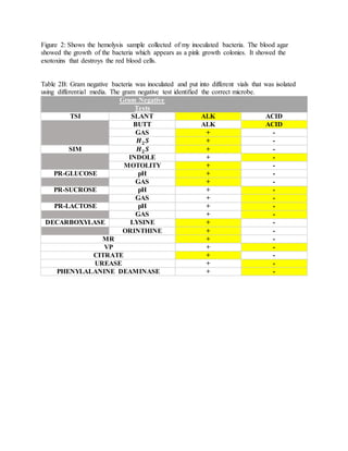
Table 2B: Gram negative bacteria was inoculated and put into different vials that was isolated
using differential media. The gram negative test identified the correct microbe.
Gram Negative
Tests
TSI SLANT ALK ACID
BUTT ALK ACID
GAS + -
𝑯 𝟐 𝑺 + -
SIM 𝑯 𝟐 𝑺 + -
INDOLE + -
MOTOLITY + -
PR-GLUCOSE pH + -
GAS + -
PR-SUCROSE pH + -
GAS + -
PR-LACTOSE pH + -
GAS + -
DECARBOXYLASE LYSINE + -
ORINTHINE + -
MR + -
VP + -
CITRATE + -
UREASE + -
PHENYLALANINE DEAMINASE + -
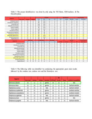
Table 3: The proper identification was done by only using the TSI Slants, SIM medium. & The
Decarboxylase.
Table 4: The following table was identified by conducting the appropriate gram stain results
fallowed by the catalase test, oxidase test and the Hemolysis test.

More Related Content

What's hot

Medical Microbiology Laboratory (biochemical tests - i)
Medical Microbiology Laboratory (biochemical tests - i)Medical Microbiology Laboratory (biochemical tests - i)
Medical Microbiology Laboratory (biochemical tests - i)Hussein Al-tameemi
 
Identification of gram positive and gram negative bacteria
Identification of gram positive and gram negative bacteriaIdentification of gram positive and gram negative bacteria
Identification of gram positive and gram negative bacteriaMicrobiology
 
Automated system for bacterial identification
Automated system for bacterial identificationAutomated system for bacterial identification
Automated system for bacterial identificationDEEKSHANT KUMAR
 
BLOOD CULTURE AS AN IMPORTANT DIAGNOSTIC TOOL IN MEDICAL MICROBIOLOGY
BLOOD CULTURE  AS AN IMPORTANT DIAGNOSTIC  TOOL IN MEDICAL MICROBIOLOGYBLOOD CULTURE  AS AN IMPORTANT DIAGNOSTIC  TOOL IN MEDICAL MICROBIOLOGY
BLOOD CULTURE AS AN IMPORTANT DIAGNOSTIC TOOL IN MEDICAL MICROBIOLOGYChelsea Osayande
 
Biochemical reactions
Biochemical reactionsBiochemical reactions
Biochemical reactionsSakeena Asmi
 
Specimen collection and transport Dr.Ashna Ajimsha
Specimen collection and transport  Dr.Ashna AjimshaSpecimen collection and transport  Dr.Ashna Ajimsha
Specimen collection and transport Dr.Ashna AjimshaAshna Ajimsha
 
Gram stain by manoj
Gram stain by manojGram stain by manoj
Gram stain by manojManoj Mahato
 
Sample collection for bacterial isolation, characterization and antibiotic se...
Sample collection for bacterial isolation, characterization and antibiotic se...Sample collection for bacterial isolation, characterization and antibiotic se...
Sample collection for bacterial isolation, characterization and antibiotic se...Bhoj Raj Singh
 
media and its types
media and its typesmedia and its types
media and its typesMicrobiology
 
Sugar fermentation tests, Cetrimide agar medium, Hugh Leifson medium
Sugar fermentation tests, Cetrimide agar medium, Hugh Leifson medium Sugar fermentation tests, Cetrimide agar medium, Hugh Leifson medium
Sugar fermentation tests, Cetrimide agar medium, Hugh Leifson medium Shivam kumar Sriwas
 
Stock culture mahadi ppt
Stock culture mahadi pptStock culture mahadi ppt
Stock culture mahadi pptDido Medo
 
Enterobacteriaceae basic properties
Enterobacteriaceae basic propertiesEnterobacteriaceae basic properties
Enterobacteriaceae basic propertiesAlia Najiha
 

What's hot (20)

Test Review
Test ReviewTest Review
Test Review
 
Api 20 e
Api 20 eApi 20 e
Api 20 e
 
Basics in Identification of Bacteria by Dr.T.V.Rao MD
Basics in Identification of Bacteria by Dr.T.V.Rao MDBasics in Identification of Bacteria by Dr.T.V.Rao MD
Basics in Identification of Bacteria by Dr.T.V.Rao MD
 
Medical Microbiology Laboratory (biochemical tests - i)
Medical Microbiology Laboratory (biochemical tests - i)Medical Microbiology Laboratory (biochemical tests - i)
Medical Microbiology Laboratory (biochemical tests - i)
 
Identification of gram positive and gram negative bacteria
Identification of gram positive and gram negative bacteriaIdentification of gram positive and gram negative bacteria
Identification of gram positive and gram negative bacteria
 
Automated system for bacterial identification
Automated system for bacterial identificationAutomated system for bacterial identification
Automated system for bacterial identification
 
BLOOD CULTURE AS AN IMPORTANT DIAGNOSTIC TOOL IN MEDICAL MICROBIOLOGY
BLOOD CULTURE  AS AN IMPORTANT DIAGNOSTIC  TOOL IN MEDICAL MICROBIOLOGYBLOOD CULTURE  AS AN IMPORTANT DIAGNOSTIC  TOOL IN MEDICAL MICROBIOLOGY
BLOOD CULTURE AS AN IMPORTANT DIAGNOSTIC TOOL IN MEDICAL MICROBIOLOGY
 
Biochemical reactions
Biochemical reactionsBiochemical reactions
Biochemical reactions
 
Specimen collection and transport Dr.Ashna Ajimsha
Specimen collection and transport  Dr.Ashna AjimshaSpecimen collection and transport  Dr.Ashna Ajimsha
Specimen collection and transport Dr.Ashna Ajimsha
 
Fungal media
Fungal mediaFungal media
Fungal media
 
Gram stain by manoj
Gram stain by manojGram stain by manoj
Gram stain by manoj
 
Sample collection for bacterial isolation, characterization and antibiotic se...
Sample collection for bacterial isolation, characterization and antibiotic se...Sample collection for bacterial isolation, characterization and antibiotic se...
Sample collection for bacterial isolation, characterization and antibiotic se...
 
Enterobacteriaceae
EnterobacteriaceaeEnterobacteriaceae
Enterobacteriaceae
 
media and its types
media and its typesmedia and its types
media and its types
 
Sugar fermentation tests, Cetrimide agar medium, Hugh Leifson medium
Sugar fermentation tests, Cetrimide agar medium, Hugh Leifson medium Sugar fermentation tests, Cetrimide agar medium, Hugh Leifson medium
Sugar fermentation tests, Cetrimide agar medium, Hugh Leifson medium
 
Biochemical test of bacteria
Biochemical test of bacteriaBiochemical test of bacteria
Biochemical test of bacteria
 
Stock culture mahadi ppt
Stock culture mahadi pptStock culture mahadi ppt
Stock culture mahadi ppt
 
identification of bacteria
identification of bacteriaidentification of bacteria
identification of bacteria
 
Mc leod
Mc leodMc leod
Mc leod
 
Enterobacteriaceae basic properties
Enterobacteriaceae basic propertiesEnterobacteriaceae basic properties
Enterobacteriaceae basic properties
 

Viewers also liked

Daufon Nano Silver Test Report
Daufon Nano Silver Test ReportDaufon Nano Silver Test Report
Daufon Nano Silver Test Reportdaufon
 
Fruit Fly Genetics lab report
Fruit Fly Genetics lab reportFruit Fly Genetics lab report
Fruit Fly Genetics lab reportJessica Olivares
 
How to write a 7th grade Lab Report
How to write a 7th grade Lab ReportHow to write a 7th grade Lab Report
How to write a 7th grade Lab ReportTman
 
Experimental Design and the Bacteria lab
Experimental Design and the Bacteria lab  Experimental Design and the Bacteria lab
Experimental Design and the Bacteria lab niall highland
 
Acid fast staining procedure for staining mycobacteria
Acid fast staining procedure for staining mycobacteriaAcid fast staining procedure for staining mycobacteria
Acid fast staining procedure for staining mycobacteriaBruno Mmassy
 
Module 5a biochemical activities
Module 5a   biochemical activitiesModule 5a   biochemical activities
Module 5a biochemical activitiesHuang Yu-Wen
 
How to use a dichotomous key (2) (5)
How to use a dichotomous key (2) (5)How to use a dichotomous key (2) (5)
How to use a dichotomous key (2) (5)kinsey2trace
 
Ib biology lab_report_sample
Ib biology lab_report_sampleIb biology lab_report_sample
Ib biology lab_report_samplenpopova
 
5th lab 1st part biochemical tests (1)
5th lab 1st part biochemical tests (1)5th lab 1st part biochemical tests (1)
5th lab 1st part biochemical tests (1)Areej Abu Hanieh
 
Module5a biochemicalactivities-120620065355-phpapp01
Module5a biochemicalactivities-120620065355-phpapp01Module5a biochemicalactivities-120620065355-phpapp01
Module5a biochemicalactivities-120620065355-phpapp01viancksislove
 
Biology - Chp 18 - Dichotomous Key - PowerPoint
Biology - Chp 18 - Dichotomous Key - PowerPointBiology - Chp 18 - Dichotomous Key - PowerPoint
Biology - Chp 18 - Dichotomous Key - PowerPointMel Anthony Pepito
 
Classification and Keys
Classification and KeysClassification and Keys
Classification and KeysMrs Parker
 
Lab Report: Isolation of Pure Culture, Gram-staining, and Microscopic Observa...
Lab Report: Isolation of Pure Culture, Gram-staining, and Microscopic Observa...Lab Report: Isolation of Pure Culture, Gram-staining, and Microscopic Observa...
Lab Report: Isolation of Pure Culture, Gram-staining, and Microscopic Observa...Annisa Hayatunnufus
 
Biochemical tests in clinical medicine lect1
Biochemical tests in clinical medicine lect1Biochemical tests in clinical medicine lect1
Biochemical tests in clinical medicine lect1MUDASSAR ANWER
 

Viewers also liked (20)

UNKNOWN BAC
UNKNOWN BACUNKNOWN BAC
UNKNOWN BAC
 
Identification of bacteria, Bacterial identification, Lab identification of b...
Identification of bacteria, Bacterial identification, Lab identification of b...Identification of bacteria, Bacterial identification, Lab identification of b...
Identification of bacteria, Bacterial identification, Lab identification of b...
 
Poster My 1m^3 24 x 36-1
Poster My 1m^3 24 x 36-1Poster My 1m^3 24 x 36-1
Poster My 1m^3 24 x 36-1
 
Daufon Nano Silver Test Report
Daufon Nano Silver Test ReportDaufon Nano Silver Test Report
Daufon Nano Silver Test Report
 
Fruit Fly Genetics lab report
Fruit Fly Genetics lab reportFruit Fly Genetics lab report
Fruit Fly Genetics lab report
 
Unknown bacteria
Unknown bacteriaUnknown bacteria
Unknown bacteria
 
How to write a 7th grade Lab Report
How to write a 7th grade Lab ReportHow to write a 7th grade Lab Report
How to write a 7th grade Lab Report
 
Experimental Design and the Bacteria lab
Experimental Design and the Bacteria lab  Experimental Design and the Bacteria lab
Experimental Design and the Bacteria lab
 
Lab Study Guide
Lab Study GuideLab Study Guide
Lab Study Guide
 
Acid fast staining procedure for staining mycobacteria
Acid fast staining procedure for staining mycobacteriaAcid fast staining procedure for staining mycobacteria
Acid fast staining procedure for staining mycobacteria
 
Module 5a biochemical activities
Module 5a   biochemical activitiesModule 5a   biochemical activities
Module 5a biochemical activities
 
How to use a dichotomous key (2) (5)
How to use a dichotomous key (2) (5)How to use a dichotomous key (2) (5)
How to use a dichotomous key (2) (5)
 
Dichotomous key
Dichotomous keyDichotomous key
Dichotomous key
 
Ib biology lab_report_sample
Ib biology lab_report_sampleIb biology lab_report_sample
Ib biology lab_report_sample
 
5th lab 1st part biochemical tests (1)
5th lab 1st part biochemical tests (1)5th lab 1st part biochemical tests (1)
5th lab 1st part biochemical tests (1)
 
Module5a biochemicalactivities-120620065355-phpapp01
Module5a biochemicalactivities-120620065355-phpapp01Module5a biochemicalactivities-120620065355-phpapp01
Module5a biochemicalactivities-120620065355-phpapp01
 
Biology - Chp 18 - Dichotomous Key - PowerPoint
Biology - Chp 18 - Dichotomous Key - PowerPointBiology - Chp 18 - Dichotomous Key - PowerPoint
Biology - Chp 18 - Dichotomous Key - PowerPoint
 
Classification and Keys
Classification and KeysClassification and Keys
Classification and Keys
 
Lab Report: Isolation of Pure Culture, Gram-staining, and Microscopic Observa...
Lab Report: Isolation of Pure Culture, Gram-staining, and Microscopic Observa...Lab Report: Isolation of Pure Culture, Gram-staining, and Microscopic Observa...
Lab Report: Isolation of Pure Culture, Gram-staining, and Microscopic Observa...
 
Biochemical tests in clinical medicine lect1
Biochemical tests in clinical medicine lect1Biochemical tests in clinical medicine lect1
Biochemical tests in clinical medicine lect1
 

Similar to Identifying Unknown Bacteria Lab

Food Standards Australia New Zealand
Food Standards Australia New ZealandFood Standards Australia New Zealand
Food Standards Australia New ZealandDeborah Gastineau
 
Practical 40 biochemical test microbiology.pdf
Practical 40 biochemical test microbiology.pdfPractical 40 biochemical test microbiology.pdf
Practical 40 biochemical test microbiology.pdfssuser668f10
 
Gram Positive Bacteria
Gram Positive BacteriaGram Positive Bacteria
Gram Positive BacteriaLaura Martin
 
Materials​ ​and ​ ​methods Describe ​ ​the ​ ​staph ​ ​st
Materials​ ​and ​ ​methods  Describe ​ ​the ​ ​staph ​ ​stMaterials​ ​and ​ ​methods  Describe ​ ​the ​ ​staph ​ ​st
Materials​ ​and ​ ​methods Describe ​ ​the ​ ​staph ​ ​stkarenahmanny4c
 
Characterization of Bacteria Isolated from Tropical Soils of Puerto Rico
Characterization of Bacteria Isolated from Tropical Soils of Puerto Rico Characterization of Bacteria Isolated from Tropical Soils of Puerto Rico
Characterization of Bacteria Isolated from Tropical Soils of Puerto Rico ramoncolon7
 
Gram, Gram And C. Gram
Gram, Gram And C. GramGram, Gram And C. Gram
Gram, Gram And C. GramKaty Allen
 
7.ann&2.ramon bacteriareportofficial
7.ann&2.ramon bacteriareportofficial7.ann&2.ramon bacteriareportofficial
7.ann&2.ramon bacteriareportofficialamgonzalezpineiro
 
Coagulase Negative Staphylococcus.docx
Coagulase Negative Staphylococcus.docxCoagulase Negative Staphylococcus.docx
Coagulase Negative Staphylococcus.docx4934bk
 
Gram positive and gram negitive bacteria
Gram positive and gram negitive bacteriaGram positive and gram negitive bacteria
Gram positive and gram negitive bacteriaCollege of Agriculture
 
Peck- Lab Report MMMB
Peck- Lab Report MMMBPeck- Lab Report MMMB
Peck- Lab Report MMMBJessica Peck
 
Clinical Micro.pptx
Clinical Micro.pptxClinical Micro.pptx
Clinical Micro.pptxssuser957fe2
 
Bacteria Classification By Gram Staining Essay
Bacteria Classification By Gram Staining EssayBacteria Classification By Gram Staining Essay
Bacteria Classification By Gram Staining EssayChristy Hunt
 
Gram Staining Paper
Gram Staining PaperGram Staining Paper
Gram Staining PaperJulie Brown
 
Microbiology Unknown Report
Microbiology Unknown ReportMicrobiology Unknown Report
Microbiology Unknown ReportMelanie Erickson
 
Identification of gram positive and gram negative bacteria
Identification of gram positive and gram negative bacteriaIdentification of gram positive and gram negative bacteria
Identification of gram positive and gram negative bacteriaMicrobiology
 
Identification of gram positive and gram negative bacteria
Identification of gram positive and gram negative bacteriaIdentification of gram positive and gram negative bacteria
Identification of gram positive and gram negative bacteriaMicrobiology
 

Similar to Identifying Unknown Bacteria Lab (20)

Staphylococcus Aureus
Staphylococcus AureusStaphylococcus Aureus
Staphylococcus Aureus
 
Food Standards Australia New Zealand
Food Standards Australia New ZealandFood Standards Australia New Zealand
Food Standards Australia New Zealand
 
Practical 40 biochemical test microbiology.pdf
Practical 40 biochemical test microbiology.pdfPractical 40 biochemical test microbiology.pdf
Practical 40 biochemical test microbiology.pdf
 
MRSA_Research_Poster
MRSA_Research_PosterMRSA_Research_Poster
MRSA_Research_Poster
 
Microbiology
MicrobiologyMicrobiology
Microbiology
 
Gram Positive Bacteria
Gram Positive BacteriaGram Positive Bacteria
Gram Positive Bacteria
 
Materials​ ​and ​ ​methods Describe ​ ​the ​ ​staph ​ ​st
Materials​ ​and ​ ​methods  Describe ​ ​the ​ ​staph ​ ​stMaterials​ ​and ​ ​methods  Describe ​ ​the ​ ​staph ​ ​st
Materials​ ​and ​ ​methods Describe ​ ​the ​ ​staph ​ ​st
 
Characterization of Bacteria Isolated from Tropical Soils of Puerto Rico
Characterization of Bacteria Isolated from Tropical Soils of Puerto Rico Characterization of Bacteria Isolated from Tropical Soils of Puerto Rico
Characterization of Bacteria Isolated from Tropical Soils of Puerto Rico
 
Gram, Gram And C. Gram
Gram, Gram And C. GramGram, Gram And C. Gram
Gram, Gram And C. Gram
 
7.ann&2.ramon bacteriareportofficial
7.ann&2.ramon bacteriareportofficial7.ann&2.ramon bacteriareportofficial
7.ann&2.ramon bacteriareportofficial
 
Coagulase Negative Staphylococcus.docx
Coagulase Negative Staphylococcus.docxCoagulase Negative Staphylococcus.docx
Coagulase Negative Staphylococcus.docx
 
Gram positive and gram negitive bacteria
Gram positive and gram negitive bacteriaGram positive and gram negitive bacteria
Gram positive and gram negitive bacteria
 
Peck- Lab Report MMMB
Peck- Lab Report MMMBPeck- Lab Report MMMB
Peck- Lab Report MMMB
 
Clinical Micro.pptx
Clinical Micro.pptxClinical Micro.pptx
Clinical Micro.pptx
 
Bacteria Classification By Gram Staining Essay
Bacteria Classification By Gram Staining EssayBacteria Classification By Gram Staining Essay
Bacteria Classification By Gram Staining Essay
 
Gram Staining Paper
Gram Staining PaperGram Staining Paper
Gram Staining Paper
 
Microbiology Unknown Report
Microbiology Unknown ReportMicrobiology Unknown Report
Microbiology Unknown Report
 
Identification of gram positive and gram negative bacteria
Identification of gram positive and gram negative bacteriaIdentification of gram positive and gram negative bacteria
Identification of gram positive and gram negative bacteria
 
Identification of gram positive and gram negative bacteria
Identification of gram positive and gram negative bacteriaIdentification of gram positive and gram negative bacteria
Identification of gram positive and gram negative bacteria
 
Strep. Agalactiae
Strep. AgalactiaeStrep. Agalactiae
Strep. Agalactiae
 

Identifying Unknown Bacteria Lab

  • 1. Report for “Unknown” Identification Project Jessica Olivares Francisco Alarcon-Chaidez 12/02/2015 Negative and Positive Identification Of Unknown Bacterial Strains Report Introduction/Purpose The objective of this lab was to classify the unknown bacterial strains provided utilizing a number of biochemical procedures. In this lab each student was assigned two different strands of bacteria, one strand being a Gram negative bacteria and the other a Gram positive bacteria. Classifying and knowing the bacteria we had was done through a series of biochemical test. This lab project took 2 weeks of lab experimentation. Depending on each test the unknown strain transitioned to another test whether it was a positive result or a negative result we would go from there to another test and soon to finally identifying both of our bacteria’s. By following the procedures and methods of inoculation the results I obtained showed that my unknown strains were positive bacteria enterococcus faecalis, and negative bacteria Salmonella typhimurium. Materials Types of media used included broth tube of mixed culture unknown bacteria strain #92. Used selective and differential media, for our unknown bacteria, such as slants, broth, and semisolid agar plates to identify negative and positive bacteria. To describe the morphology such as the growth, color, texture, and shape of our unknown strain we inoculated our bacteria into a differential media grown on a TSA plate. We used selective media MacConkey Agar and Mannitol Salt Agar for isolating and inhibit growth of the gram negative bacteria and gram positive bacteria. MacConkey agar is used for the isolation of gram-negative bacteria and variates lactose fermenting from lactose non-fermenting gram-negative bacteria we also used the gram staining technique to view the selected bacteria under the microscope. Catalase test was used to identify catalase negative bacteria the results were facultative in the production of bubbles and bacteria that test negative do not require oxygen as a terminal electron acceptor which anaerobes only ferment. Likewise following this we used the oxidase test as well. Following this we also used Gram positive test to identify unknown such as the identification of Hemolysis, Bile Esculin, DNase and the salt broth test. Gram-negative bacteria have a very thin peptidoglycan layer that is in between an inner cell membrane and a bacterial outer membrane fallowing the gram positive test we also conducted many of the gram negative test used to identify the correct gram negative sample. Methods The lab project took 2 weeks of observation and identification of our unknown strains which began from Tuesday 11/10/2015-Thursday 11/19/2015 and consisted of four days of working with the bacteria sample. On the first day each student picked up an unknown mixed broth tube culture that
  • 2. was selected to work for two weeks, my unknown bacteria was number 92. This sample was then inoculated by streaking for the isolation on a TSA agar plate. We then put the inoculated plate media into the incubator for the next lab period. On the second lab day we took our TSA plate and inoculated the TSA plate into two mini Nutrient Agar plates to store for next the following week, the same day we collected our results from our TSA agar plate and we streaked our growth of bacteria from our TSA plates to be used for the gram staining technique in order to isolate both of the organisms. We then had to inoculate the two species to identify if our bacteria’s were Gram negative or Gram positive because from there we would follow charts that were specific for Gram negative or Gram positive bacteria. Gram staining is a method of determining bacterial species into two groups, it distinguishes bacteria by the chemical and physical properties of their cell walls which detects the peptidoglycan, Gram positive results in a purple/blue color while a Gram negative results in a pink/red color. Gram staining takes roughly around 5-8 minutes if done correctly. After gram staining we also conducted the catalase test which used a small sample of our unknown bacteria this test for a catalase enzyme I added a drop of hydrogen peroxide and positive bubbling occurs to differentiate between staphylococcus which is catalase positive and streptococcus which is catalase negative, an oxidase test was also conducted the same day. After initial gram staining was done TSA plate was used to also streak MAC Agar plates and MSA plates to test for positive and negative bacteria. We then disposed the TSA plate and incubated the mini NA plates for the next lab period. On day three we used are MAC and MSA bacterial growth results to gather the test needed to identify both the species. We used the identification report sheet to figure out what test we needed to conduct for the negative and positive bacteria. We again inoculated the media appropriately using the mini NA plates that were made from the last lab period. After everything had been properly followed and each test was conducted accordingly we then incubated all the test we needed and disposed the 2 NA plates because we no longer needed for the experiment. On the fourth and last day of this lab project we gathered all of our results from the incubator and recorded them into our data sheets for proper identification. We then examined all of our biochemical test and filled out our report sheet. After, we cleaned up our entire work station and disposed all our media and we finished by using our identification chart to match our results with the identity of our microbe. Finally we checked with our professor to see if our results were correct. Results and Conclusions From my results I obtained my unknown sample #92 Enterococcus faecalis as my positive bacteria and Salmonella typhimurium as my negative bacteria. Below I have provide a data sheet that illustrates my observations which also shows the conducted test and results of both of my unknown species.
  • 3. Table 1: The following table has a list of my data recording that were observed during the lab inoculation of the two species. For identification of culture (morphology-growth, color, texture, shape.Etc.) Unknown ID Report Grown on TSA Completely filled with bacterial growth colonies cannot yet be distinguished which is gram positive or gram negative species. Grown on MAC (inhibits negative bacteria) Pink to red growth with or without bile precipitate, some colonies were barely pink. Grown on MSA (inhibits positive bacteria) Yellow growth and halo showed up as positive bacteria. Viewed under microscope (gram stain) Gram-positive bacteria species inoculated to identify if our bacteria’s were Gram negative or Gram positive bacteria. Chemical and physical properties of their cell walls detected the peptidoglycan, Gram positive results in a purple/blue color while a Gram negative results in a pink/red color. Gram-positive bacteria appeared a bit more red than pink Gram-negative bacteria appeared a really deep purple color
  • 4. Table 2: By conducting the preliminary test I first streaked the media on a glass slide. And did tconducted the gram stains for the species. The catalase test created no bubbles which ment ngative sample bacteria. Table 2A: The Hemolysis test is where a bacteria is inoculated onto blood agar which uses bacteria sample to test how much hemolysins the bacteria produces which is an exotoxin that destroys red blood cells (RBCs); and in the case of my bacteria it tested out gamma. Figure 1: Shows the DNase sample of my inoculated bacteria. The DNase agar contains nutrients for the bacteria or DNA and uses methyl green as an indicator. : Preliminary Test Gram Stain + - Catalase + - Oxidase + - Gram Positive Test Hemolysis α β γ 6.5% Salt Broth + - Bile Esculin + - DNase + -
  • 5. Figure 2: Shows the hemolysis sample collected of my inoculated bacteria. The blood agar showed the growth of the bacteria which appears as a pink growth colonies. It showed the exotoxins that destroys the red blood cells. Table 2B: Gram negative bacteria was inoculated and put into different vials that was isolated using differential media. The gram negative test identified the correct microbe. Gram Negative Tests TSI SLANT ALK ACID BUTT ALK ACID GAS + - 𝑯 𝟐 𝑺 + - SIM 𝑯 𝟐 𝑺 + - INDOLE + - MOTOLITY + - PR-GLUCOSE pH + - GAS + - PR-SUCROSE pH + - GAS + - PR-LACTOSE pH + - GAS + - DECARBOXYLASE LYSINE + - ORINTHINE + - MR + - VP + - CITRATE + - UREASE + - PHENYLALANINE DEAMINASE + -
  • 6. Table 3: The proper identification was done by only using the TSI Slants, SIM medium. & The Decarboxylase. Table 4: The following table was identified by conducting the appropriate gram stain results fallowed by the catalase test, oxidase test and the Hemolysis test.