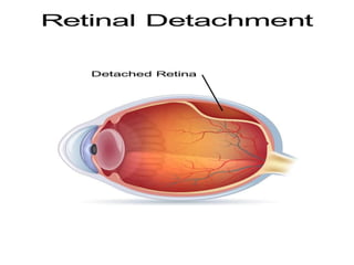
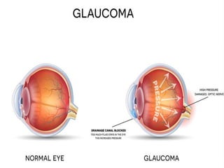
Retinal Detachment And
Glaucoma
Retinal detachment
Objectives
• What Is Retinal detachment. Types of Retinal
detachment.
• Epidemiology of Retinal detachment.
• Etiology and pathophysiology of retinal
detachment.
• Sign and symptoms and diagnosis of retinal
detachment.
• How is Retinal detachment Treated.
• Medical, Surgical and Nursing Management of
Retinal detachment.
Definition
• Retinal detachment is a serious eye condition
that happens when your retina a layer of
tissue at the back of your eye that processes
light pulls away from the tissue around it.
Types
• Retinal Detachment Causes and Types
• There are three main types of retinal
detachment:
• Rhegmatogenous. This is the most common
kind. It happens because of a retinal tear. Age
usually causes it, as the vitreous gel that fills
your eyeball pulls away from your retina. You
can also have it because of an eye injury,
surgery, or nearsightedness.
Cont..
• Tractional. This type happens when scar tissue
pulls on your retina, usually because diabetes has
damaged the blood vessels in the back of your
eye.
• Exudative. This kind happens when fluid builds
up behind your retina, but there’s no tear. The
fluid pushes your retina away from the tissue
behind it. Common causes include leaking blood
vessels and swelling because of conditions such
as an injury, inflammation, or age-related macular
degeneration.
Epidemiology
• The incidence of retinal detachment in otherwise
normal eyes is around 5 new cases in 100,000
persons per year. Detachment is more frequent in
middle-aged or elderly populations, with rates of
around 20 in 100,000 per year. The lifetime risk in
normal individuals is about 1 in 300.
• Although retinal detachment usually occurs in
just one eye, there is a 15% chance of it
developing in the other eye
Etiology
• Trauma
• Advanced diabetes
• Shrinkage of the jelly-like vitreous that fills the
inside of the eye
• Myopia
• Degenerative disorders
• Inflammation and infections
• Scarring and fibrous material due to retinopathy
and hemorrhages
• Ocular tumors
Pathophysiology
• Due to etiological factors (a torn or break in
retina)
• Vitreous fluid or serous fluid leaks in between the
layers of retina or behind the retinal layers
• Detachment of retinal layer
• Retina can peel away from the underlying layer of
blood vessels
• Lack of oxygenation in tissues of retina
• Vision disturbances
Sign & Symptoms
• Retinal detachment itself is painless. But warning
signs almost always appear before it occurs or
has advanced, such as:
• The sudden appearance of many floaters — tiny
specks that seem to drift through your field of
vision
• Flashes of light in one or both eyes (photopsia)
• Blurred vision
• Gradually reduced side (peripheral) vision
• A curtain-like shadow over your visual field
Diagnosis
• Retinal detachment can be examined by:
• Retinal examination.
• Ultrasound imaging.
• Fluorescein Angiography
• Tonometry
• Ophthalmoscopy
• Refraction Test
• Color Vision Test
• Visual Acuity
• Slit-lamp Examination
Treatment
• Your treatment may involve one or more of these
procedures:
• injecting a bubble of gas into your eye to push
the retina against the back of your eye
(pneumatic retinopexy).
• Laser (thermal) or freezing (cryopexy).
• Pneumatic retinopexy.
• Scleral buckle.
• Vitrectomy.
Medical Managements
Mydriatic, cycloplegic
Photocoagulation of retnial break
External beam radiation therapy or brachytherapy with
a plaque may be used for choroidal melanoma.
Metastatic lesions respond to chemotherapy or
localized radiation therapy.
Choroidal hemangiomas may respond to laser
photocoagulation or plaque brachytherapy.
Retinoblastomas may be shrunk with chemotherapy
and then treated locally with heat, laser, or
cryotherapy.
Surgical management
Retinal detachment:
• pneumatic retinopexy
• scleral buckling
• vitrectomy
Cont..
• Pneumatic retinopexy. This works well for a tear that’s
small and easy to close. Your doctor injects a tiny gas
bubble into your vitreous gel. It presses against the upper
part of your retina, closing the tear. You’ll need to hold your
head in a certain position for several days to keep the
bubble in the right place.
• Scleral buckle. Your doctor sews a silicone band (buckle)
around the white of your eye (called the sclera). This
pushes it toward the tear or detachment until it heals. This
band is invisible and is permanently attached.
• Vitrectomy. This surgery repairs large tears or detachment.
Your doctor removes the vitreous gel and replaces it with a
gas bubble or oil. A Vitrectomy also might require you to
hold your head in one position for some time.
Nursing Management
• Nursing Diagnosis:
• Disturbed sensory perception (visual).
• Anxiety.
• Risk for injury.
Nursing Management
• Interventions:
• Provide emotional support to the patient who may be
distraught at the potential loss of vision.
• Prepare the patient for surgery by cleaning his face and
giving him antibiotics and eyedrops, as ordered.
• Teach the patient about the role of the retina and why
floaters, flashes of light, and decreased vision occur.
• Allow the patient and family to discuss their concerns.
Complications
• Any surgical procedure has some risks. Surgery
for a detached retina can lead to:
• Infection
• Bleeding
• Higher pressure inside your eye (glaucoma)
• Fogging of the lens in your eye (cataract)
Preventions
• Get to your eye doctor right away if you see
new floaters, flashing lights, or any other
changes in your vision.
• Use protective eye wear to prevent eye
trauma.
• Control of blood sugar in diabetic patients.
• Frequent visits to eye specialist.
Objectives
• What Is Glaucoma.
• Epidemiology of glaucoma.
• What are the main causes of glaucoma.
• What Are the Symptoms of Glaucoma.
• How Is Glaucoma Diagnosed.
• How Is Glaucoma Treated.
• Medical Surgical and Nursing Management of
Glaucoma.
Introduction
• Definition:
• Glaucoma is an eye disease that can damage
your optic nerve. The optic nerve supplies
visual information to your brain from your
eyes.
• It gets worse over time. It's often linked to a
buildup of pressure inside your eye. Glaucoma
tends to run in families. You usually don’t get
it until later in life.
Epidemiology
• Globally, there are an estimated 60 million people
with glaucomatous optic neuropathy and an
estimated 8.4 million people who are blind as the
result of glaucoma. These numbers are set to
increase to 80 million and 11.2 million by 2020.
Glaucoma is the second leading cause of
blindness globally. The highest prevalence of
open-angle glaucoma occurs in Africans, and the
highest prevalence of angle-closure glaucoma
occurs in the Inuit. Population-based screening
for open-angle glaucoma is not recommended.
Screening for angle-closure may be feasible
Etiology
• Having high internal eye pressure (intraocular
pressure)
• Being over age 60.
• Being black, Asian or Hispanic.
• Having a family history of glaucoma.
• Having certain medical conditions, such as
diabetes, heart disease, high blood pressure and
sickle cell anemia.
• Having corneas that are thin in the center.
Pathophysiology
• The primary site of damage is the optic nerve
leading to loss of vision.
• In open angle glaucoma, the angle between the
cornea and iris is open.
• In this type the drainage system slowly get
clogged overtime and thus gradual increase in
pressure on optic nerve, results in dec. of
peripheral vision, as the pressure increase even
more, continuous damage to optic nerve, which
eventually leads to loss of central vision.
Cont..
• In closed angle glaucoma the angle between the iris
and cornea is too small, that means the passage way
for aqueous humor outflow is too narrow and this is
result of lens been pushed against the iris, result of this
leads to blockage of drainage system.
• This is most serious type of glaucoma in which rapid
pressure build up in the eye which can cause onset of
eye pain and redness, blurry vision, headache.
• This may occur due to dilation of lens or pupil which
cause the iris pushing forward and close the angle.
Clinical Manifestations
• Glaucoma is typically characterized by:
• IOP > 21 mmHg
• Visual field loss
• Glaucomatous retinal nerve damage
• Glaucomatous cupping (Increase in IOP pushes the
optic disc back forming an cup size).
• In close angle glaucoma the pressure lies between 50
to 80 mm Hg. And cause cornea edema.
• The pupil is vertically oval & un reactive to light and
accommodation.
Sign & Symptoms
• The most common type of glaucoma is primary open-angle glaucoma. It
has no signs or symptoms except gradual vision loss. For that reason, it’s
important that you go to yearly comprehensive eye exams so your
ophthalmologist, or eye specialist, can monitor any changes in your vision.
• Acute-angle closure glaucoma, which is also known as narrow-angle
glaucoma, is a medical emergency.it has following sign & symptoms:
• severe eye pain
• nausea
• vomiting
• redness in your eye
• sudden vision disturbances
• seeing colored rings around lights
• sudden blurred vision
Diagnosis
• Tonometry: measure intraocular pressure.
• Tonography: measure the outflow of aqueous
humor from the eye.
• Gonioscopy: is used to estimate width of the
anterior chamber angle.
• Perimetry: diagnosis of scotoma (blind spot).
• Visual field testing
• Looking for optic nerve damage, glaucomatous
cupping by imaging.
Treatment
• The treatment options for early glaucoma
have expanded in recent years and fall into
three categories:
• medications
• laser, and incisional surgery
• Medications or laser are both considered first-
line treatments. It is not imperative that you
start with medications and then proceed to
laser treatment.
Medical Management
• Most glaucoma medications are administered topically
but the absorption may occurs systemically as it passes
through the lacrimal drainage system.
• It can be overcome by applying a digital pressure on
the lacrimal sac for three minutes so that to enhance
the drug contact time with the eye is prolonged.
• Glaucoma medications should be avoided in pregnancy
if possible, with systemic carbonic anhydrase inhibitors
perhaps carrying the greatest risk due to teratogenicity
concerns.
Major Groups of Drugs Treating
Glaucoma
• Prostaglandin Analogues
• ß Blockers
• Carbonic Anhydrase Inhibitors
• Alpha 2 Agonist
• Miotics
• Combined Therapy
• Osmotic Agents
Surgical management
• Laser surgeries
• Trabeculotomy and goniotomy
• Penetrating filtering surgeriestrabeculectomy
• Non penetrating filtering surgeries
• Cyclo destructive procedures
• Artificial drainage implants
Nursing Management
• ASSESSMENT:
• History or presence of risk factor: positive
family history, tumor of eye, hemorrhage,
uveitis, trauma etc.
• Physical examination based on those in
general assessment of the eye may indicate:
blurred vision, decreased light perception
redness cloudy appearance etc.
DIAGNOSIS
• Acute pain related to increased IOP and
surgical complications as evidenced by patient
verbalization or facial expression of the
patient.
• GOAL: The pain of patient will be reduced.
INTERVENTIONS
• Monitor vital signs of the patient.
• Monitor the degree of eye pain very 30 min
during the acute phase.
• Monitor visual acuity at any time before
hatching ophthalmic agent for glaucoma.
• Maintain the bed rest in semi- fowler position.
• Give analgesic prescription and evaluation of
its effectiveness.
Glaucoma and Retinal detachment.pptx     ..

Glaucoma and Retinal detachment.pptx ..

  • 1.
  • 2.
  • 3.
    Objectives • What IsRetinal detachment. Types of Retinal detachment. • Epidemiology of Retinal detachment. • Etiology and pathophysiology of retinal detachment. • Sign and symptoms and diagnosis of retinal detachment. • How is Retinal detachment Treated. • Medical, Surgical and Nursing Management of Retinal detachment.
  • 4.
    Definition • Retinal detachmentis a serious eye condition that happens when your retina a layer of tissue at the back of your eye that processes light pulls away from the tissue around it.
  • 5.
    Types • Retinal DetachmentCauses and Types • There are three main types of retinal detachment: • Rhegmatogenous. This is the most common kind. It happens because of a retinal tear. Age usually causes it, as the vitreous gel that fills your eyeball pulls away from your retina. You can also have it because of an eye injury, surgery, or nearsightedness.
  • 6.
    Cont.. • Tractional. Thistype happens when scar tissue pulls on your retina, usually because diabetes has damaged the blood vessels in the back of your eye. • Exudative. This kind happens when fluid builds up behind your retina, but there’s no tear. The fluid pushes your retina away from the tissue behind it. Common causes include leaking blood vessels and swelling because of conditions such as an injury, inflammation, or age-related macular degeneration.
  • 7.
    Epidemiology • The incidenceof retinal detachment in otherwise normal eyes is around 5 new cases in 100,000 persons per year. Detachment is more frequent in middle-aged or elderly populations, with rates of around 20 in 100,000 per year. The lifetime risk in normal individuals is about 1 in 300. • Although retinal detachment usually occurs in just one eye, there is a 15% chance of it developing in the other eye
  • 8.
    Etiology • Trauma • Advanceddiabetes • Shrinkage of the jelly-like vitreous that fills the inside of the eye • Myopia • Degenerative disorders • Inflammation and infections • Scarring and fibrous material due to retinopathy and hemorrhages • Ocular tumors
  • 9.
    Pathophysiology • Due toetiological factors (a torn or break in retina) • Vitreous fluid or serous fluid leaks in between the layers of retina or behind the retinal layers • Detachment of retinal layer • Retina can peel away from the underlying layer of blood vessels • Lack of oxygenation in tissues of retina • Vision disturbances
  • 10.
    Sign & Symptoms •Retinal detachment itself is painless. But warning signs almost always appear before it occurs or has advanced, such as: • The sudden appearance of many floaters — tiny specks that seem to drift through your field of vision • Flashes of light in one or both eyes (photopsia) • Blurred vision • Gradually reduced side (peripheral) vision • A curtain-like shadow over your visual field
  • 11.
    Diagnosis • Retinal detachmentcan be examined by: • Retinal examination. • Ultrasound imaging. • Fluorescein Angiography • Tonometry • Ophthalmoscopy • Refraction Test • Color Vision Test • Visual Acuity • Slit-lamp Examination
  • 12.
    Treatment • Your treatmentmay involve one or more of these procedures: • injecting a bubble of gas into your eye to push the retina against the back of your eye (pneumatic retinopexy). • Laser (thermal) or freezing (cryopexy). • Pneumatic retinopexy. • Scleral buckle. • Vitrectomy.
  • 13.
    Medical Managements Mydriatic, cycloplegic Photocoagulationof retnial break External beam radiation therapy or brachytherapy with a plaque may be used for choroidal melanoma. Metastatic lesions respond to chemotherapy or localized radiation therapy. Choroidal hemangiomas may respond to laser photocoagulation or plaque brachytherapy. Retinoblastomas may be shrunk with chemotherapy and then treated locally with heat, laser, or cryotherapy.
  • 14.
    Surgical management Retinal detachment: •pneumatic retinopexy • scleral buckling • vitrectomy
  • 15.
    Cont.. • Pneumatic retinopexy.This works well for a tear that’s small and easy to close. Your doctor injects a tiny gas bubble into your vitreous gel. It presses against the upper part of your retina, closing the tear. You’ll need to hold your head in a certain position for several days to keep the bubble in the right place. • Scleral buckle. Your doctor sews a silicone band (buckle) around the white of your eye (called the sclera). This pushes it toward the tear or detachment until it heals. This band is invisible and is permanently attached. • Vitrectomy. This surgery repairs large tears or detachment. Your doctor removes the vitreous gel and replaces it with a gas bubble or oil. A Vitrectomy also might require you to hold your head in one position for some time.
  • 16.
    Nursing Management • NursingDiagnosis: • Disturbed sensory perception (visual). • Anxiety. • Risk for injury.
  • 17.
    Nursing Management • Interventions: •Provide emotional support to the patient who may be distraught at the potential loss of vision. • Prepare the patient for surgery by cleaning his face and giving him antibiotics and eyedrops, as ordered. • Teach the patient about the role of the retina and why floaters, flashes of light, and decreased vision occur. • Allow the patient and family to discuss their concerns.
  • 18.
    Complications • Any surgicalprocedure has some risks. Surgery for a detached retina can lead to: • Infection • Bleeding • Higher pressure inside your eye (glaucoma) • Fogging of the lens in your eye (cataract)
  • 19.
    Preventions • Get toyour eye doctor right away if you see new floaters, flashing lights, or any other changes in your vision. • Use protective eye wear to prevent eye trauma. • Control of blood sugar in diabetic patients. • Frequent visits to eye specialist.
  • 21.
    Objectives • What IsGlaucoma. • Epidemiology of glaucoma. • What are the main causes of glaucoma. • What Are the Symptoms of Glaucoma. • How Is Glaucoma Diagnosed. • How Is Glaucoma Treated. • Medical Surgical and Nursing Management of Glaucoma.
  • 22.
    Introduction • Definition: • Glaucomais an eye disease that can damage your optic nerve. The optic nerve supplies visual information to your brain from your eyes. • It gets worse over time. It's often linked to a buildup of pressure inside your eye. Glaucoma tends to run in families. You usually don’t get it until later in life.
  • 23.
    Epidemiology • Globally, thereare an estimated 60 million people with glaucomatous optic neuropathy and an estimated 8.4 million people who are blind as the result of glaucoma. These numbers are set to increase to 80 million and 11.2 million by 2020. Glaucoma is the second leading cause of blindness globally. The highest prevalence of open-angle glaucoma occurs in Africans, and the highest prevalence of angle-closure glaucoma occurs in the Inuit. Population-based screening for open-angle glaucoma is not recommended. Screening for angle-closure may be feasible
  • 24.
    Etiology • Having highinternal eye pressure (intraocular pressure) • Being over age 60. • Being black, Asian or Hispanic. • Having a family history of glaucoma. • Having certain medical conditions, such as diabetes, heart disease, high blood pressure and sickle cell anemia. • Having corneas that are thin in the center.
  • 25.
    Pathophysiology • The primarysite of damage is the optic nerve leading to loss of vision. • In open angle glaucoma, the angle between the cornea and iris is open. • In this type the drainage system slowly get clogged overtime and thus gradual increase in pressure on optic nerve, results in dec. of peripheral vision, as the pressure increase even more, continuous damage to optic nerve, which eventually leads to loss of central vision.
  • 26.
    Cont.. • In closedangle glaucoma the angle between the iris and cornea is too small, that means the passage way for aqueous humor outflow is too narrow and this is result of lens been pushed against the iris, result of this leads to blockage of drainage system. • This is most serious type of glaucoma in which rapid pressure build up in the eye which can cause onset of eye pain and redness, blurry vision, headache. • This may occur due to dilation of lens or pupil which cause the iris pushing forward and close the angle.
  • 27.
    Clinical Manifestations • Glaucomais typically characterized by: • IOP > 21 mmHg • Visual field loss • Glaucomatous retinal nerve damage • Glaucomatous cupping (Increase in IOP pushes the optic disc back forming an cup size). • In close angle glaucoma the pressure lies between 50 to 80 mm Hg. And cause cornea edema. • The pupil is vertically oval & un reactive to light and accommodation.
  • 28.
    Sign & Symptoms •The most common type of glaucoma is primary open-angle glaucoma. It has no signs or symptoms except gradual vision loss. For that reason, it’s important that you go to yearly comprehensive eye exams so your ophthalmologist, or eye specialist, can monitor any changes in your vision. • Acute-angle closure glaucoma, which is also known as narrow-angle glaucoma, is a medical emergency.it has following sign & symptoms: • severe eye pain • nausea • vomiting • redness in your eye • sudden vision disturbances • seeing colored rings around lights • sudden blurred vision
  • 29.
    Diagnosis • Tonometry: measureintraocular pressure. • Tonography: measure the outflow of aqueous humor from the eye. • Gonioscopy: is used to estimate width of the anterior chamber angle. • Perimetry: diagnosis of scotoma (blind spot). • Visual field testing • Looking for optic nerve damage, glaucomatous cupping by imaging.
  • 30.
    Treatment • The treatmentoptions for early glaucoma have expanded in recent years and fall into three categories: • medications • laser, and incisional surgery • Medications or laser are both considered first- line treatments. It is not imperative that you start with medications and then proceed to laser treatment.
  • 31.
    Medical Management • Mostglaucoma medications are administered topically but the absorption may occurs systemically as it passes through the lacrimal drainage system. • It can be overcome by applying a digital pressure on the lacrimal sac for three minutes so that to enhance the drug contact time with the eye is prolonged. • Glaucoma medications should be avoided in pregnancy if possible, with systemic carbonic anhydrase inhibitors perhaps carrying the greatest risk due to teratogenicity concerns.
  • 32.
    Major Groups ofDrugs Treating Glaucoma • Prostaglandin Analogues • ß Blockers • Carbonic Anhydrase Inhibitors • Alpha 2 Agonist • Miotics • Combined Therapy • Osmotic Agents
  • 33.
    Surgical management • Lasersurgeries • Trabeculotomy and goniotomy • Penetrating filtering surgeriestrabeculectomy • Non penetrating filtering surgeries • Cyclo destructive procedures • Artificial drainage implants
  • 34.
    Nursing Management • ASSESSMENT: •History or presence of risk factor: positive family history, tumor of eye, hemorrhage, uveitis, trauma etc. • Physical examination based on those in general assessment of the eye may indicate: blurred vision, decreased light perception redness cloudy appearance etc.
  • 35.
    DIAGNOSIS • Acute painrelated to increased IOP and surgical complications as evidenced by patient verbalization or facial expression of the patient. • GOAL: The pain of patient will be reduced.
  • 36.
    INTERVENTIONS • Monitor vitalsigns of the patient. • Monitor the degree of eye pain very 30 min during the acute phase. • Monitor visual acuity at any time before hatching ophthalmic agent for glaucoma. • Maintain the bed rest in semi- fowler position. • Give analgesic prescription and evaluation of its effectiveness.