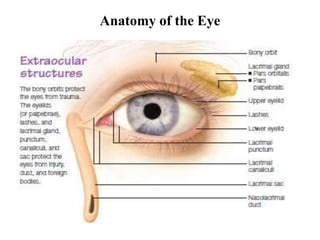
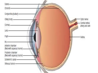
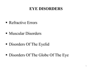
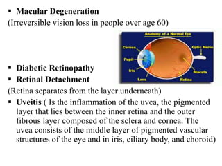
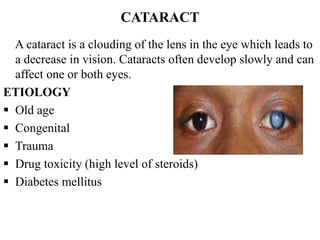
DISORDERS(INFECTIONS/INFLAMMATIONS)
OF THE EYE
by
Shahla Arshad
Lecturer
INS-KMU
Anatomy of the Eye
Eyelids
Each upper eyelid should cover the top quarter of the iris so
the eyes look similar.
Conjunctivae and sclera
The conjunctivae should be clear and shiny. The sclera should be white .
Pupils
Each pupil should be round and equal in size .
Retina
The retina should have a uniform color and be free from scars and
pigmentation
Macula
The part of the eye most sensitive to light. It appears as a darker
structure, free from blood vessels
5
EYE DISORDERS
 Refractive Errors
 Muscular Disorders
 Disorders Of The Eyelid
 Disorders Of The Globe Of The Eye
Refractive Errors of Eye
Refractive error means that the shape of your eye does not
bend light correctly, resulting in a blurred image. The main
types of refractive errors are:
 Myopia (Nearsightedness)
 Hyperopia (Farsightedness)
 Presbyopia (loss of near vision with age)
 Astigmatism (causes blurred vision)
MUSCULAR DISORDERS
 Nystagmus: (is a condition in which the eyes make repetitive,
uncontrolled movements. These involuntary eye movements can occur
from side to side, up and down, or in a circular pattern)
 Strabismus (Crossed Eyes)
DISORDERS OF THE EYE LID
 Hordeolum (Stye)
 Chalazion (Meibomian Cyst)
 Blepharitis
 Entropion (The eyelid usually the lower lid folds
inward)
 Ectropion (lower eyelid turns outward, away from
your eye)
 Conjunctivitis (Pink Eye)
DISORDERS OF THE GLOBE OF THE EYE
 Keratitis (inflammation of the cornea of the eye)
 Corneal Abrasion Or Ulcer
 Scleritis (inflammation of sclera)
 Cataract
 Glaucoma
 Macular Degeneration
(Irreversible vision loss in people over age 60)
 Diabetic Retinopathy
 Retinal Detachment
(Retina separates from the layer underneath)
 Uveitis ( Is the inflammation of the uvea, the pigmented
layer that lies between the inner retina and the outer
fibrous layer composed of the sclera and cornea. The
uvea consists of the middle layer of pigmented vascular
structures of the eye and in iris, ciliary body, and choroid)
HORDEOLUM (STYE)
Is a common disorder of the eyelid or Inflammatory
infection of the hair follicle of the eye lid
ETIOLOGY
 Staphylococcal infection
 Usually associated with Blepharitis
SYMPTOMS AND SIGNS
 Occurs on the outside
 Pain/swelling/redness/pus
 patient feels something in the eye
DIAGNOSIS
 Visual exam
 Culture if needed
TREATMENT
Medical therapy for Hordeolum includes
 Eyelid hygiene (lid scrubs)
 Hot compress to alleviate pain
 Massages of the lesions for 10 minutes 4 times per day.
 Topical or systemic antibiotics
CHALAZION (MEIBOMIAN CYST)
 A chalazion is a benign, painless bump or nodule inside
the upper or lower eyelid. These cyst-like nodules form
around an oil gland (meibomian) within the eyelid,
resulting in red, swollen eyelids. chalazion is Collection
of fluid or soft mass cyst.
ETIOLOGY
 Blockage of meibomian gland
SYMPTOMS AND SIGNS
 Pea size cyst
 painless slow swelling of the inner part of eye lid
Could become infected
DIAGNOSIS
 Visual Examination
TREATMENT
 small ones usually disappear spontaneously after a
month or two
 Warm compresses. Soak a clean washcloth in hot
water and hold it to your eyelid for 10–15 minutes at
a time, 3–5 times a day.
 Antibiotics
 large ones usually need surgical removal
 Do not squeeze or try to pop a stye or chalazion.
BLEPHARITIS
Blepharitis is inflammation of the of the margins of the
eyelids. Blepharitis usually involves the part of the eyelid where the
eyelashes grow and affects both eyelids. it is commonly occurs when
tiny oil glands located near the base of the eyelashes become clogged.
ETIOLOGY
 Staphylococcus bacterial infection
 Allergies, smoke, dust, chemicals,
seborrhea, stye, chalazions.
SYMPTOMS AND SIGNS
 Persistent redness & crusting on eyelids
 Itching / burning sensation
 Feeling something in the eye
 Ulcers can cause eye lashes to fall out
 Scales can get into eye causing conjunctivitis
DIAGNOSIS
 visual examination
TREATMENT
 Apply a warm compress over your closed eye for several
minutes to loosen the crusty deposits on your eyelids.
 Immediately afterward, use a washcloth moistened with warm
water and a few drops of diluted baby shampoo to wash away
any oily debris or scales at the base of your eyelashes.
• Medications that fight against infection including eye drops,
creams and ointments. If you don't respond to topical
antibiotics, you may suggest an oral antibiotic.
• Steroid eye drops or ointments may help control inflammation.
• Blepharitis caused by seborrheic dermatitis, rosacea or other
diseases may be controlled by treating the underlying disease.
BLEPHAROPTOSIS (PTOSIS)
weakness of eye muscle. Blepharoptosis is an abnormal
low-lying upper eyelid margin that raises eyelid
(superior rectus, superior oblique)
ETIOLOGY
 Caused by aging
 Trauma or any eye injury.
 Diabetes mellitus
 Muscular dystrophy
 Myasthenia gravis
 Brain tumors
 Caused by more serious conditions, such as a stroke, brain
tumor, or cancer of the nerves or muscles.
 Side effect after certain eye surgery
SYMPTOMS AND SIGNS
 Drooping eye
 Blocks vision
DIAGNOSIS
 Ophthalmic examination
 Blood test to rule out underlying disease
TREATMENT
 Eye glasses with raised eyelid support
 Treat underlying disease
 Surgery (strengthen muscles)
CONJUNCTIVITIS (PINK EYE)
An inflammation or swelling of the conjunctiva
ETIOLOGY
 Viral / bacterial
 Irritants (allergies, chemicals, UV light)
SYMPTOMS AND SIGNS
 Redness / swelling / itching
 Tearing when exposed to light
 Pus if infectious
 “Contagious” with contaminated
hands.
DIAGNOSIS
 Ophthalmic examination
 Culture discharge
TREATMENT
 Protect your eyes from dirt and other irritating substances.
 Avoid the use of makeup.
 Remove contact lenses if you wear them.
 Warm compress 3-4 times daily (10-15 min.)
 If bacterial (antibiotic eye drops or ointment ). Bacterial conjunctivitis
may improve after three or four days of treatment,
 Eye drops may help relieve itching and burning from the irritating
substances causing your pink eye.
 If viral- self limiting
Applying Eye Drop Medicine
STEP : 1
Tilt your head back. Using your middle finger, gently press the
corner of the eye by the side of the nose.
STEP : 2
Use your index finger to pull down the lower lid.
Then apply the eye drop medicine.
STEP: 3
After applying the eye drop, let go of your lower lid. Close the
eye and keep the middle finger in place for at least two minutes.
If you’re applying more than one type of drop, wait at least 15
minutes for the next application. Use a tissue to wipe away
excess drops on eyelids.
CATARACT
A cataract is a clouding of the lens in the eye which leads to
a decrease in vision. Cataracts often develop slowly and can
affect one or both eyes.
ETIOLOGY
 Old age
 Congenital
 Trauma
 Drug toxicity (high level of steroids)
 Diabetes mellitus
SYMPTOMS AND SIGNS
 Cloudy / white opaque area of the lens
 Reduce visual acuity
 Blurring of vision
 Photosensitivity
 Trouble seeing at night
DIAGNOSIS
 Visual examination
TREATMENT
 The symptoms of early cataract may be improved with new
eyeglasses.
 Surgery is the only effective treatment. Surgery involves
removing the cloudy lens and replacing it with an artificial
lens.
 Intra-capsular phacoemulsification
(involves breakage of cataract then aspiration)
 Extra-capsular phacoemulsification:
(artificial lens replacement)
GLAUCOMA
Glaucoma is an eye disease that is often associated with
elevated intraocular pressure, in which damage to the
eye (optic) nerve can lead to loss of vision and even
blindness. Glaucoma is the leading cause of irreversible
blindness in the world.
Chronic Open-Angle Glaucoma
Increased intraocular pressure due to a malfunction in
eyes aqueous humor drainage system - can lead to optic
nerve damage
ETIOLOGY
 Trauma
 Overuse of steroids
SYMPTOMS AND SIGNS
 Gradual loss of peripheral vision.
 If untreated - eventually complete vision loss
DIAGNOSIS
 Ophthalmic examination
 Tonometry (pressure measure)
TREATMENT
 Medication that helps decrease aqueous humor
production or opens drainage system
 Laser to open drainage
 Surgery (bypass)
Normal IOP 15 if IOP is 21 or 22
it means patient will develop
Glaucoma
Acute Angle-Closure Glaucoma
 Complete blockage of aqueous humor drainage
system
ETIOLOGY
 Trauma
SYMPTOMS AND SIGNS
 Blurred vision
 Severe eye pain
 Redness of the eye
 Nausea & vomiting
 Photophobia
 Hazy cornea
 If untreated --> blindness
DIAGNOSIS
 Goniolens (special lens to view the opening)
TREATMENT
 LASER IRIDOTOMY (creation of a hole in the iris between the
anterior and posterior chamber)
 Medications to reduce pressure
ENT.pptx                                ..

ENT.pptx ..

  • 1.
  • 2.
  • 4.
    Eyelids Each upper eyelidshould cover the top quarter of the iris so the eyes look similar. Conjunctivae and sclera The conjunctivae should be clear and shiny. The sclera should be white . Pupils Each pupil should be round and equal in size . Retina The retina should have a uniform color and be free from scars and pigmentation Macula The part of the eye most sensitive to light. It appears as a darker structure, free from blood vessels
  • 5.
    5 EYE DISORDERS  RefractiveErrors  Muscular Disorders  Disorders Of The Eyelid  Disorders Of The Globe Of The Eye
  • 6.
    Refractive Errors ofEye Refractive error means that the shape of your eye does not bend light correctly, resulting in a blurred image. The main types of refractive errors are:  Myopia (Nearsightedness)  Hyperopia (Farsightedness)  Presbyopia (loss of near vision with age)  Astigmatism (causes blurred vision)
  • 7.
    MUSCULAR DISORDERS  Nystagmus:(is a condition in which the eyes make repetitive, uncontrolled movements. These involuntary eye movements can occur from side to side, up and down, or in a circular pattern)  Strabismus (Crossed Eyes) DISORDERS OF THE EYE LID  Hordeolum (Stye)  Chalazion (Meibomian Cyst)  Blepharitis
  • 8.
     Entropion (Theeyelid usually the lower lid folds inward)  Ectropion (lower eyelid turns outward, away from your eye)  Conjunctivitis (Pink Eye)
  • 9.
    DISORDERS OF THEGLOBE OF THE EYE  Keratitis (inflammation of the cornea of the eye)  Corneal Abrasion Or Ulcer  Scleritis (inflammation of sclera)  Cataract  Glaucoma
  • 10.
     Macular Degeneration (Irreversiblevision loss in people over age 60)  Diabetic Retinopathy  Retinal Detachment (Retina separates from the layer underneath)  Uveitis ( Is the inflammation of the uvea, the pigmented layer that lies between the inner retina and the outer fibrous layer composed of the sclera and cornea. The uvea consists of the middle layer of pigmented vascular structures of the eye and in iris, ciliary body, and choroid)
  • 11.
    HORDEOLUM (STYE) Is acommon disorder of the eyelid or Inflammatory infection of the hair follicle of the eye lid ETIOLOGY  Staphylococcal infection  Usually associated with Blepharitis SYMPTOMS AND SIGNS  Occurs on the outside  Pain/swelling/redness/pus  patient feels something in the eye
  • 12.
    DIAGNOSIS  Visual exam Culture if needed TREATMENT Medical therapy for Hordeolum includes  Eyelid hygiene (lid scrubs)  Hot compress to alleviate pain  Massages of the lesions for 10 minutes 4 times per day.  Topical or systemic antibiotics
  • 13.
    CHALAZION (MEIBOMIAN CYST) A chalazion is a benign, painless bump or nodule inside the upper or lower eyelid. These cyst-like nodules form around an oil gland (meibomian) within the eyelid, resulting in red, swollen eyelids. chalazion is Collection of fluid or soft mass cyst. ETIOLOGY  Blockage of meibomian gland SYMPTOMS AND SIGNS  Pea size cyst  painless slow swelling of the inner part of eye lid Could become infected
  • 14.
    DIAGNOSIS  Visual Examination TREATMENT small ones usually disappear spontaneously after a month or two  Warm compresses. Soak a clean washcloth in hot water and hold it to your eyelid for 10–15 minutes at a time, 3–5 times a day.  Antibiotics  large ones usually need surgical removal  Do not squeeze or try to pop a stye or chalazion.
  • 15.
    BLEPHARITIS Blepharitis is inflammationof the of the margins of the eyelids. Blepharitis usually involves the part of the eyelid where the eyelashes grow and affects both eyelids. it is commonly occurs when tiny oil glands located near the base of the eyelashes become clogged. ETIOLOGY  Staphylococcus bacterial infection  Allergies, smoke, dust, chemicals, seborrhea, stye, chalazions. SYMPTOMS AND SIGNS  Persistent redness & crusting on eyelids  Itching / burning sensation  Feeling something in the eye  Ulcers can cause eye lashes to fall out  Scales can get into eye causing conjunctivitis
  • 16.
    DIAGNOSIS  visual examination TREATMENT Apply a warm compress over your closed eye for several minutes to loosen the crusty deposits on your eyelids.  Immediately afterward, use a washcloth moistened with warm water and a few drops of diluted baby shampoo to wash away any oily debris or scales at the base of your eyelashes. • Medications that fight against infection including eye drops, creams and ointments. If you don't respond to topical antibiotics, you may suggest an oral antibiotic. • Steroid eye drops or ointments may help control inflammation. • Blepharitis caused by seborrheic dermatitis, rosacea or other diseases may be controlled by treating the underlying disease.
  • 17.
    BLEPHAROPTOSIS (PTOSIS) weakness ofeye muscle. Blepharoptosis is an abnormal low-lying upper eyelid margin that raises eyelid (superior rectus, superior oblique) ETIOLOGY  Caused by aging  Trauma or any eye injury.  Diabetes mellitus  Muscular dystrophy  Myasthenia gravis  Brain tumors  Caused by more serious conditions, such as a stroke, brain tumor, or cancer of the nerves or muscles.  Side effect after certain eye surgery
  • 18.
    SYMPTOMS AND SIGNS Drooping eye  Blocks vision DIAGNOSIS  Ophthalmic examination  Blood test to rule out underlying disease TREATMENT  Eye glasses with raised eyelid support  Treat underlying disease  Surgery (strengthen muscles)
  • 19.
    CONJUNCTIVITIS (PINK EYE) Aninflammation or swelling of the conjunctiva ETIOLOGY  Viral / bacterial  Irritants (allergies, chemicals, UV light) SYMPTOMS AND SIGNS  Redness / swelling / itching  Tearing when exposed to light  Pus if infectious  “Contagious” with contaminated hands.
  • 20.
    DIAGNOSIS  Ophthalmic examination Culture discharge TREATMENT  Protect your eyes from dirt and other irritating substances.  Avoid the use of makeup.  Remove contact lenses if you wear them.  Warm compress 3-4 times daily (10-15 min.)  If bacterial (antibiotic eye drops or ointment ). Bacterial conjunctivitis may improve after three or four days of treatment,  Eye drops may help relieve itching and burning from the irritating substances causing your pink eye.  If viral- self limiting
  • 21.
    Applying Eye DropMedicine STEP : 1 Tilt your head back. Using your middle finger, gently press the corner of the eye by the side of the nose. STEP : 2 Use your index finger to pull down the lower lid. Then apply the eye drop medicine. STEP: 3 After applying the eye drop, let go of your lower lid. Close the eye and keep the middle finger in place for at least two minutes. If you’re applying more than one type of drop, wait at least 15 minutes for the next application. Use a tissue to wipe away excess drops on eyelids.
  • 22.
    CATARACT A cataract isa clouding of the lens in the eye which leads to a decrease in vision. Cataracts often develop slowly and can affect one or both eyes. ETIOLOGY  Old age  Congenital  Trauma  Drug toxicity (high level of steroids)  Diabetes mellitus
  • 23.
    SYMPTOMS AND SIGNS Cloudy / white opaque area of the lens  Reduce visual acuity  Blurring of vision  Photosensitivity  Trouble seeing at night DIAGNOSIS  Visual examination TREATMENT  The symptoms of early cataract may be improved with new eyeglasses.  Surgery is the only effective treatment. Surgery involves removing the cloudy lens and replacing it with an artificial lens.
  • 24.
     Intra-capsular phacoemulsification (involvesbreakage of cataract then aspiration)  Extra-capsular phacoemulsification: (artificial lens replacement)
  • 25.
    GLAUCOMA Glaucoma is aneye disease that is often associated with elevated intraocular pressure, in which damage to the eye (optic) nerve can lead to loss of vision and even blindness. Glaucoma is the leading cause of irreversible blindness in the world. Chronic Open-Angle Glaucoma Increased intraocular pressure due to a malfunction in eyes aqueous humor drainage system - can lead to optic nerve damage ETIOLOGY  Trauma  Overuse of steroids
  • 26.
    SYMPTOMS AND SIGNS Gradual loss of peripheral vision.  If untreated - eventually complete vision loss DIAGNOSIS  Ophthalmic examination  Tonometry (pressure measure) TREATMENT  Medication that helps decrease aqueous humor production or opens drainage system  Laser to open drainage  Surgery (bypass)
  • 27.
    Normal IOP 15if IOP is 21 or 22 it means patient will develop Glaucoma
  • 28.
    Acute Angle-Closure Glaucoma Complete blockage of aqueous humor drainage system ETIOLOGY  Trauma
  • 29.
    SYMPTOMS AND SIGNS Blurred vision  Severe eye pain  Redness of the eye  Nausea & vomiting  Photophobia  Hazy cornea  If untreated --> blindness DIAGNOSIS  Goniolens (special lens to view the opening) TREATMENT  LASER IRIDOTOMY (creation of a hole in the iris between the anterior and posterior chamber)  Medications to reduce pressure