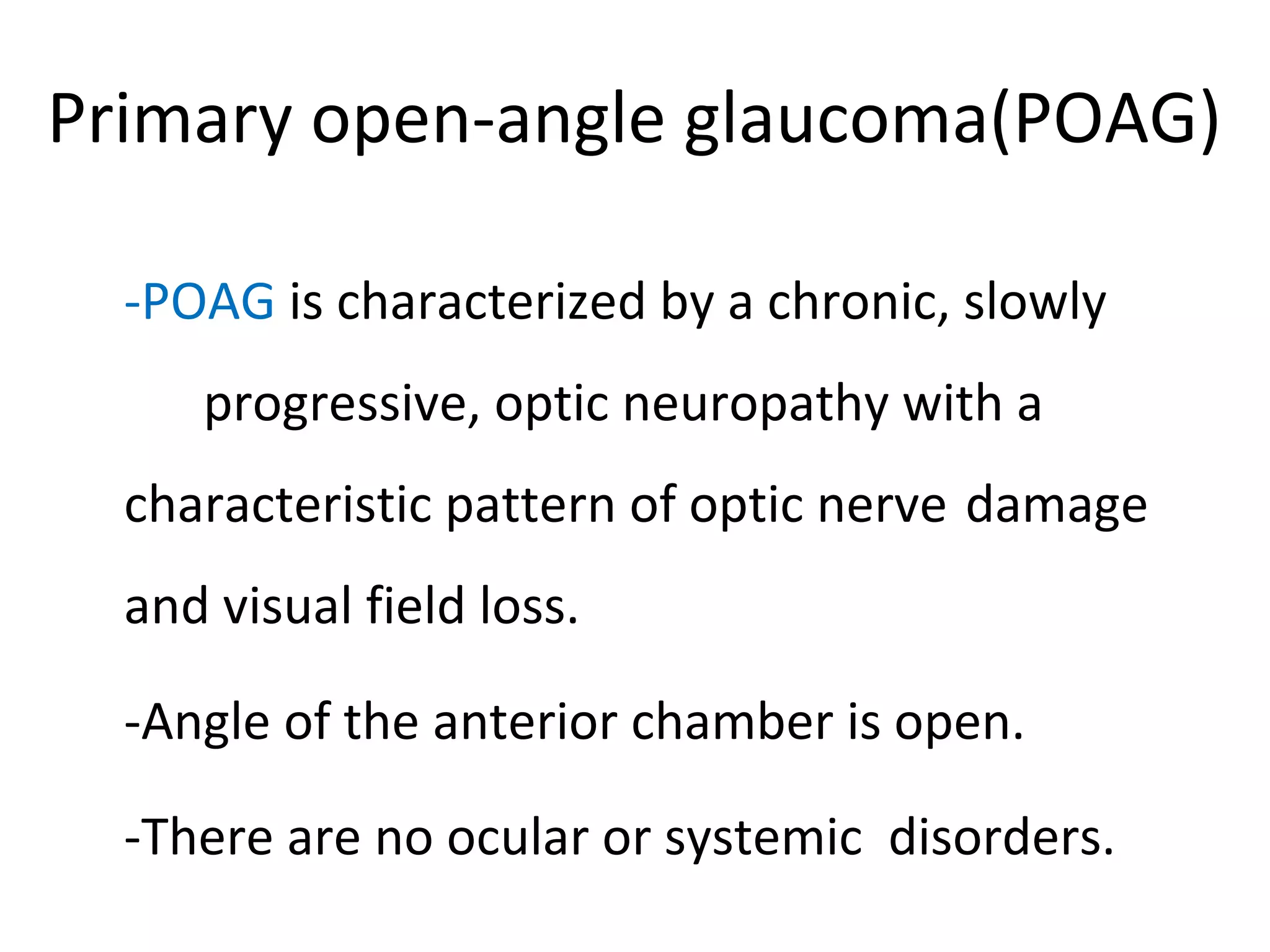
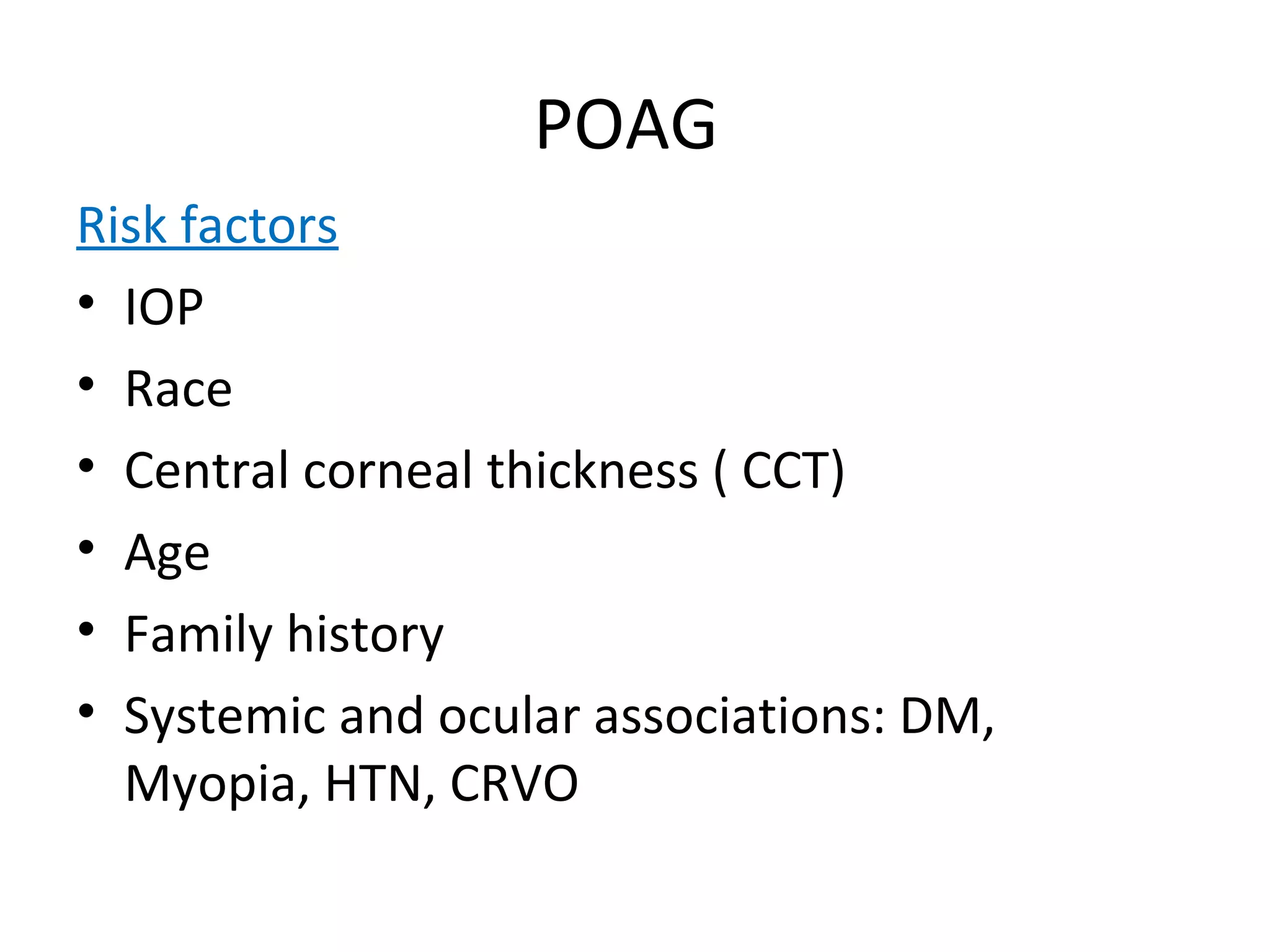
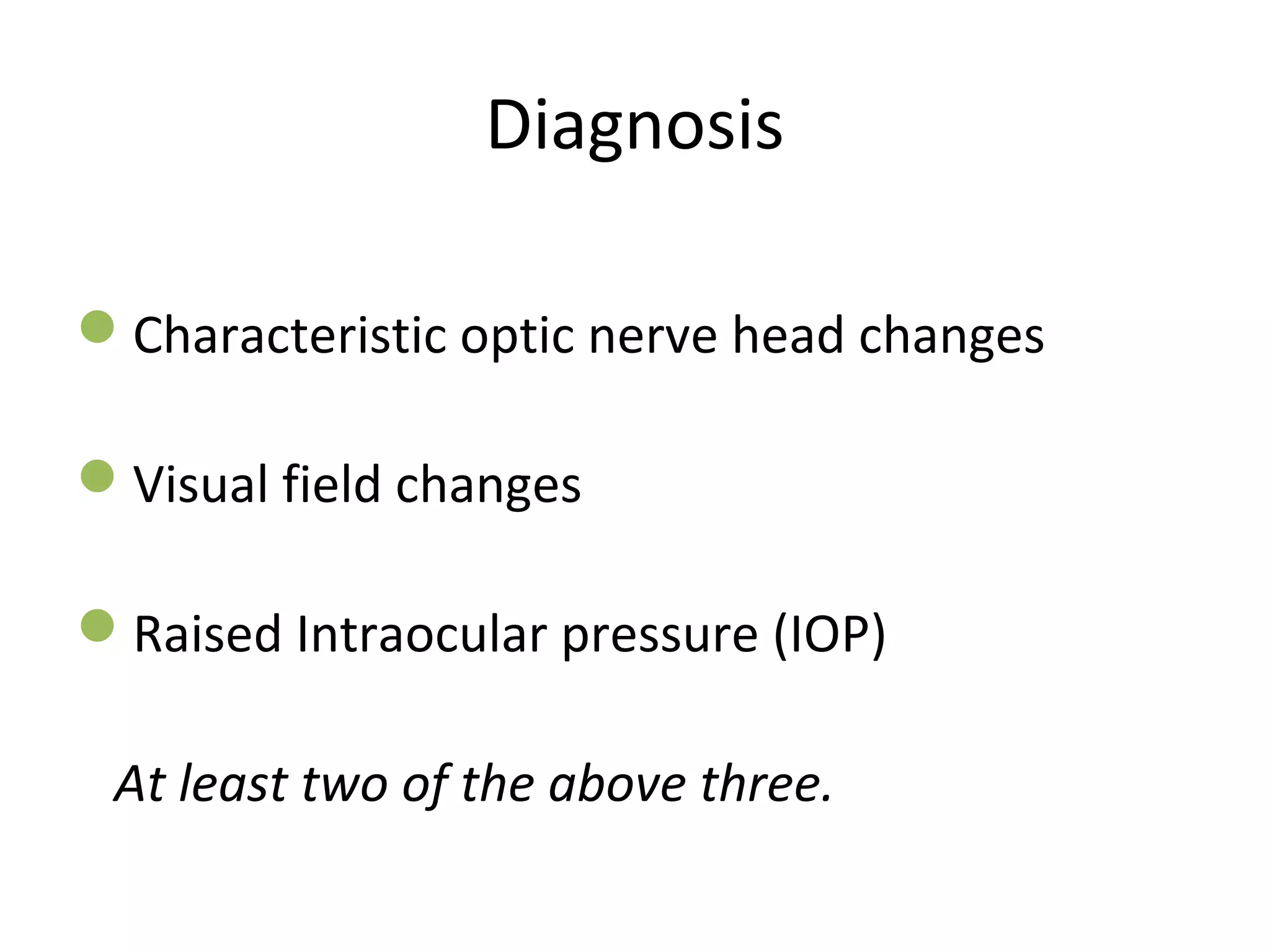
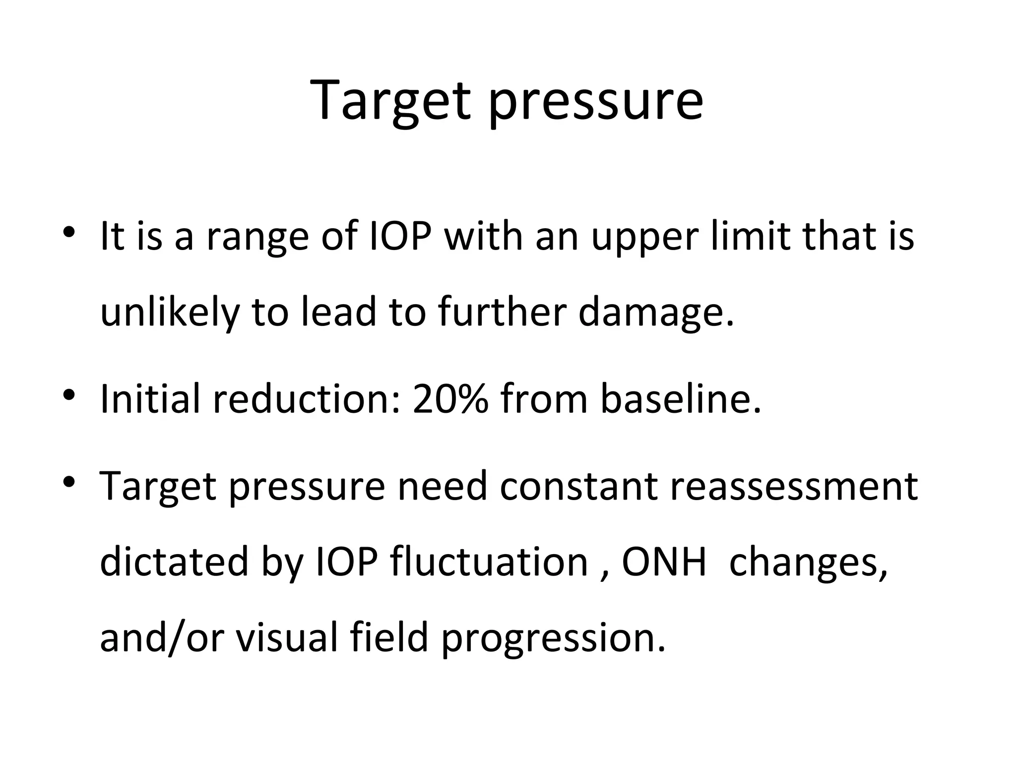
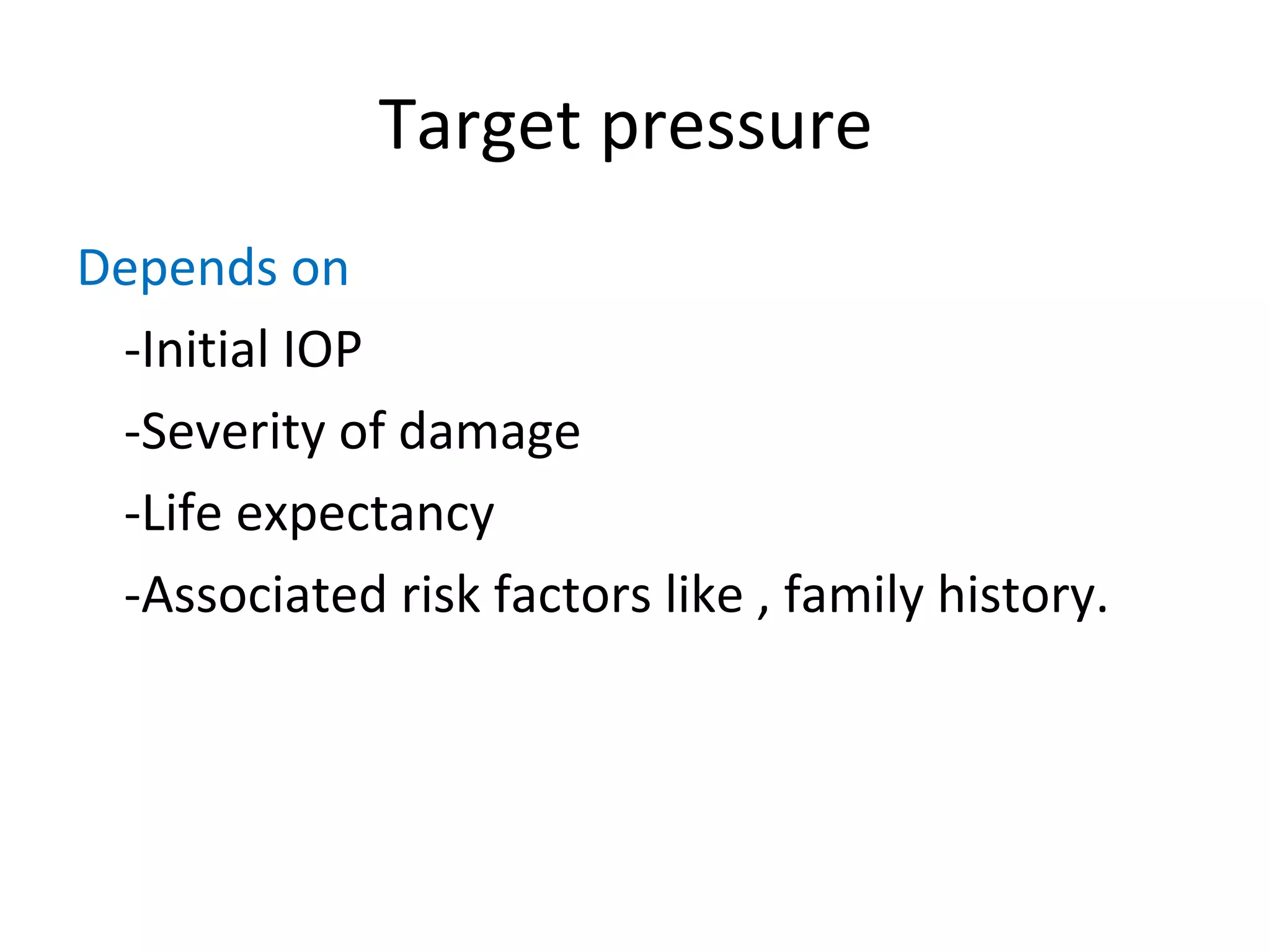
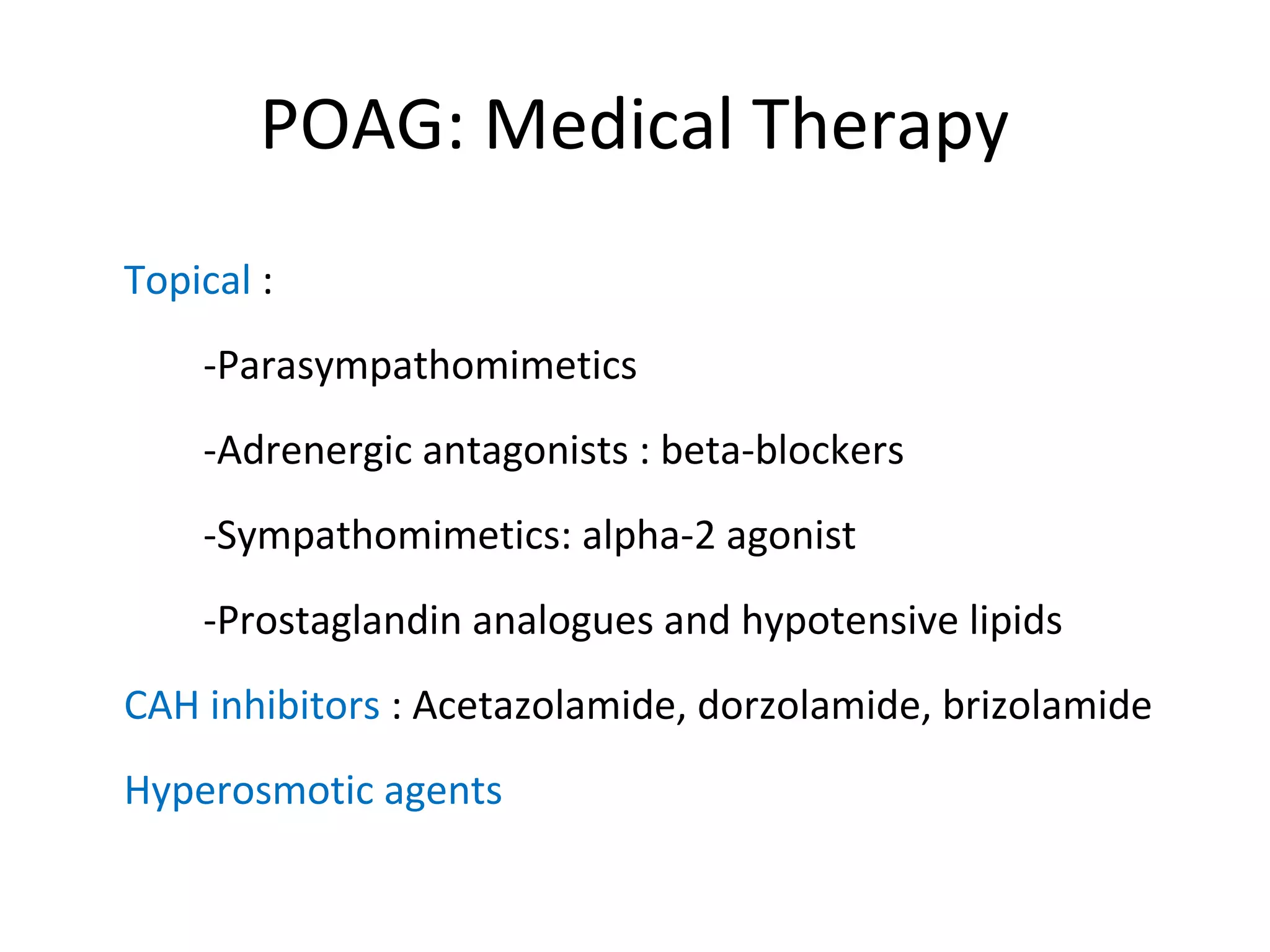
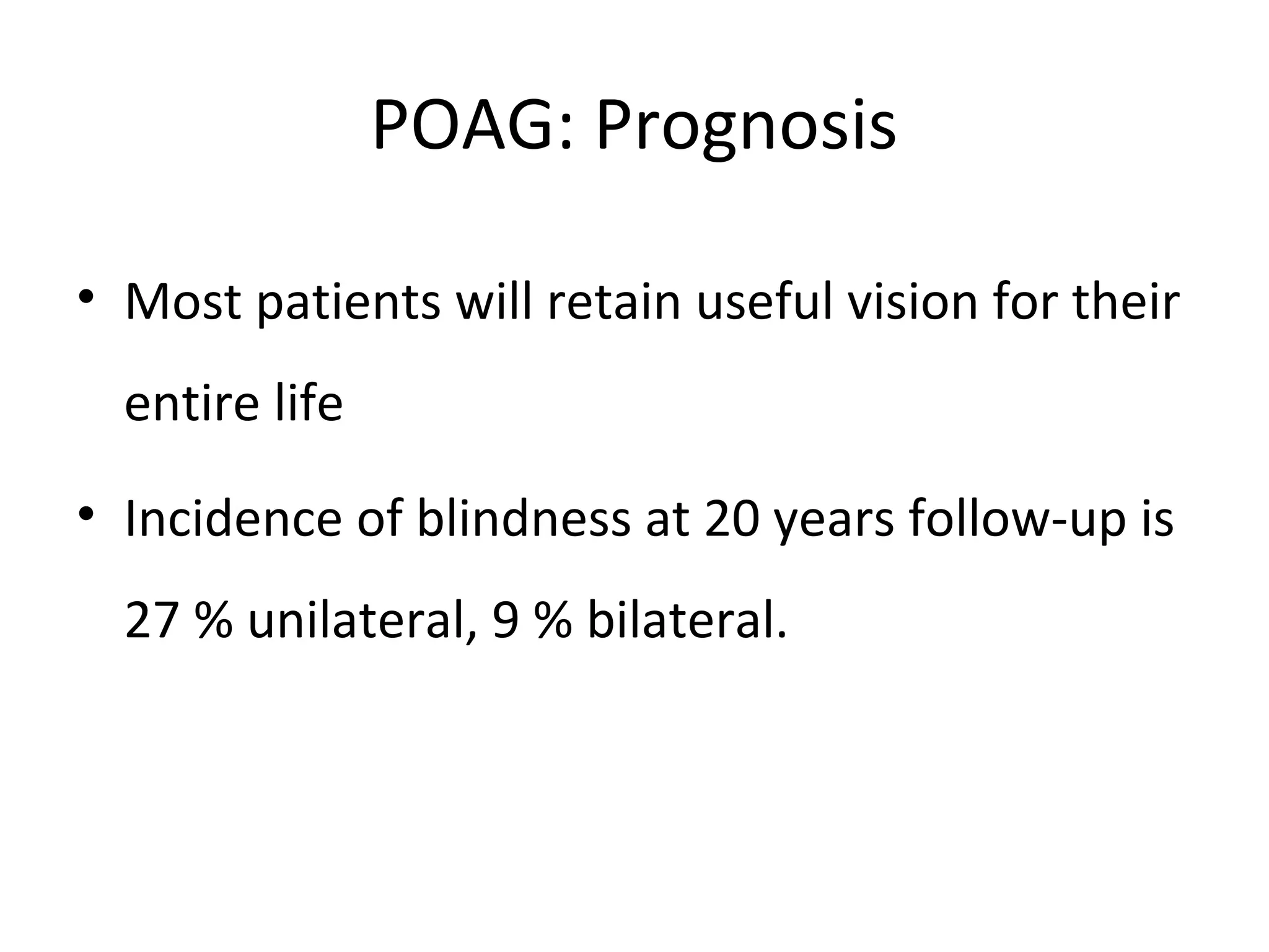
Primary Open-Angle Glaucoma
Professor K N Jha,MS
Learning Aim
• Definition of Open-angle glaucoma
• Primary open-angle glaucoma (POAG)
• Clinical features (IOP, fundus, and field
changes) of POAG
• Treatment of POAG
Primary open-angle glaucoma(POAG)
-POAG is characterized by a chronic, slowly
progressive, optic neuropathy with a
characteristic pattern of optic nerve damage
and visual field loss.
-Angle of the anterior chamber is open.
-There are no ocular or systemic disorders.
POAG
Risk factors
• IOP
• Race
• Central corneal thickness ( CCT)
• Age
• Family history
• Systemic and ocular associations: DM,
Myopia, HTN, CRVO
POAG: Contributory factors
• Reduced perfusion of the optic nerve head
- Inadequate auto-regulation in blood vessels
- Mechanical compression of capillaries
• Abnormality of axonal , or ganglion cell
metabolism
• Disorders of the extracellular matrix of lamina
cribrosa.
Pathogenesis
• Increased resistance to aqueous outflow
• Mechanical changes due to raised IOP
- Decreased axoplasmic flow
• Optic nerve head vascular perfusion
- Inadequate autoregulation in blood
vessels
- Mechanical compression of capillaries
• Glaucomatous Optic atrophy
POAG: Clinical features
• Onset : Insidious,
• Slowly progressive, and painless
• Bilateral: initially may manifest in one eye.
• Visual acuity remains unaffected until late
• Diagnosis : IOP, visual fields, and optic disc
appearance.
• Gonioscopy: open angles
• Associations: myopia, DM, CVS disease, CRVO
Diagnosis
Characteristic optic nerve head changes
Visual field changes
Raised Intraocular pressure (IOP)
At least two of the above three.
Optic disc(ONH) changes in glaucoma
• Increased vertical cup : disc ratio
• Asymmetry of cups between two eyes
• Notching and pallor of neuroretinal rim(NRR)
• Disc hemorrhage
• Baring of circumlinear vessels
• Peripapillary retinal atrophy
Optic Nerve head changes
Normal fundus oculi Glaucomatous cupping
Visual field changes in glaucoma
• Relative paracentral scotoma: smaller/
dimmer targets are not visualized.
• Nasal step: appearance of horizontal shelf in
nasal visual field
• Seidel scotoma: starting from one pole of
blind spot and arches over macula without
reaching horizontal meridian nasally.
• Arcuate scotoma
• Double arcuate or ring scotoma
• End-stage or near total field defect
POAG : Management
• Early detection and routine screening
• Meticulous documentation: IOP, optic nerve
damage and risk factors
Primary open-angle glaucoma
Treatment goal
• Modify and slow progression of optic nerve
damage.
• To lower IOP
Target pressure
• It is a range of IOP with an upper limit that is
unlikely to lead to further damage.
• Initial reduction: 20% from baseline.
• Target pressure need constant reassessment
dictated by IOP fluctuation , ONH changes,
and/or visual field progression.
Target pressure
Depends on
-Initial IOP
-Severity of damage
-Life expectancy
-Associated risk factors like , family history.
POAG: Management
• Patient education
• Cost and Compliance
• Medical or surgical therapy
• Progression and follow-up
POAG: Management
Modalities of treatment
Drug therapy
Laser
Surgery
POAG: Medical Therapy
Topical :
-Parasympathomimetics
-Adrenergic antagonists : beta-blockers
-Sympathomimetics: alpha-2 agonist
-Prostaglandin analogues and hypotensive lipids
CAH inhibitors : Acetazolamide, dorzolamide, brizolamide
Hyperosmotic agents
Primary open-angle glaucoma
• Laser therapy: Argon-laser trabeculoplasty
• Glaucoma surgery: Trabeculectomy
POAG: Prognosis
• Most patients will retain useful vision for their
entire life
• Incidence of blindness at 20 years follow-up is
27 % unilateral, 9 % bilateral.
Primary open-angle glaucoma
Summary:
• Clinical features
• Fundus changes
• Field changes
• Diagnosis
• Treatment
• Follow-up

Glaucoma 3 primary open angle glaucoma,dr.k.n.jha, 03.11.16

  • 1.
  • 2.
    Learning Aim • Definitionof Open-angle glaucoma • Primary open-angle glaucoma (POAG) • Clinical features (IOP, fundus, and field changes) of POAG • Treatment of POAG
  • 3.
    Primary open-angle glaucoma(POAG) -POAGis characterized by a chronic, slowly progressive, optic neuropathy with a characteristic pattern of optic nerve damage and visual field loss. -Angle of the anterior chamber is open. -There are no ocular or systemic disorders.
  • 4.
    POAG Risk factors • IOP •Race • Central corneal thickness ( CCT) • Age • Family history • Systemic and ocular associations: DM, Myopia, HTN, CRVO
  • 5.
    POAG: Contributory factors •Reduced perfusion of the optic nerve head - Inadequate auto-regulation in blood vessels - Mechanical compression of capillaries • Abnormality of axonal , or ganglion cell metabolism • Disorders of the extracellular matrix of lamina cribrosa.
  • 6.
    Pathogenesis • Increased resistanceto aqueous outflow • Mechanical changes due to raised IOP - Decreased axoplasmic flow • Optic nerve head vascular perfusion - Inadequate autoregulation in blood vessels - Mechanical compression of capillaries • Glaucomatous Optic atrophy
  • 7.
    POAG: Clinical features •Onset : Insidious, • Slowly progressive, and painless • Bilateral: initially may manifest in one eye. • Visual acuity remains unaffected until late • Diagnosis : IOP, visual fields, and optic disc appearance. • Gonioscopy: open angles • Associations: myopia, DM, CVS disease, CRVO
  • 8.
    Diagnosis Characteristic optic nervehead changes Visual field changes Raised Intraocular pressure (IOP) At least two of the above three.
  • 9.
    Optic disc(ONH) changesin glaucoma • Increased vertical cup : disc ratio • Asymmetry of cups between two eyes • Notching and pallor of neuroretinal rim(NRR) • Disc hemorrhage • Baring of circumlinear vessels • Peripapillary retinal atrophy
  • 10.
    Optic Nerve headchanges Normal fundus oculi Glaucomatous cupping
  • 11.
    Visual field changesin glaucoma • Relative paracentral scotoma: smaller/ dimmer targets are not visualized. • Nasal step: appearance of horizontal shelf in nasal visual field • Seidel scotoma: starting from one pole of blind spot and arches over macula without reaching horizontal meridian nasally. • Arcuate scotoma • Double arcuate or ring scotoma • End-stage or near total field defect
  • 13.
    POAG : Management •Early detection and routine screening • Meticulous documentation: IOP, optic nerve damage and risk factors
  • 14.
    Primary open-angle glaucoma Treatmentgoal • Modify and slow progression of optic nerve damage. • To lower IOP
  • 15.
    Target pressure • Itis a range of IOP with an upper limit that is unlikely to lead to further damage. • Initial reduction: 20% from baseline. • Target pressure need constant reassessment dictated by IOP fluctuation , ONH changes, and/or visual field progression.
  • 16.
    Target pressure Depends on -InitialIOP -Severity of damage -Life expectancy -Associated risk factors like , family history.
  • 17.
    POAG: Management • Patienteducation • Cost and Compliance • Medical or surgical therapy • Progression and follow-up
  • 18.
    POAG: Management Modalities oftreatment Drug therapy Laser Surgery
  • 19.
    POAG: Medical Therapy Topical: -Parasympathomimetics -Adrenergic antagonists : beta-blockers -Sympathomimetics: alpha-2 agonist -Prostaglandin analogues and hypotensive lipids CAH inhibitors : Acetazolamide, dorzolamide, brizolamide Hyperosmotic agents
  • 20.
    Primary open-angle glaucoma •Laser therapy: Argon-laser trabeculoplasty • Glaucoma surgery: Trabeculectomy
  • 21.
    POAG: Prognosis • Mostpatients will retain useful vision for their entire life • Incidence of blindness at 20 years follow-up is 27 % unilateral, 9 % bilateral.
  • 22.
    Primary open-angle glaucoma Summary: •Clinical features • Fundus changes • Field changes • Diagnosis • Treatment • Follow-up