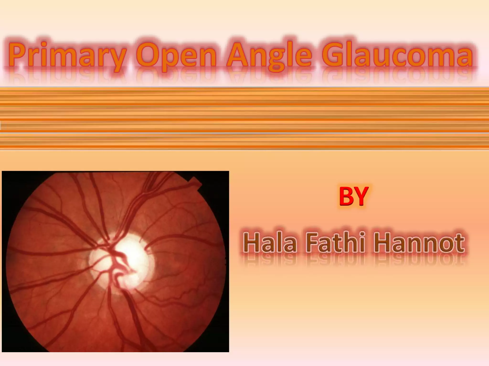
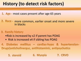
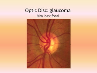
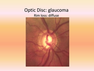
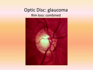
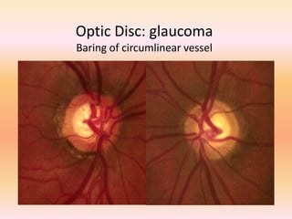
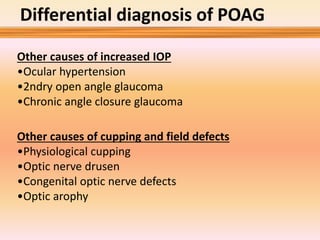
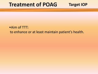
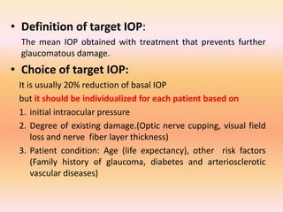
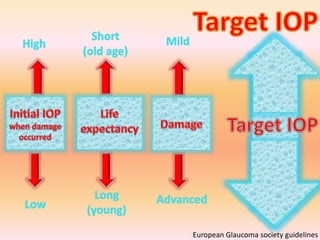
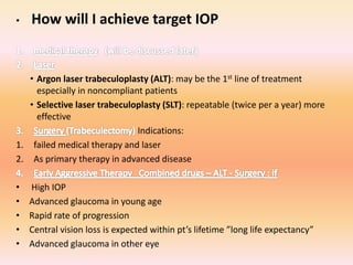
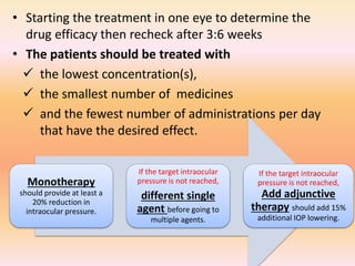
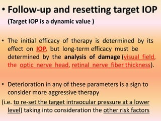
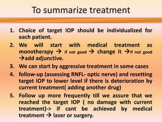
BY
1
2
3
4
5
6
7
Important N.B
• Primary open angle glaucoma is age relaed
trabecular sclerosis
• Primary open angle glaucoma is diagnosis of
exclusion
• Primary glaucoma is a bilateral disease which
is asymmetrical and progressive
History (to detect risk factors)
most cases present after age 65 years
•Risk is increased by x2 if parent has POAG
• Risk is increased x4 if sibling has POAG
more common, earlier onset and more severe
in blacks
Examination
Visual acuity and refraction-color
vision
Important to DD
from other causes
of optic neuropathy
Slit lamp
Looking for back of the cornea-
iris- angle depth
pachymetry
Patients with less than 555 U had
3 times greater risk of POAG
(To confirm diagnosis):
to detect risk factors, exclude 2ndry causes:
• Raised IOP (we can’t diagnose glaucoma by
measuring IOP alone ,there is low and normal
tension glaucoma, also there are 2% of
population over age of 40 have IOP more than
24 mmHg
• Diurnal fluctuations in IOP more than 5 mmHg
• Asymmetry in IOP measurement between the
2 eyes of 5 mmHg or more
Tonometry & IOP phasing
• Angle examination is important to exclude
other causes of 2ndry glaucoma(PDS-PXE-
angle recession-chronic type of angle closure
glaucoma-neovascularization detection)
Gonioscopy
Fundus examination
Optic Disc: normal variation
The ISNT Rule
• Retinal nerve fibre layer
Nerve fiber layer dropout
• Optic nerve head
1. Focal notching of neuroretinal rim - Thining of
infero-temporal Rim (ISNT rule is not preserved)
2. Vertical elongation of cup
3. Asymmetry of cupping between 2 eyes
• Non specific signs of glaucomatous damage
1. Baring of circumlinear blood vessels
2. Splinter haemorrhages
• Optic nerve head
1. Generalized enlargement of cup
2. Vertical elongation of cup
3. Asymmetry of cupping between 2 eyes
• Peripapillary changes
• Non specific signs of glaucomatous damage
1. Exposure of lamina cribrosa
2. Bayoneting (double angulation of blood vessel)
Optic Disc: glaucoma
Rim loss: focal
Optic Disc: glaucoma
Rim loss: diffuse
Optic Disc: glaucoma
Rim loss: combined
Optic Disc: glaucoma
Baring of circumlinear vessel
Optic Disc: glaucoma
Haemorrhage
• When Visual acuity is out of proportion to
cupping
• When colour vision is lost especially red and
green
• When optic nerve pallor is out of proportion
to degree of cupping
• When there is fundus abnormalities like optic
nerve pit, drusen, tiltes disc,..
Investigations
1. Focal visual field defects respecting the horizonal meridian
including
 Nasal step-
 Baring of blind spot-
 Paracentral scotoms-
 Arcuate defects-
 Altitudinal defects
2. Generalized depression
Visual field defects greater than expected
Visual field patterns not typical to glaucoma e.g defects respect
vertical midline-hemianopic defects-enlarged blind spot
• Neural retinal rim
• Cup area
Normal
Glaucoma
ONH parameters & RNFL thickness maps
• RNFL TSNIT graph
• RNFL Thickness
Map
Imaging of the ONH
3-D , true optic nerve topography
only surface topography
Differential diagnosis of POAG
Other causes of increased IOP
•Ocular hypertension
•2ndry open angle glaucoma
•Chronic angle closure glaucoma
Other causes of cupping and field defects
•Physiological cupping
•Optic nerve drusen
•Congenital optic nerve defects
•Optic arophy
Treatment of POAG Target IOP
•Aim of TTT:
to enhance or at least maintain patient’s health.
• Definition of target IOP:
The mean IOP obtained with treatment that prevents further
glaucomatous damage.
• Choice of target IOP:
It is usually 20% reduction of basal IOP
but it should be individualized for each patient based on
1. initial intraocular pressure
2. Degree of existing damage.(Optic nerve cupping, visual field
loss and nerve fiber layer thickness)
3. Patient condition: Age (life expectancy), other risk factors
(Family history of glaucoma, diabetes and arteriosclerotic
vascular diseases)
European Glaucoma society guidelines
• How will I achieve target IOP
• Argon laser trabeculoplasty (ALT): may be the 1st line of treatment
especially in noncompliant patients
• Selective laser trabeculoplasty (SLT): repeatable (twice per a year) more
effective
Indications:
1. failed medical therapy and laser
2. As primary therapy in advanced disease
• High IOP
• Advanced glaucoma in young age
• Rapid rate of progression
• Central vision loss is expected within pt’s lifetime ”long life expectancy”
• Advanced glaucoma in other eye
Medical therapy
• Starting the treatment in one eye to determine the
drug efficacy then recheck after 3:6 weeks
• The patients should be treated with
 the lowest concentration(s),
 the smallest number of medicines
 and the fewest number of administrations per day
that have the desired effect.
Monotherapy
should provide at least a
20% reduction in
intraocular pressure.
If the target intraocular
pressure is not reached,
different single
agent before going to
multiple agents.
If the target intraocular
pressure is not reached,
Add adjunctive
therapy should add 15%
additional IOP lowering.
1. First line therapy (monotherapy):
- Beta blockers
- Alpha2- agonist brimonidine
- Prostaglandin analog latanoprost
2. Second line medications:
- Topical carbonic anhydrase inhibitors
- Alpha2- agonist brimonidine
3. Third and fourth line agents:
- pro-drug dipiverin - epinephrine
- pilocarpine
- oral carbonic anhydrase inhibitors
Choosing initial and combination medical therapy for
glaucoma:
Mechanism of action of anti-glaucomatous drugs
• Follow-up and resetting target IOP
(Target IOP is a dynamic value )
• The initial efficacy of therapy is determined by its
effect on IOP, but long-term efficacy must be
determined by the analysis of damage (visual field,
the optic nerve head, retinal nerve fiber thickness).
• Deterioration in any of these parameters is a sign to
consider more aggressive therapy
(i.e. to re-set the target intraocular pressure at a lower
level) taking into consideration the other risk factors
To summarize treatment
1. Choice of target IOP should be individualized for
each patient.
2. We will start with medical treatment as
monotherapy  if not good  change it if not good
add adjunctive.
3. We can start by aggressive treatment in some cases
4. follow-up (assessing RNFL- optic nerve) and resetting
target IOP to lower level if there is deterioration by
current treatment( adding another drug)
5. Follow up more frequently till we assure that we
reached the target IOP ( no damage with current
treatment)-> if cant be achieved by medical
treatment  laser or surgery.
primary open angle glaucoma by Hala Fathi Hannot

primary open angle glaucoma by Hala Fathi Hannot

  • 1.
  • 3.
  • 4.
    Important N.B • Primaryopen angle glaucoma is age relaed trabecular sclerosis • Primary open angle glaucoma is diagnosis of exclusion • Primary glaucoma is a bilateral disease which is asymmetrical and progressive
  • 5.
    History (to detectrisk factors) most cases present after age 65 years •Risk is increased by x2 if parent has POAG • Risk is increased x4 if sibling has POAG more common, earlier onset and more severe in blacks
  • 6.
    Examination Visual acuity andrefraction-color vision Important to DD from other causes of optic neuropathy Slit lamp Looking for back of the cornea- iris- angle depth pachymetry Patients with less than 555 U had 3 times greater risk of POAG (To confirm diagnosis): to detect risk factors, exclude 2ndry causes:
  • 7.
    • Raised IOP(we can’t diagnose glaucoma by measuring IOP alone ,there is low and normal tension glaucoma, also there are 2% of population over age of 40 have IOP more than 24 mmHg • Diurnal fluctuations in IOP more than 5 mmHg • Asymmetry in IOP measurement between the 2 eyes of 5 mmHg or more Tonometry & IOP phasing
  • 8.
    • Angle examinationis important to exclude other causes of 2ndry glaucoma(PDS-PXE- angle recession-chronic type of angle closure glaucoma-neovascularization detection) Gonioscopy
  • 9.
  • 10.
    Optic Disc: normalvariation The ISNT Rule
  • 11.
    • Retinal nervefibre layer Nerve fiber layer dropout • Optic nerve head 1. Focal notching of neuroretinal rim - Thining of infero-temporal Rim (ISNT rule is not preserved) 2. Vertical elongation of cup 3. Asymmetry of cupping between 2 eyes • Non specific signs of glaucomatous damage 1. Baring of circumlinear blood vessels 2. Splinter haemorrhages
  • 12.
    • Optic nervehead 1. Generalized enlargement of cup 2. Vertical elongation of cup 3. Asymmetry of cupping between 2 eyes • Peripapillary changes • Non specific signs of glaucomatous damage 1. Exposure of lamina cribrosa 2. Bayoneting (double angulation of blood vessel)
  • 13.
  • 14.
  • 15.
  • 16.
    Optic Disc: glaucoma Baringof circumlinear vessel
  • 17.
  • 18.
    • When Visualacuity is out of proportion to cupping • When colour vision is lost especially red and green • When optic nerve pallor is out of proportion to degree of cupping • When there is fundus abnormalities like optic nerve pit, drusen, tiltes disc,..
  • 19.
    Investigations 1. Focal visualfield defects respecting the horizonal meridian including  Nasal step-  Baring of blind spot-  Paracentral scotoms-  Arcuate defects-  Altitudinal defects 2. Generalized depression Visual field defects greater than expected Visual field patterns not typical to glaucoma e.g defects respect vertical midline-hemianopic defects-enlarged blind spot
  • 21.
    • Neural retinalrim • Cup area Normal Glaucoma ONH parameters & RNFL thickness maps • RNFL TSNIT graph • RNFL Thickness Map Imaging of the ONH 3-D , true optic nerve topography
  • 22.
  • 23.
    Differential diagnosis ofPOAG Other causes of increased IOP •Ocular hypertension •2ndry open angle glaucoma •Chronic angle closure glaucoma Other causes of cupping and field defects •Physiological cupping •Optic nerve drusen •Congenital optic nerve defects •Optic arophy
  • 24.
    Treatment of POAGTarget IOP •Aim of TTT: to enhance or at least maintain patient’s health.
  • 25.
    • Definition oftarget IOP: The mean IOP obtained with treatment that prevents further glaucomatous damage. • Choice of target IOP: It is usually 20% reduction of basal IOP but it should be individualized for each patient based on 1. initial intraocular pressure 2. Degree of existing damage.(Optic nerve cupping, visual field loss and nerve fiber layer thickness) 3. Patient condition: Age (life expectancy), other risk factors (Family history of glaucoma, diabetes and arteriosclerotic vascular diseases)
  • 26.
  • 27.
    • How willI achieve target IOP • Argon laser trabeculoplasty (ALT): may be the 1st line of treatment especially in noncompliant patients • Selective laser trabeculoplasty (SLT): repeatable (twice per a year) more effective Indications: 1. failed medical therapy and laser 2. As primary therapy in advanced disease • High IOP • Advanced glaucoma in young age • Rapid rate of progression • Central vision loss is expected within pt’s lifetime ”long life expectancy” • Advanced glaucoma in other eye
  • 28.
  • 29.
    • Starting thetreatment in one eye to determine the drug efficacy then recheck after 3:6 weeks • The patients should be treated with  the lowest concentration(s),  the smallest number of medicines  and the fewest number of administrations per day that have the desired effect. Monotherapy should provide at least a 20% reduction in intraocular pressure. If the target intraocular pressure is not reached, different single agent before going to multiple agents. If the target intraocular pressure is not reached, Add adjunctive therapy should add 15% additional IOP lowering.
  • 30.
    1. First linetherapy (monotherapy): - Beta blockers - Alpha2- agonist brimonidine - Prostaglandin analog latanoprost 2. Second line medications: - Topical carbonic anhydrase inhibitors - Alpha2- agonist brimonidine 3. Third and fourth line agents: - pro-drug dipiverin - epinephrine - pilocarpine - oral carbonic anhydrase inhibitors Choosing initial and combination medical therapy for glaucoma:
  • 31.
    Mechanism of actionof anti-glaucomatous drugs
  • 32.
    • Follow-up andresetting target IOP (Target IOP is a dynamic value ) • The initial efficacy of therapy is determined by its effect on IOP, but long-term efficacy must be determined by the analysis of damage (visual field, the optic nerve head, retinal nerve fiber thickness). • Deterioration in any of these parameters is a sign to consider more aggressive therapy (i.e. to re-set the target intraocular pressure at a lower level) taking into consideration the other risk factors
  • 33.
    To summarize treatment 1.Choice of target IOP should be individualized for each patient. 2. We will start with medical treatment as monotherapy  if not good  change it if not good add adjunctive. 3. We can start by aggressive treatment in some cases 4. follow-up (assessing RNFL- optic nerve) and resetting target IOP to lower level if there is deterioration by current treatment( adding another drug) 5. Follow up more frequently till we assure that we reached the target IOP ( no damage with current treatment)-> if cant be achieved by medical treatment  laser or surgery.