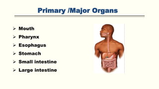
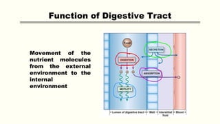
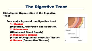
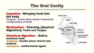
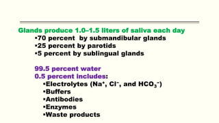
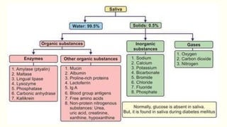
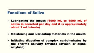
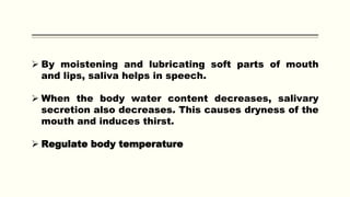
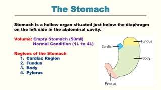
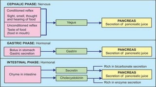
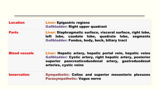
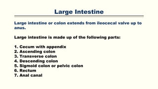
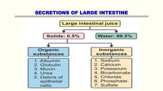
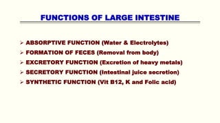
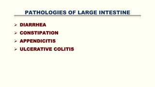
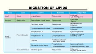
Introduction to
Digestive System
By
Dr. Faraza Javaid
INTRODUCTION
Digestive system is made up of gastrointestinal tract (GI tract)
or alimentary canal and accessory organs, which help in the
process of digestion and absorption. GI tract is a tubular
structure extending from the mouth up to anus, with a length of
about 30 feet.
Digestion
Digestion is defined as the mechanical process by which food
is broken down into simple chemical substances that can be
absorbed and used as nutrients by the body.
A normal young healthy adult consumes about 1 kg of solid diet
and about 1 to 2 liter of liquid diet every day.
Primary /Major Organs
 Mouth
 Pharynx
 Esophagus
 Stomach
 Small intestine
 Large intestine
Accessory Organs
i. Teeth
ii. Tongue
iii. Salivary glands
iv. Exocrine part of pancreas
v. Liver
vi. Gallbladder.
Movement of the
nutrient molecules
from the external
environment to the
internal
environment
Function of Digestive Tract
The Digestive Tract
Six Functions of the Digestive System
1. Ingestion
2. Mechanical processing
3. Digestion
4. Secretion
5. Absorption
6. Excretion
The Digestive Tract
Histological Organization of the Digestive
Tract
Four major layers of the digestive tract
1. Mucosa
(Digestion, Absorption and Secretion)
2. Submucosa
(Glands and Blood Supply)
3. Muscularis externa
(Circular/Longitudinal muscular Tissue)
4. Serosa (Connective Tissues)
NERVE SUPPLY TO
GASTROINTESTINAL TRACT
GI tract has two types of nerve supply:
I. Intrinsic nerve supply
II. Extrinsic nerve supply.
INTRINSIC NERVE SUPPLY –
ENTERIC NERVOUS SYSTEM
Intrinsic nerves to GI tract form the enteric nervous system that
controls all the secretions and movements of GI tract.
Enteric nervous system is present within the wall of GI tract
from esophagus to anus.
Nerve fibers of this system are interconnected and form two
major networks called:
1. Auerbach plexus
2. Meissner plexus
Auerbach Plexus/ Myenteric Nerve Plexus
Major function of this plexus is to regulate the movements of
GI tract.
Some nerve fibers of this plexus accelerate the movements by
secreting the excitatory neurotransmitter substances like
acetylcholine, serotonin and substance P.
Other fibers of this plexus inhibit the GI motility by secreting
the inhibitory neurotransmitters such as vasoactive intestinal
polypeptide (VIP), neurotensin and enkephalin.
Meissner Nerve Plexus
Function of Meissner plexus is the regulation of secretory
functions of GI tract.
EXTRINSIC NERVE SUPPLY
Extrinsic nerves that control the enteric nervous system are from
autonomic nervous system. Both sympathetic and parasympathetic
divisions of autonomic nervous system innervate the GI tract.
Sympathetic Nerve Fibers
Sympathetic nerve fibers inhibit the movements and decrease the
secretions of GI tract by secreting the neurotransmitter noradrenaline.
Parasympathetic Nerve Fibers
Parasympathetic nerve fibers accelerate the movements and increase
the secretions of GI tract.
The neurotransmitter secreted by the parasympathetic nerve fibers is
acetylcholine (Ach).
The Oral Cavity
Ingestion - Bringing food into
the body
Tongue - taste buds detect chemical
composition of food
Mastication - Chewing (physical
digestion); Teeth and Tongue
Chemical digestion – Saliva
moistens food
Amylase - breaks down starch into
maltose
Lysozyme - antibacterial agent
SALIVA
Salivary Glands
Three pairs secrete into oral cavity
1. Parotid salivary glands
2. Sublingual salivary glands
3. Submandibular salivary glands
Each pair has distinctive cellular organization
And produces saliva with different properties
Parotid Salivary Glands
Enzyme salivary amylase
(breaks down starches)
Sublingual Salivary Glands
Produce mucous secretion
Acts as a buffer and
lubricant
Submandibular Salivary Glands
Secrete buffers,
glycoproteins (mucins)
Minor Salivary Glands
 Lingual Mucus Glands
 Lingual Serous Glands
 Buccal Glands
 Labial Glands
 Palatal Glands
Glands produce 1.0–1.5 liters of saliva each day
70 percent by submandibular glands
25 percent by parotids
5 percent by sublingual glands
99.5 percent water
0.5 percent includes:
Electrolytes (Na+, Cl−, and HCO3
−)
Buffers
Antibodies
Enzymes
Waste products
Functions of Saliva
 Lubricating the mouth (1000 mL to 1500 mL of
saliva is secreted per day and it is approximately
about 1 mL/minute)
 Moistening and lubricating materials in the mouth
 Initiating digestion of complex carbohydrates by
the enzyme salivary amylase (ptyalin or alpha-
amylase)
 By moistening and lubricating soft parts of mouth
and lips, saliva helps in speech.
 When the body water content decreases, salivary
secretion also decreases. This causes dryness of the
mouth and induces thirst.
 Regulate body temperature
NERVE SUPPLY TO SALIVARY GLANDS
Parasympathetic Fibers
Parasympathetic fibers activate the acinar cells and dilate
the blood vessels of salivary glands, resulting in the
secretion of saliva with large quantity of water. The
neurotransmitter is acetylcholine.
Sympathetic Fibers
Stimulation of sympathetic fibers causes secretion of
saliva, which is thick and rich in organic constituents such
as mucus. It is because, these fibers activate the acinar
cells and cause vasoconstriction. The neurotransmitter is
noradrenaline.
Pathologies Related to Saliva
 Hyposalivation
 Hypersalivation
 Xerostomia
 Mumps
 Sjogren Syndrome
The Pharynx
A common passage
way for solid food,
liquids, and air.
Regions of the
pharynx:
Nasopharynx
Oropharynx
Laryngopharynx
Food passes through
the oropharynx to the
esophagus
The Esophagus
 A hollow muscular tube
 About 25 cm (10 in.) long and 2 cm (0.80 in.) wide
 Conveys solid food and liquids to the stomach
Swallowing
Can be initiated voluntarily
Proceeds automatically
Is divided into three phases
1. Buccal phase
2. Pharyngeal phase
3. Esophageal phase
The Stomach
Stomach is a hollow organ situated just below the diaphragm
on the left side in the abdominal cavity.
Volume: Empty Stomach (50ml)
Normal Condition (1L to 4L)
Regions of the Stomach
1. Cardiac Region
2. Fundus
3. Body
4. Pylorus
GLANDS OF STOMACH
1.Fundic glands or main gastric glands or oxyntic
glands:
Situated in body and fundus of stomach
2. Pyloric glands:
Present in the pyloric part of the stomach
3. Cardiac glands:
Located in the cardiac region of the stomach.
FUNCTIONS OF STOMACH
 MECHANICAL FUNCTION
 DIGESTIVE FUNCTION
 PROTECTIVE FUNCTION
 HEMOPOIETIC FUNCTION
 EXCRETORY FUNCTION
„PROPERTIES OF GASTRIC JUICE
STAGES OF GASTRIC SECRETION
Gastric secretion occurs in three
phases:
Cephalic Phase
Gastric Phase
Intestinal Phase
Pathologies Related to Gastric Secretion
GASTRITIS
GASTRIC ATROPHY
PEPTIC ULCER
ZOLLINGER-ELLISON SYNDROME
Pancreas
 Pancreas is a dual organ having two functions, namely
endocrine function and exocrine function.
 Endocrine function is concerned with the production of
hormones.
 The exocrine function is concerned with the secretion of
digestive juice called pancreatic juice.
PROPERTIES OF PANCREATIC JUICE
Volume : 500 to 800 mL/day
pH : Highly alkaline with a pH of 8 to 8.3
STAGES OF PANCREATIC SECRETION
Pancreatic juice is secreted in three stages like the gastric
juice:
1. Cephalic phase
2. Gastric phase
3. Intestinal phase
These three phases of pancreatic secretion correspond
with the three phases of gastric secretion.
Pathologies Related to Pancreas
 PANCREATITIS
 STEATORRHEA
LIVER & GALLBLADDER
The liver and gallbladder are the two accessory organs of
the gastrointestinal tract, which carry out a multifunctional
role that aids digestion and homeostasis.
The liver consists of several lobes and receives its blood
supply mainly from the hepatic portal vein.
The gallbladder is found inferiorly to the liver, being
involved in the storage and release of bile into
the duodenum.
Location Liver: Epigastric regions
Gallbladder: Right upper quadrant
Parts Liver: Diaphragmatic surface, visceral surface, right lobe,
left lobe, caudate lobe, quadrate lobe, segments
Gallbladder: Fundus, body, beck, biliary tract
Blood vessels Liver: Hepatic artery, hepatic portal vein, hepatic veins
Gallbladder: Cystic artery, right hepatic artery, posterior
superior pancreaticoduodenal artery, gastroduodenal
arteries, cystic veins
Innervation Sympathetic: Celiac and superior mesenteric plexuses
Parasympathetic: Vagus nerve
LIVER
Liver is made up of many lobes called hepatic lobes. Each
lobe consists of many lobules called hepatic lobules.
Hepatic lobule is the structural and functional unit of liver.
There are about 50,000 to 100,000 lobules in the liver. The
lobule is a honeycomb-like structure and it is made up of
liver cells called hepatocytes
Each lobule is surrounded by many portal triads. Each
portal triad consists of three vessels:
1. A branch of hepatic artery
2. A branch of portal vein
3. A tributary of bile duct.
BILIARY SYSTEM
Biliary system or extrahepatic biliary apparatus is
formed by gallbladder and extrahepatic bile ducts (bile
ducts outside the liver).
Right and left hepatic bile ducts which come out of
liver join to form common hepatic duct.
It unites with the cystic duct from gallbladder to form
common bile duct. All these ducts have similar
structures.
PROPERTIES AND COMPOSITION
OF BILE
Formation of Bile and
Bile Salts
 Bile acid synthesis occurs in liver cells, which
synthesize primary bile acids (cholic acid and chenodeoxycholic
acid in humans) via cytochrome P450-mediated oxidation
of cholesterol in a multi-step process.
 The rate-limiting step in synthesis is the addition of a hydroxyl
group of the 7th position of the steroid nucleus by the
enzyme cholesterol 7 alpha-hydroxylase. This enzyme is down-
regulated by cholic acid, up-regulated by cholesterol.
 In liver, bile acids are conjugated with glycine (amino acid) or
taurin (derivative of an amino acid) and form conjugated bile
acids. These bile acids combine with sodium or potassium ions
to form the salts, sodium or potassium glycocholate and sodium
or potassium taurocholate.
Formation of Bilirubin
FUNCTIONS OF BILE SALTS
1.Emulsification of Fats (Fat breakdown)
2.Absorption of Fats (Via Bile)
3.Choleretic Action (Enhance bile secretion)
4.Cholagogue Action (Gallbladder contraction)
5.Laxative Action (Stimulate peristalsis)
6.Prevention of Gallstone Formation
FUNCTIONS OF GALLBLADDER
 Storage of Bile
 Concentration of Bile
 Maintenance of Pressure in Biliary System
FUNCTIONS OF LIVER
 METABOLIC FUNCTION
 STORAGE FUNCTION
 SECRETION OF BILE
 HEAT PRODUCTION
 HEMOPOIETIC FUNCTION
INACTIVATION OF HORMONES AND DRUGS
DEFENSIVE AND DETOXIFICATION FUNCTIONS
PATHOHYSIOLOGY OF LIVER & GALL BLADDER
 JAUNDICE OR ICTERUS
 HEPATITIS
 CIRRHOSIS OF LIVER
 GALLSTONES
Small Intestine
Small intestine is the part of gastrointestinal (GI) tract,
extending between the pyloric sphincter of stomach and
ileocecal valve, which opens into large intestine. Its length
is about 6 meter.
Small intestine consists of three portions:
1. Proximal part known as duodenum
2. Middle part known as jejunum
3. Distal part known as ileum
INTESTINAL VILLI AND GLANDS
OF SMALL INTESTINE
INTESTINAL VILLI
Mucous membrane of small intestine is covered by minute projections
called villi.
MICROVILLI
Villi are lined by columnar cells, which are called enterocytes. Each
enterocyte gives rise to hair-like projections called microvilli.
CRYPTS OF LIEBERKÜHN/ INTESTINAL GLANDS
Crypts of Lieberkühn or intestinal glands are simple tubular glands of
intestine.
BRUNNER GLANDS
In addition to intestinal glands, the first part of duodenum contains
some mucus glands, which are called Brunner glands.
PROPERTIES AND COMPOSITION
OF SUCCUS ENTERICUS
Digestive Enzymes of Succus Entericus
FUNCTIONS OF SMALL INESTINE
 MECHANICAL FUNCTION
 SECRETORY FUNCTION
 HORMONAL FUNCTION
 DIGESTIVE FUNCTION
 HEMOPOIETIC FUNCTION
 ABSORPTIVE FUNCTIONS
PATHOLOGIES OF SMALL INTESTINE
 MALABSORPTION
 MALABSORPTION SYNDROME
 CROHN’S DISEASE OR ENTERITIS
 TROPICAL SPRUE
 STEATORRHEA
 CELIAC DISEASE
Large Intestine
Large intestine or colon extends from ileocecal valve up to
anus.
Large intestine is made up of the following parts:
1. Cecum with appendix
2. Ascending colon
3. Transverse colon
4. Descending colon
5. Sigmoid colon or pelvic colon
6. Rectum
7. Anal canal
SECRETIONS OF LARGE INTESTINE
FUNCTIONS OF LARGE INTESTINE
 ABSORPTIVE FUNCTION (Water & Electrolytes)
 FORMATION OF FECES (Removal from body)
 EXCRETORY FUNCTION (Excretion of heavy metals)
 SECRETORY FUNCTION (Intestinal juice secretion)
 SYNTHETIC FUNCTION (Vit B12, K and Folic acid)
PATHOLOGIES OF LARGE INTESTINE
 DIARRHEA
 CONSTIPATION
 APPENDICITIS
 ULCERATIVE COLITIS
Movements of Gastrointestinal Tract
MASTICATION
Mastication or chewing is the first mechanical process in
the gastrointestinal (GI) tract, by which the food
substances are torn or cut into small particles and crushed
or ground into a soft bolus.
Muscles of Mastication
1. Masseter muscle
2. Temporal muscle
3. Pterygoid muscles
4. Buccinator muscle.
Movements of Gastrointestinal Tract
DEGLUTITION
Deglutition or swallowing is the process by which food
moves from mouth into stomach.
Stages of Deglutition
Deglutition occurs in three stages:
I. Oral stage, when food moves from mouth to pharynx
II. Pharyngeal stage, when food moves from pharynx to
esophagus
III. Esophageal stage, when food moves from esophagus to
stomach.
Movements of Gastrointestinal Tract
MOVEMENTS OF STOMACH
Activities of smooth muscles of stomach increase during
gastric digestion (when stomach is filled with food) and
when the stomach is empty.
Types of movements in stomach:
1. Hunger contractions (Sensation to hunger)
2. Receptive relaxation (Accommodation)
3. Peristalsis (Digestive peristalsis)
Movements of Gastrointestinal Tract
MOVEMENTS OF INTESTINE
Movements of small intestine are of four types:
1. Mixing movements:
i. Segmentation movements
ii. Pendular movements
2. Propulsive movements:
i. Peristaltic movements
ii. Peristaltic rush (Irritants)
3. Peristalsis in fasting
4. Movements of villi
Movements of Gastrointestinal Tract
MOVEMENTS OF LARGE INTESTINE
Movements of large intestine are of two types:
1. Mixing movements: Segmentation contractions
2. Propulsive movements: Mass peristalsis.
PATHOLOGIES RELATED TO MOVEMETS
 Dysphagia
 Esophageal Achalasia (Food Accumulation)
 Gastroesophageal Reflux Disease (GERD)
 Gastric Dumping Syndrome (Bypass Surgery)
Gastrointestinal Hormones
Gastrointestinal (GI) hormones are the hormones secreted
in GI tract. Major function of these hormones is to regulate
the secretory activities and motility of the GI tract.
CELLS SECRETING THE HORMONES
 Enteroendocrine Cells
 Neuroendocrine Cells
 Enterochromaffin Cells
GASTROINTESTINAL HORMONES
 GASTRIN
 „
SECRETIN
 „
CHOLECYSTOKININ
 „
GLUCOSE-DEPENDENT
INSULINOTROPIC HORMONE
 „
VASOACTIVE INTESTINAL
POLYPEPTIDE
 „
GLUCAGON
 „
GLICENTIN
 GLUCAGON-LIKE POLYPEPTIDE-1
 GLUCAGON-LIKE POLYPEPTIDE-2
 SOMATOSTATIN
 PANCREATIC POLYPEPTIDE
 PEPTIDE YY
 NEUROPEPTIDE Y
 MOTILIN
 SUBSTANCE P
 GHRELIN
 OTHER GASTROINTESTINAL
HORMONES
GASTROINTESTINAL HORMONES
Digestion, Absorption and Metabolism of
Carbohydrates
1. POLYSACCHARIDES
Large polysaccharides are glycogen, amylose and amylopectin,
which are in the form of starch (glucose polymers). Glycogen is
available in non-vegetarian diet. Amylose and amylopectin are
available in vegetarian diet.
2. DISACCHARIDES
Two types of disaccharides are available in the diet.
i. Sucrose (Glucose + Fructose), which is called table sugar or
cane sugar
ii. Lactose (Glucose + Galactose), which is the sugar available in
milk.
3. MONOSACCHARIDES
Monosaccharides consumed in human diet are mostly glucose
and fructose.
DIGESTION OF CARBOHYDRATES
Carbohydrate Metabolism
Digestion, Absorption and Metabolism of
Proteins
Proteins present in common foodstuffs are:
1. Wheat: Glutenin and gliadin, which constitute
gluten
2. Milk: Casein, lactalbumin, albumin and myosin
3. Egg: Albumin and vitellin
4. Meat: Collagen, albumin and myosin.
DIGESTION OF PROTEINS
Protein Metabolism
Digestion, Absorption and Metabolism of
Lipids
Lipids are mostly consumed in the form of neutral fats,
which are also known as triglycerides. Triglycerides are
made up of glycerol nucleus and free fatty acids. Apart
from triglycerides, usual diet also contains small quantities
of cholesterol and cholesterol esters.
Dietary fats are classified into two types:
1. Saturated fats
2. Unsaturated fats
SATURATED FATS
Saturated fats are the fats which contain triglycerides formed
from only saturated fatty acids. The fatty acids having maximum
amount of hydrogen ions without any double bonds between
carbon atoms are called saturated fatty acids.
UNSATURATED FATS
Fats containing unsaturated fatty acids are known as
unsaturated fats. Unsaturated fatty acids are fatty acids formed
by dehydrogenation of saturated fatty acids.
Unsaturated fats are classified into three types:
DIGESTION OF LIPIDS
ADIPOSE TISSUE
WHITE ADIPOSE TISSUE OR WHITE FAT
White adipose tissue is distributed through the body beneath the
skin, forming subcutaneous fat. It also surrounds the internal
organs. This adipose tissue is formed by fat cells which are
unilocular, i.e. these cells contain one large vacuole filled with fat.
BROWN ADIPOSE TISSUE OR BROWN FAT
Brown adipose tissue is a specialized form of adipose tissue,
having the function opposite to that of white adipose tissue. It is
present only in certain areas of the body such as back of neck.
LIPID PROFILE
THANK YOU

GIT.pptx

  • 1.
  • 2.
    INTRODUCTION Digestive system ismade up of gastrointestinal tract (GI tract) or alimentary canal and accessory organs, which help in the process of digestion and absorption. GI tract is a tubular structure extending from the mouth up to anus, with a length of about 30 feet. Digestion Digestion is defined as the mechanical process by which food is broken down into simple chemical substances that can be absorbed and used as nutrients by the body. A normal young healthy adult consumes about 1 kg of solid diet and about 1 to 2 liter of liquid diet every day.
  • 3.
    Primary /Major Organs Mouth  Pharynx  Esophagus  Stomach  Small intestine  Large intestine
  • 4.
    Accessory Organs i. Teeth ii.Tongue iii. Salivary glands iv. Exocrine part of pancreas v. Liver vi. Gallbladder.
  • 5.
    Movement of the nutrientmolecules from the external environment to the internal environment Function of Digestive Tract
  • 6.
    The Digestive Tract SixFunctions of the Digestive System 1. Ingestion 2. Mechanical processing 3. Digestion 4. Secretion 5. Absorption 6. Excretion
  • 7.
    The Digestive Tract HistologicalOrganization of the Digestive Tract Four major layers of the digestive tract 1. Mucosa (Digestion, Absorption and Secretion) 2. Submucosa (Glands and Blood Supply) 3. Muscularis externa (Circular/Longitudinal muscular Tissue) 4. Serosa (Connective Tissues)
  • 8.
    NERVE SUPPLY TO GASTROINTESTINALTRACT GI tract has two types of nerve supply: I. Intrinsic nerve supply II. Extrinsic nerve supply.
  • 9.
    INTRINSIC NERVE SUPPLY– ENTERIC NERVOUS SYSTEM Intrinsic nerves to GI tract form the enteric nervous system that controls all the secretions and movements of GI tract. Enteric nervous system is present within the wall of GI tract from esophagus to anus. Nerve fibers of this system are interconnected and form two major networks called: 1. Auerbach plexus 2. Meissner plexus
  • 10.
    Auerbach Plexus/ MyentericNerve Plexus Major function of this plexus is to regulate the movements of GI tract. Some nerve fibers of this plexus accelerate the movements by secreting the excitatory neurotransmitter substances like acetylcholine, serotonin and substance P. Other fibers of this plexus inhibit the GI motility by secreting the inhibitory neurotransmitters such as vasoactive intestinal polypeptide (VIP), neurotensin and enkephalin. Meissner Nerve Plexus Function of Meissner plexus is the regulation of secretory functions of GI tract.
  • 11.
    EXTRINSIC NERVE SUPPLY Extrinsicnerves that control the enteric nervous system are from autonomic nervous system. Both sympathetic and parasympathetic divisions of autonomic nervous system innervate the GI tract. Sympathetic Nerve Fibers Sympathetic nerve fibers inhibit the movements and decrease the secretions of GI tract by secreting the neurotransmitter noradrenaline. Parasympathetic Nerve Fibers Parasympathetic nerve fibers accelerate the movements and increase the secretions of GI tract. The neurotransmitter secreted by the parasympathetic nerve fibers is acetylcholine (Ach).
  • 12.
    The Oral Cavity Ingestion- Bringing food into the body Tongue - taste buds detect chemical composition of food Mastication - Chewing (physical digestion); Teeth and Tongue Chemical digestion – Saliva moistens food Amylase - breaks down starch into maltose Lysozyme - antibacterial agent
  • 13.
    SALIVA Salivary Glands Three pairssecrete into oral cavity 1. Parotid salivary glands 2. Sublingual salivary glands 3. Submandibular salivary glands Each pair has distinctive cellular organization And produces saliva with different properties
  • 14.
    Parotid Salivary Glands Enzymesalivary amylase (breaks down starches) Sublingual Salivary Glands Produce mucous secretion Acts as a buffer and lubricant Submandibular Salivary Glands Secrete buffers, glycoproteins (mucins)
  • 16.
    Minor Salivary Glands Lingual Mucus Glands  Lingual Serous Glands  Buccal Glands  Labial Glands  Palatal Glands
  • 17.
    Glands produce 1.0–1.5liters of saliva each day 70 percent by submandibular glands 25 percent by parotids 5 percent by sublingual glands 99.5 percent water 0.5 percent includes: Electrolytes (Na+, Cl−, and HCO3 −) Buffers Antibodies Enzymes Waste products
  • 19.
    Functions of Saliva Lubricating the mouth (1000 mL to 1500 mL of saliva is secreted per day and it is approximately about 1 mL/minute)  Moistening and lubricating materials in the mouth  Initiating digestion of complex carbohydrates by the enzyme salivary amylase (ptyalin or alpha- amylase)
  • 20.
     By moisteningand lubricating soft parts of mouth and lips, saliva helps in speech.  When the body water content decreases, salivary secretion also decreases. This causes dryness of the mouth and induces thirst.  Regulate body temperature
  • 21.
    NERVE SUPPLY TOSALIVARY GLANDS Parasympathetic Fibers Parasympathetic fibers activate the acinar cells and dilate the blood vessels of salivary glands, resulting in the secretion of saliva with large quantity of water. The neurotransmitter is acetylcholine. Sympathetic Fibers Stimulation of sympathetic fibers causes secretion of saliva, which is thick and rich in organic constituents such as mucus. It is because, these fibers activate the acinar cells and cause vasoconstriction. The neurotransmitter is noradrenaline.
  • 22.
    Pathologies Related toSaliva  Hyposalivation  Hypersalivation  Xerostomia  Mumps  Sjogren Syndrome
  • 23.
    The Pharynx A commonpassage way for solid food, liquids, and air. Regions of the pharynx: Nasopharynx Oropharynx Laryngopharynx Food passes through the oropharynx to the esophagus
  • 24.
    The Esophagus  Ahollow muscular tube  About 25 cm (10 in.) long and 2 cm (0.80 in.) wide  Conveys solid food and liquids to the stomach Swallowing Can be initiated voluntarily Proceeds automatically Is divided into three phases 1. Buccal phase 2. Pharyngeal phase 3. Esophageal phase
  • 25.
    The Stomach Stomach isa hollow organ situated just below the diaphragm on the left side in the abdominal cavity. Volume: Empty Stomach (50ml) Normal Condition (1L to 4L) Regions of the Stomach 1. Cardiac Region 2. Fundus 3. Body 4. Pylorus
  • 26.
    GLANDS OF STOMACH 1.Fundicglands or main gastric glands or oxyntic glands: Situated in body and fundus of stomach 2. Pyloric glands: Present in the pyloric part of the stomach 3. Cardiac glands: Located in the cardiac region of the stomach.
  • 27.
    FUNCTIONS OF STOMACH MECHANICAL FUNCTION  DIGESTIVE FUNCTION  PROTECTIVE FUNCTION  HEMOPOIETIC FUNCTION  EXCRETORY FUNCTION
  • 28.
  • 30.
    STAGES OF GASTRICSECRETION Gastric secretion occurs in three phases: Cephalic Phase Gastric Phase Intestinal Phase
  • 34.
    Pathologies Related toGastric Secretion GASTRITIS GASTRIC ATROPHY PEPTIC ULCER ZOLLINGER-ELLISON SYNDROME
  • 35.
    Pancreas  Pancreas isa dual organ having two functions, namely endocrine function and exocrine function.  Endocrine function is concerned with the production of hormones.  The exocrine function is concerned with the secretion of digestive juice called pancreatic juice. PROPERTIES OF PANCREATIC JUICE Volume : 500 to 800 mL/day pH : Highly alkaline with a pH of 8 to 8.3
  • 38.
    STAGES OF PANCREATICSECRETION Pancreatic juice is secreted in three stages like the gastric juice: 1. Cephalic phase 2. Gastric phase 3. Intestinal phase These three phases of pancreatic secretion correspond with the three phases of gastric secretion.
  • 40.
    Pathologies Related toPancreas  PANCREATITIS  STEATORRHEA
  • 41.
    LIVER & GALLBLADDER Theliver and gallbladder are the two accessory organs of the gastrointestinal tract, which carry out a multifunctional role that aids digestion and homeostasis. The liver consists of several lobes and receives its blood supply mainly from the hepatic portal vein. The gallbladder is found inferiorly to the liver, being involved in the storage and release of bile into the duodenum.
  • 42.
    Location Liver: Epigastricregions Gallbladder: Right upper quadrant Parts Liver: Diaphragmatic surface, visceral surface, right lobe, left lobe, caudate lobe, quadrate lobe, segments Gallbladder: Fundus, body, beck, biliary tract Blood vessels Liver: Hepatic artery, hepatic portal vein, hepatic veins Gallbladder: Cystic artery, right hepatic artery, posterior superior pancreaticoduodenal artery, gastroduodenal arteries, cystic veins Innervation Sympathetic: Celiac and superior mesenteric plexuses Parasympathetic: Vagus nerve
  • 43.
    LIVER Liver is madeup of many lobes called hepatic lobes. Each lobe consists of many lobules called hepatic lobules. Hepatic lobule is the structural and functional unit of liver. There are about 50,000 to 100,000 lobules in the liver. The lobule is a honeycomb-like structure and it is made up of liver cells called hepatocytes Each lobule is surrounded by many portal triads. Each portal triad consists of three vessels: 1. A branch of hepatic artery 2. A branch of portal vein 3. A tributary of bile duct.
  • 44.
    BILIARY SYSTEM Biliary systemor extrahepatic biliary apparatus is formed by gallbladder and extrahepatic bile ducts (bile ducts outside the liver). Right and left hepatic bile ducts which come out of liver join to form common hepatic duct. It unites with the cystic duct from gallbladder to form common bile duct. All these ducts have similar structures.
  • 45.
  • 46.
    Formation of Bileand Bile Salts
  • 47.
     Bile acidsynthesis occurs in liver cells, which synthesize primary bile acids (cholic acid and chenodeoxycholic acid in humans) via cytochrome P450-mediated oxidation of cholesterol in a multi-step process.  The rate-limiting step in synthesis is the addition of a hydroxyl group of the 7th position of the steroid nucleus by the enzyme cholesterol 7 alpha-hydroxylase. This enzyme is down- regulated by cholic acid, up-regulated by cholesterol.  In liver, bile acids are conjugated with glycine (amino acid) or taurin (derivative of an amino acid) and form conjugated bile acids. These bile acids combine with sodium or potassium ions to form the salts, sodium or potassium glycocholate and sodium or potassium taurocholate.
  • 48.
  • 50.
    FUNCTIONS OF BILESALTS 1.Emulsification of Fats (Fat breakdown) 2.Absorption of Fats (Via Bile) 3.Choleretic Action (Enhance bile secretion) 4.Cholagogue Action (Gallbladder contraction) 5.Laxative Action (Stimulate peristalsis) 6.Prevention of Gallstone Formation
  • 51.
    FUNCTIONS OF GALLBLADDER Storage of Bile  Concentration of Bile  Maintenance of Pressure in Biliary System
  • 52.
    FUNCTIONS OF LIVER METABOLIC FUNCTION  STORAGE FUNCTION  SECRETION OF BILE  HEAT PRODUCTION  HEMOPOIETIC FUNCTION INACTIVATION OF HORMONES AND DRUGS DEFENSIVE AND DETOXIFICATION FUNCTIONS
  • 53.
    PATHOHYSIOLOGY OF LIVER& GALL BLADDER  JAUNDICE OR ICTERUS  HEPATITIS  CIRRHOSIS OF LIVER  GALLSTONES
  • 54.
    Small Intestine Small intestineis the part of gastrointestinal (GI) tract, extending between the pyloric sphincter of stomach and ileocecal valve, which opens into large intestine. Its length is about 6 meter. Small intestine consists of three portions: 1. Proximal part known as duodenum 2. Middle part known as jejunum 3. Distal part known as ileum
  • 55.
    INTESTINAL VILLI ANDGLANDS OF SMALL INTESTINE INTESTINAL VILLI Mucous membrane of small intestine is covered by minute projections called villi. MICROVILLI Villi are lined by columnar cells, which are called enterocytes. Each enterocyte gives rise to hair-like projections called microvilli. CRYPTS OF LIEBERKÜHN/ INTESTINAL GLANDS Crypts of Lieberkühn or intestinal glands are simple tubular glands of intestine. BRUNNER GLANDS In addition to intestinal glands, the first part of duodenum contains some mucus glands, which are called Brunner glands.
  • 56.
  • 58.
    Digestive Enzymes ofSuccus Entericus
  • 59.
    FUNCTIONS OF SMALLINESTINE  MECHANICAL FUNCTION  SECRETORY FUNCTION  HORMONAL FUNCTION  DIGESTIVE FUNCTION  HEMOPOIETIC FUNCTION  ABSORPTIVE FUNCTIONS
  • 60.
    PATHOLOGIES OF SMALLINTESTINE  MALABSORPTION  MALABSORPTION SYNDROME  CROHN’S DISEASE OR ENTERITIS  TROPICAL SPRUE  STEATORRHEA  CELIAC DISEASE
  • 61.
    Large Intestine Large intestineor colon extends from ileocecal valve up to anus. Large intestine is made up of the following parts: 1. Cecum with appendix 2. Ascending colon 3. Transverse colon 4. Descending colon 5. Sigmoid colon or pelvic colon 6. Rectum 7. Anal canal
  • 62.
  • 63.
    FUNCTIONS OF LARGEINTESTINE  ABSORPTIVE FUNCTION (Water & Electrolytes)  FORMATION OF FECES (Removal from body)  EXCRETORY FUNCTION (Excretion of heavy metals)  SECRETORY FUNCTION (Intestinal juice secretion)  SYNTHETIC FUNCTION (Vit B12, K and Folic acid)
  • 64.
    PATHOLOGIES OF LARGEINTESTINE  DIARRHEA  CONSTIPATION  APPENDICITIS  ULCERATIVE COLITIS
  • 65.
    Movements of GastrointestinalTract MASTICATION Mastication or chewing is the first mechanical process in the gastrointestinal (GI) tract, by which the food substances are torn or cut into small particles and crushed or ground into a soft bolus. Muscles of Mastication 1. Masseter muscle 2. Temporal muscle 3. Pterygoid muscles 4. Buccinator muscle.
  • 66.
    Movements of GastrointestinalTract DEGLUTITION Deglutition or swallowing is the process by which food moves from mouth into stomach. Stages of Deglutition Deglutition occurs in three stages: I. Oral stage, when food moves from mouth to pharynx II. Pharyngeal stage, when food moves from pharynx to esophagus III. Esophageal stage, when food moves from esophagus to stomach.
  • 67.
    Movements of GastrointestinalTract MOVEMENTS OF STOMACH Activities of smooth muscles of stomach increase during gastric digestion (when stomach is filled with food) and when the stomach is empty. Types of movements in stomach: 1. Hunger contractions (Sensation to hunger) 2. Receptive relaxation (Accommodation) 3. Peristalsis (Digestive peristalsis)
  • 68.
    Movements of GastrointestinalTract MOVEMENTS OF INTESTINE Movements of small intestine are of four types: 1. Mixing movements: i. Segmentation movements ii. Pendular movements 2. Propulsive movements: i. Peristaltic movements ii. Peristaltic rush (Irritants) 3. Peristalsis in fasting 4. Movements of villi
  • 69.
    Movements of GastrointestinalTract MOVEMENTS OF LARGE INTESTINE Movements of large intestine are of two types: 1. Mixing movements: Segmentation contractions 2. Propulsive movements: Mass peristalsis.
  • 70.
    PATHOLOGIES RELATED TOMOVEMETS  Dysphagia  Esophageal Achalasia (Food Accumulation)  Gastroesophageal Reflux Disease (GERD)  Gastric Dumping Syndrome (Bypass Surgery)
  • 71.
    Gastrointestinal Hormones Gastrointestinal (GI)hormones are the hormones secreted in GI tract. Major function of these hormones is to regulate the secretory activities and motility of the GI tract. CELLS SECRETING THE HORMONES  Enteroendocrine Cells  Neuroendocrine Cells  Enterochromaffin Cells
  • 72.
    GASTROINTESTINAL HORMONES  GASTRIN „ SECRETIN  „ CHOLECYSTOKININ  „ GLUCOSE-DEPENDENT INSULINOTROPIC HORMONE  „ VASOACTIVE INTESTINAL POLYPEPTIDE  „ GLUCAGON  „ GLICENTIN  GLUCAGON-LIKE POLYPEPTIDE-1  GLUCAGON-LIKE POLYPEPTIDE-2  SOMATOSTATIN  PANCREATIC POLYPEPTIDE  PEPTIDE YY  NEUROPEPTIDE Y  MOTILIN  SUBSTANCE P  GHRELIN  OTHER GASTROINTESTINAL HORMONES
  • 73.
  • 74.
  • 75.
  • 76.
    Digestion, Absorption andMetabolism of Carbohydrates 1. POLYSACCHARIDES Large polysaccharides are glycogen, amylose and amylopectin, which are in the form of starch (glucose polymers). Glycogen is available in non-vegetarian diet. Amylose and amylopectin are available in vegetarian diet. 2. DISACCHARIDES Two types of disaccharides are available in the diet. i. Sucrose (Glucose + Fructose), which is called table sugar or cane sugar ii. Lactose (Glucose + Galactose), which is the sugar available in milk. 3. MONOSACCHARIDES Monosaccharides consumed in human diet are mostly glucose and fructose.
  • 77.
  • 78.
  • 81.
    Digestion, Absorption andMetabolism of Proteins Proteins present in common foodstuffs are: 1. Wheat: Glutenin and gliadin, which constitute gluten 2. Milk: Casein, lactalbumin, albumin and myosin 3. Egg: Albumin and vitellin 4. Meat: Collagen, albumin and myosin.
  • 82.
  • 83.
  • 86.
    Digestion, Absorption andMetabolism of Lipids Lipids are mostly consumed in the form of neutral fats, which are also known as triglycerides. Triglycerides are made up of glycerol nucleus and free fatty acids. Apart from triglycerides, usual diet also contains small quantities of cholesterol and cholesterol esters. Dietary fats are classified into two types: 1. Saturated fats 2. Unsaturated fats
  • 87.
    SATURATED FATS Saturated fatsare the fats which contain triglycerides formed from only saturated fatty acids. The fatty acids having maximum amount of hydrogen ions without any double bonds between carbon atoms are called saturated fatty acids. UNSATURATED FATS Fats containing unsaturated fatty acids are known as unsaturated fats. Unsaturated fatty acids are fatty acids formed by dehydrogenation of saturated fatty acids. Unsaturated fats are classified into three types:
  • 88.
  • 89.
    ADIPOSE TISSUE WHITE ADIPOSETISSUE OR WHITE FAT White adipose tissue is distributed through the body beneath the skin, forming subcutaneous fat. It also surrounds the internal organs. This adipose tissue is formed by fat cells which are unilocular, i.e. these cells contain one large vacuole filled with fat. BROWN ADIPOSE TISSUE OR BROWN FAT Brown adipose tissue is a specialized form of adipose tissue, having the function opposite to that of white adipose tissue. It is present only in certain areas of the body such as back of neck.
  • 90.
  • 91.