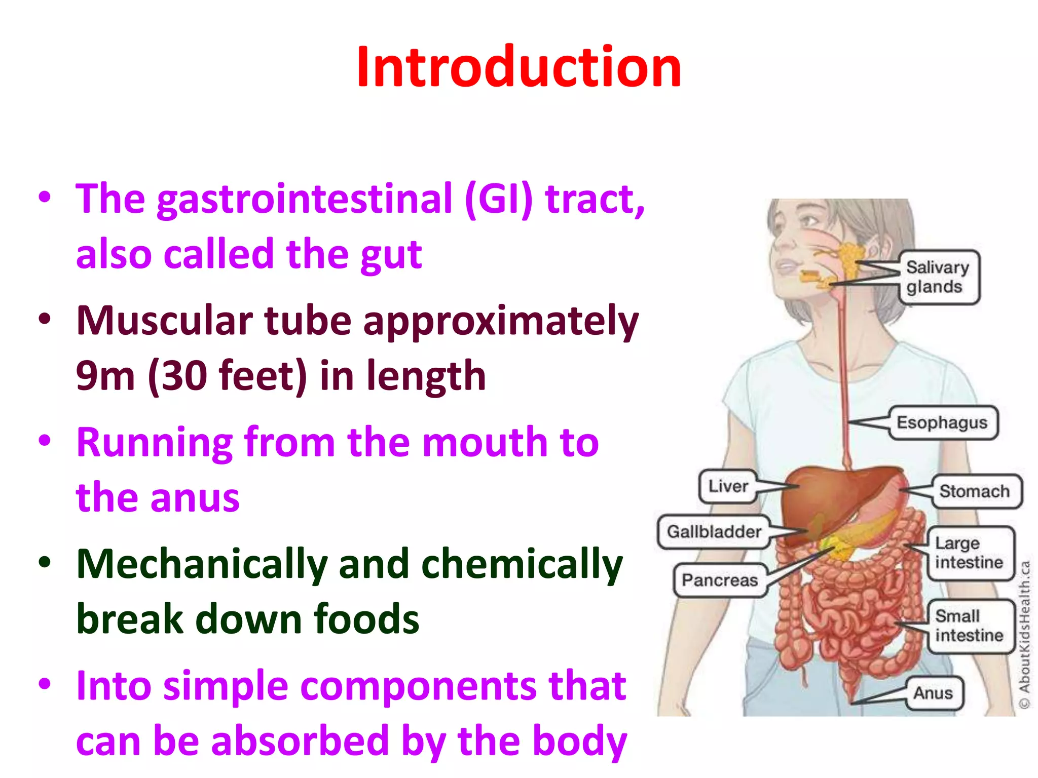
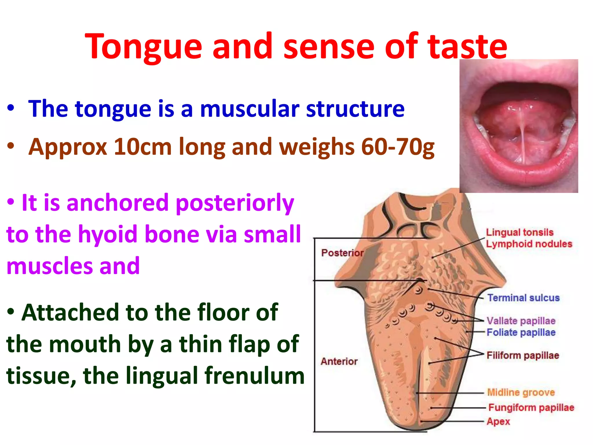
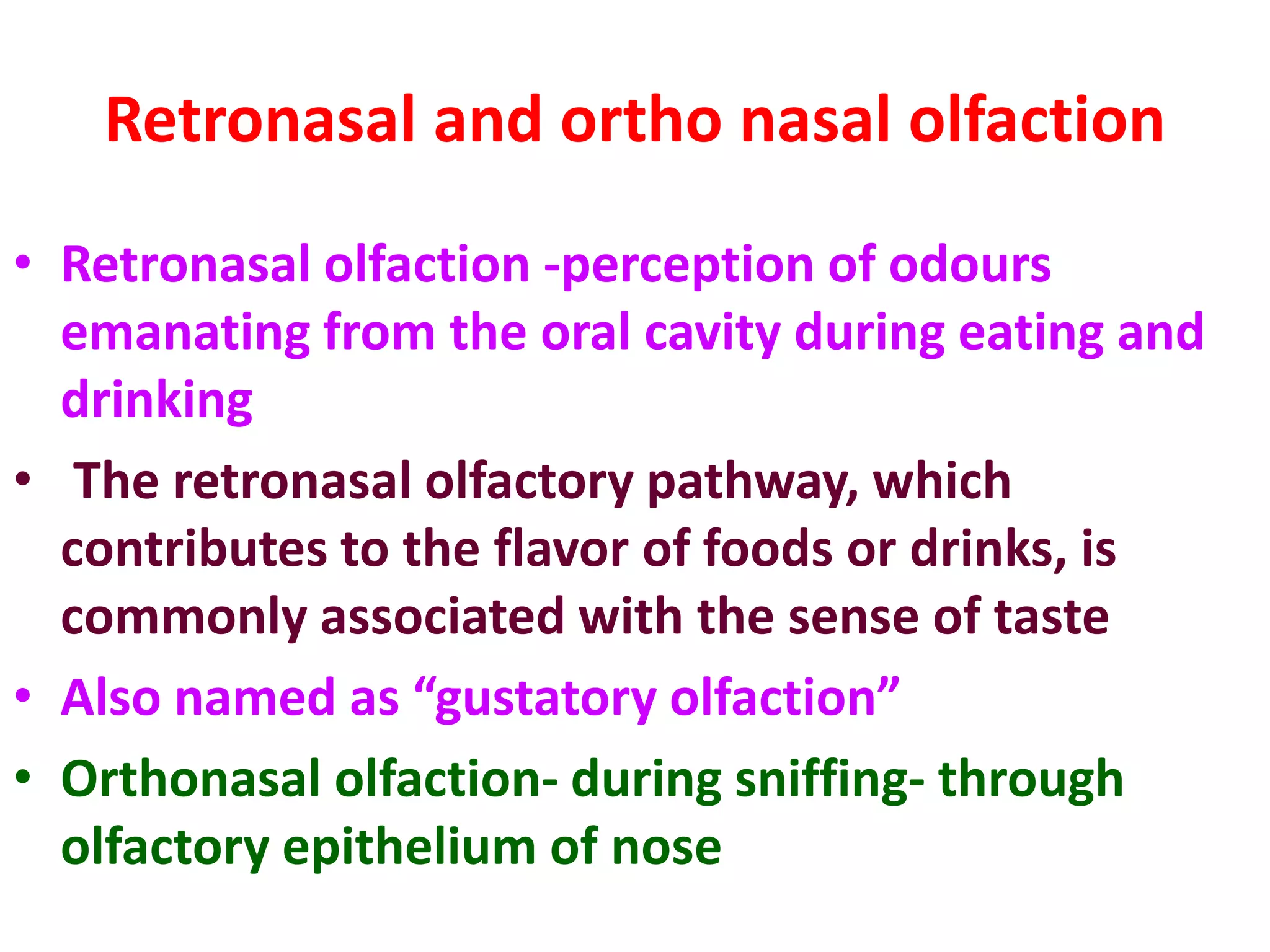
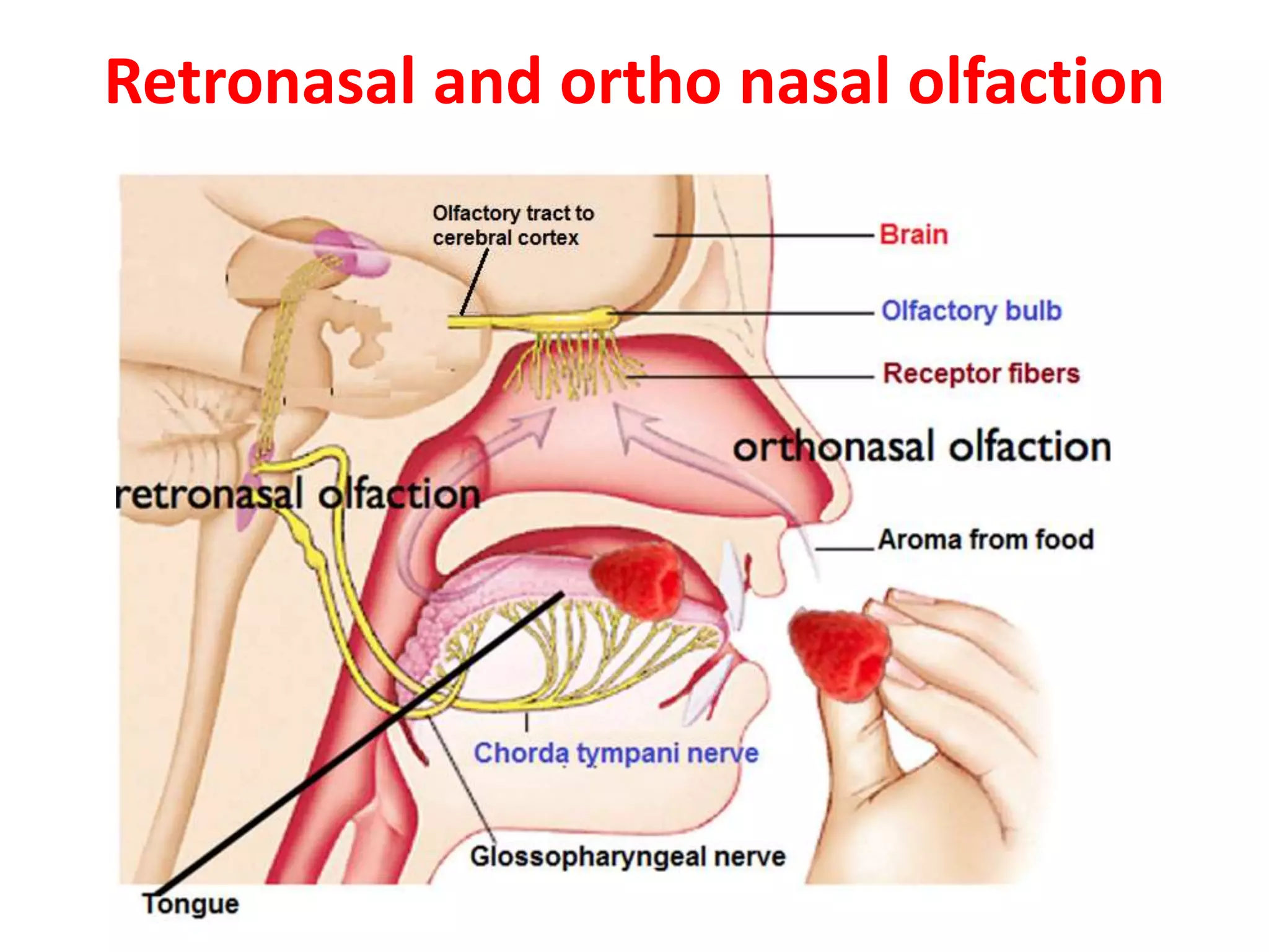
1) The gastrointestinal tract is approximately 9 meters long and runs from the mouth to the anus, mechanically and chemically breaking down food.
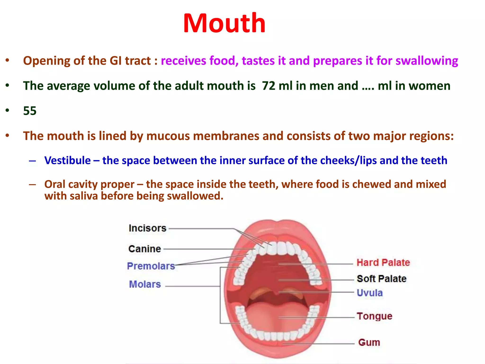
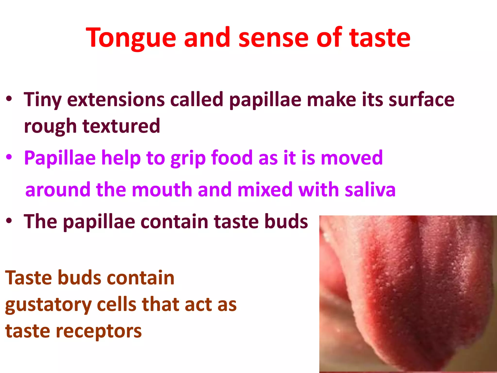
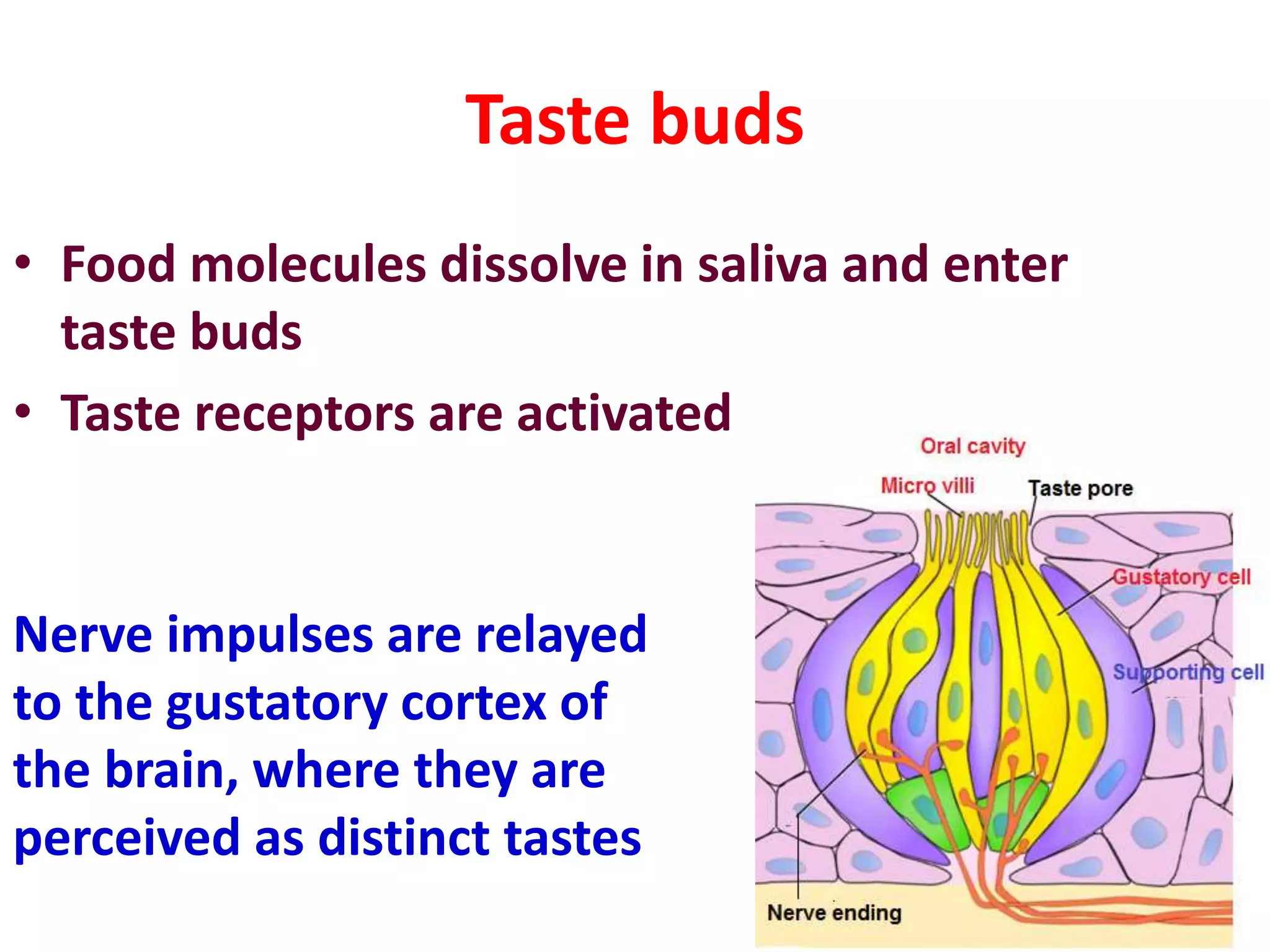
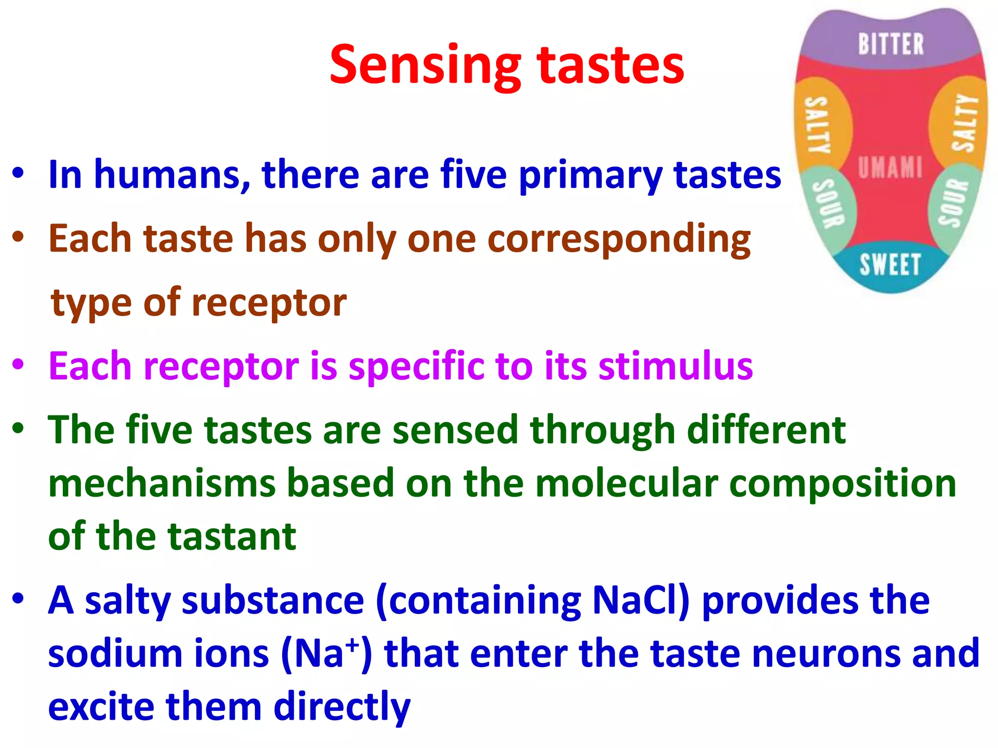
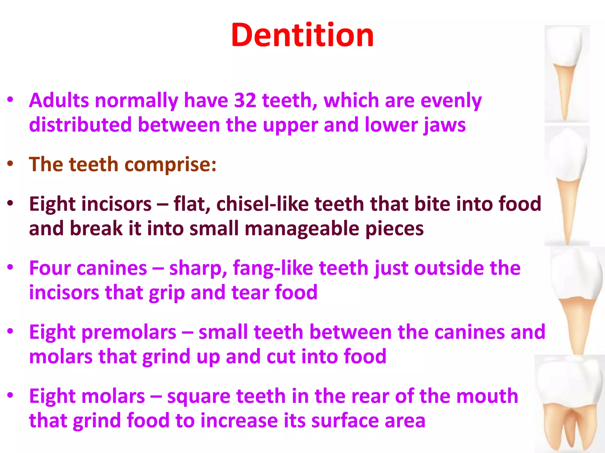
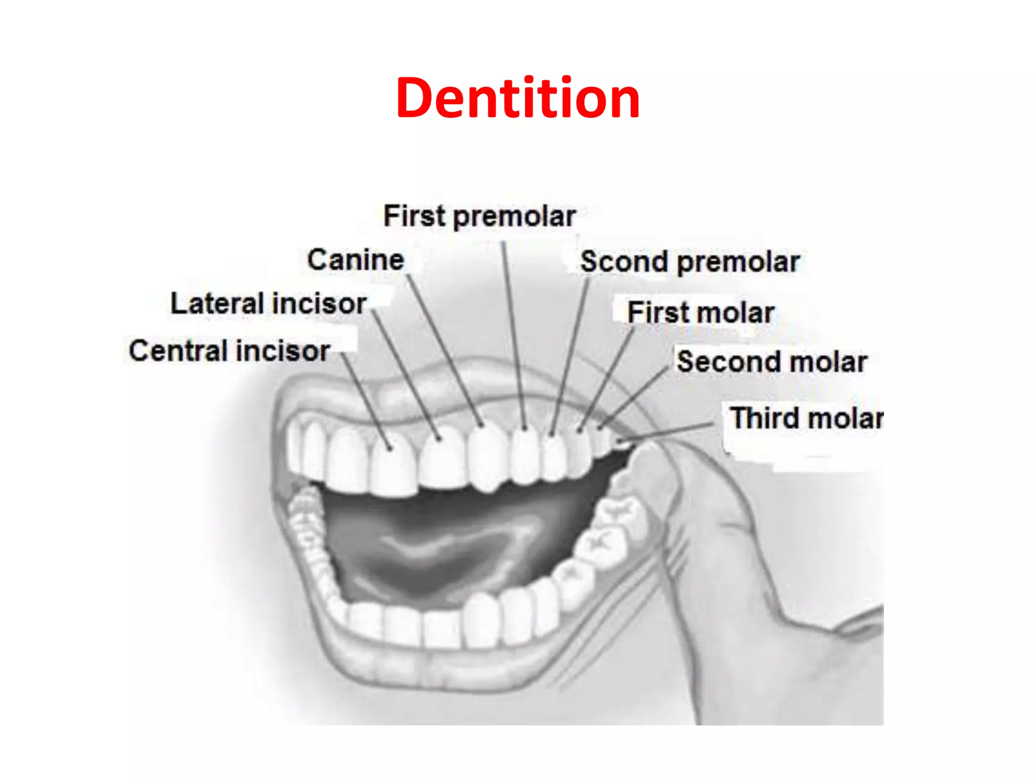
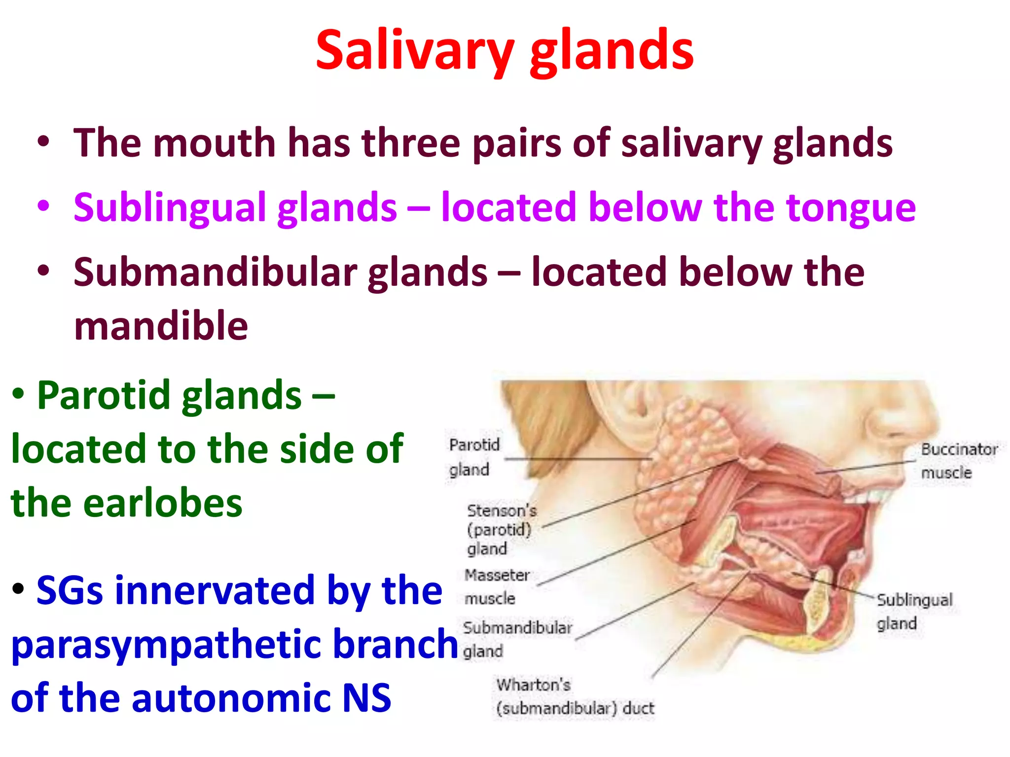
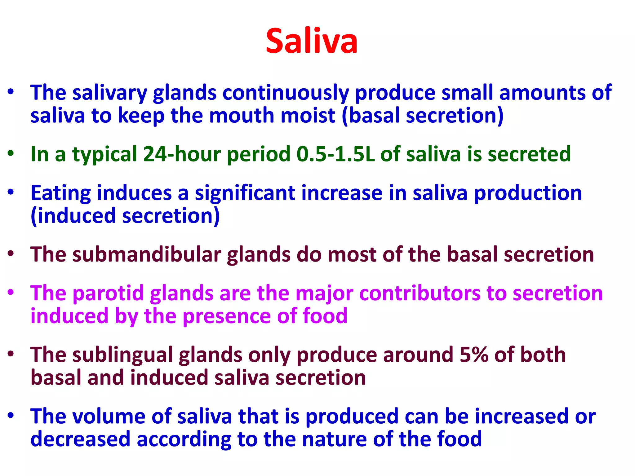
2) The mouth contains taste buds that detect the five basic tastes and contains salivary glands that produce saliva to moisten food for swallowing.
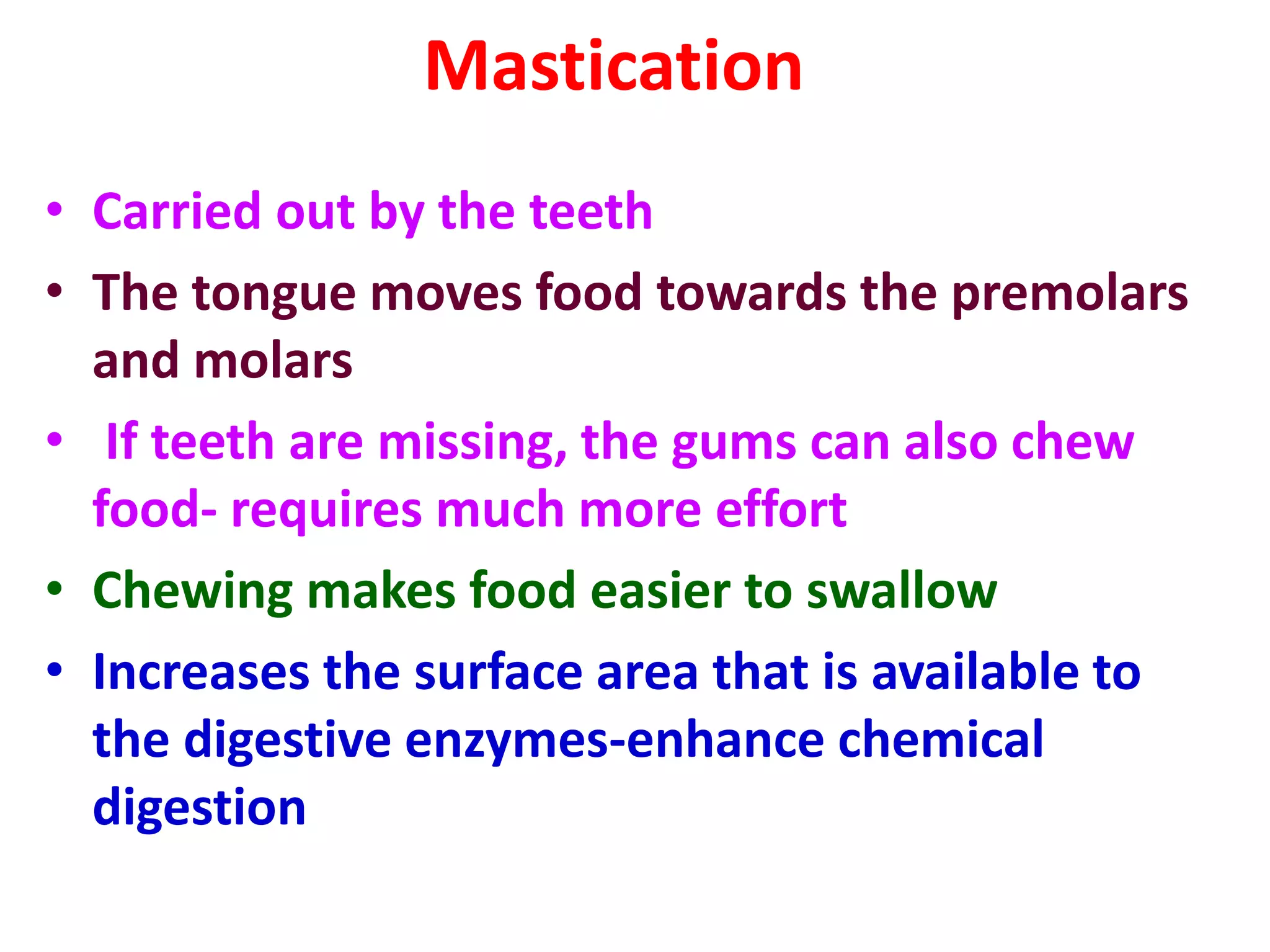
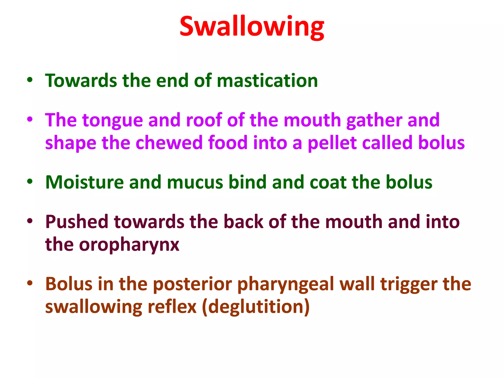
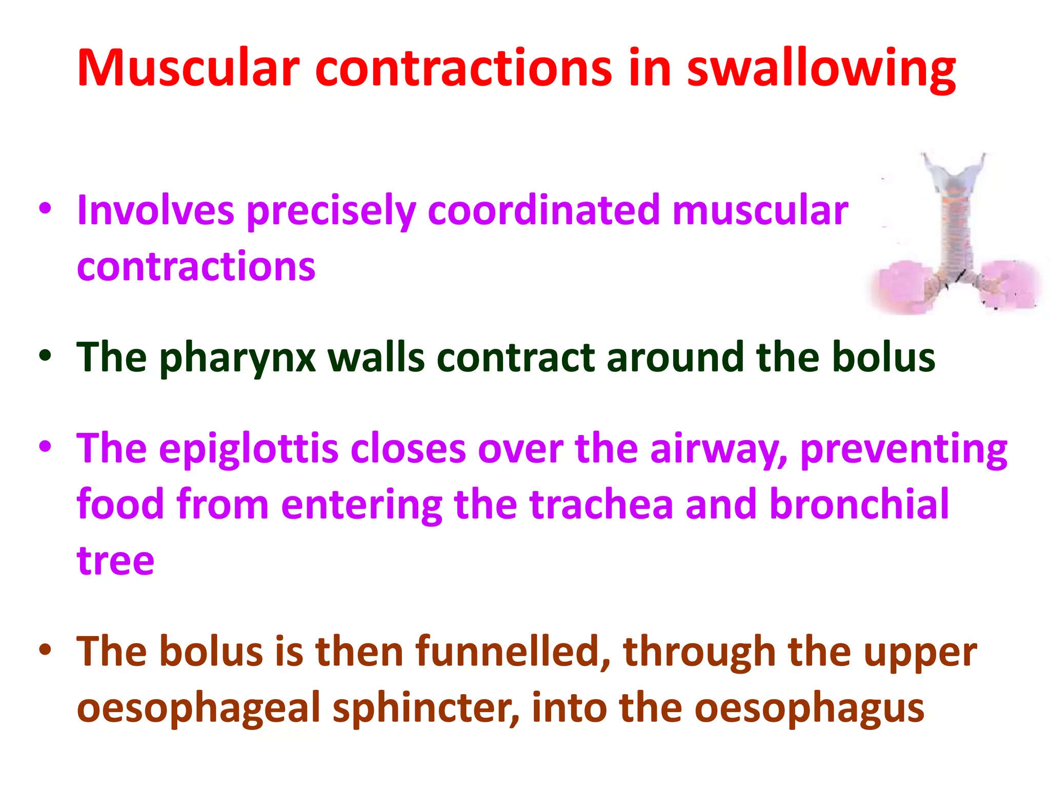
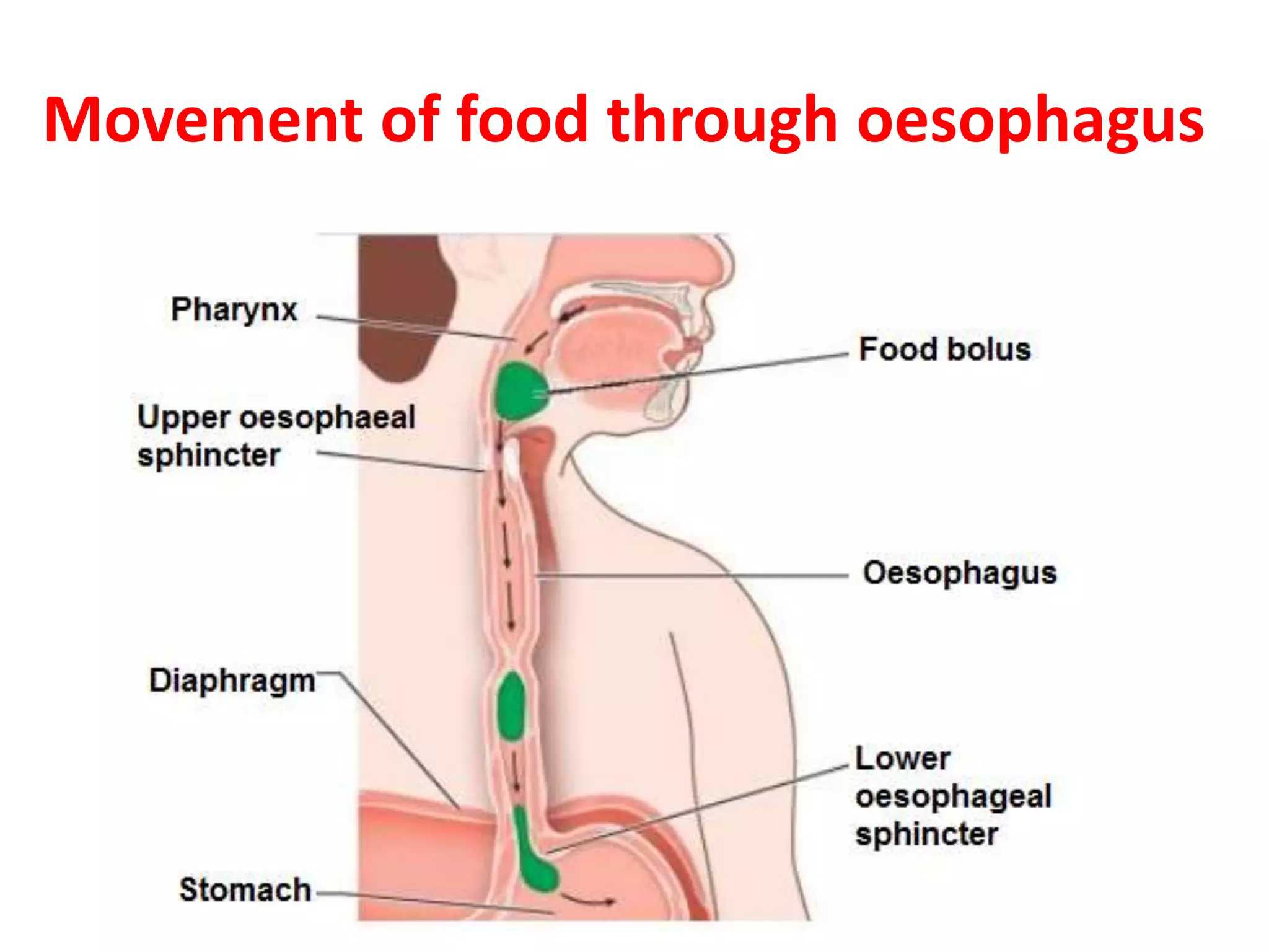
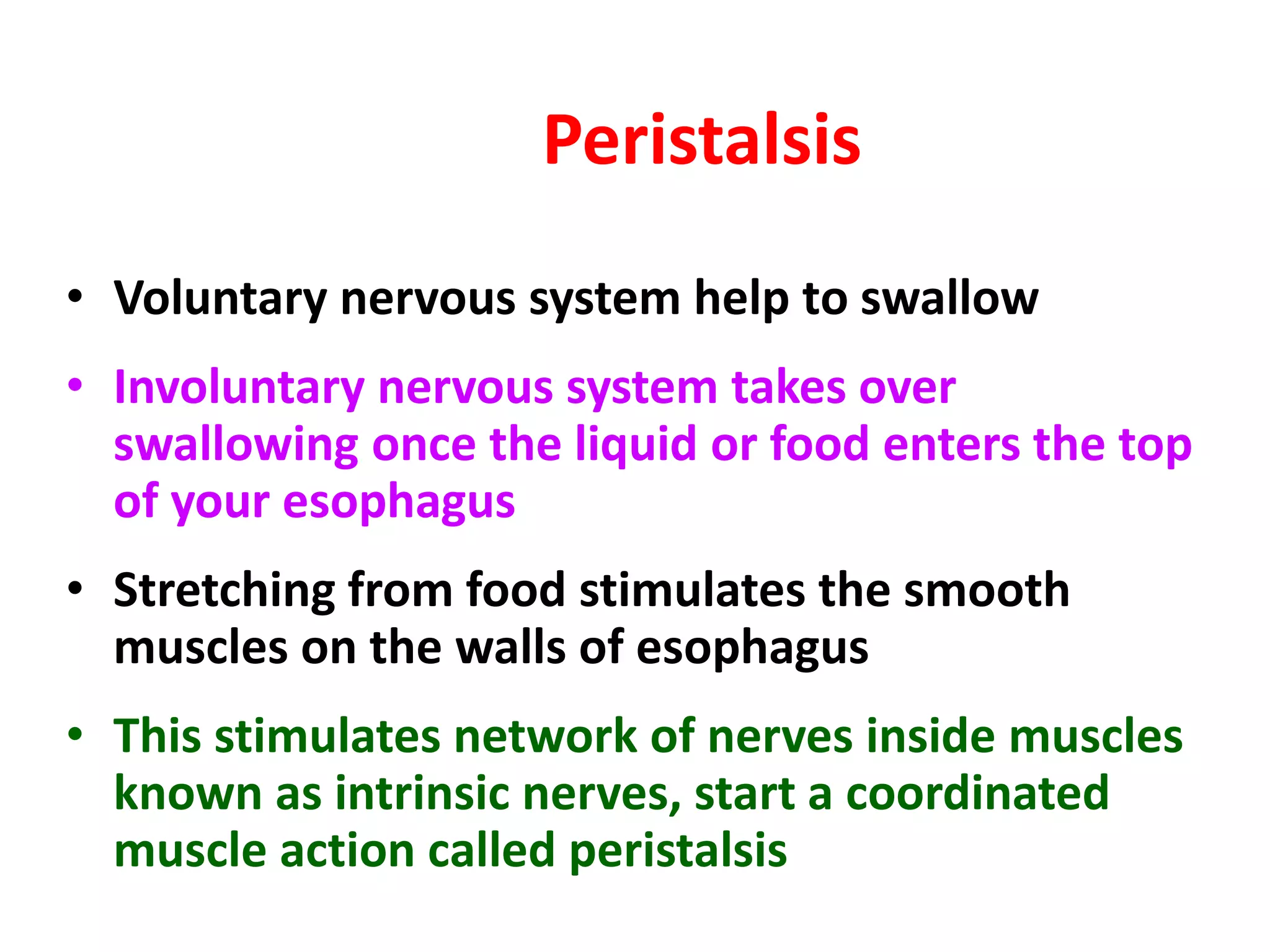
3) Chewing and swallowing propel food through the esophagus and into the stomach through peristalsis, where further digestion will occur.