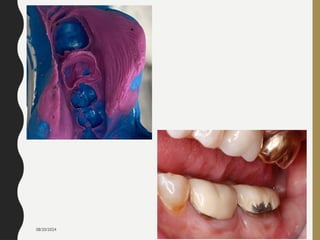
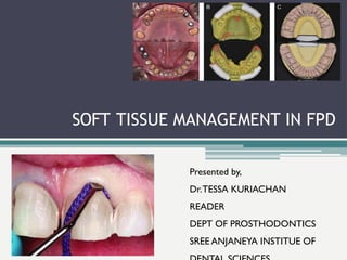
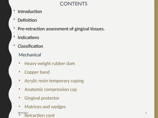
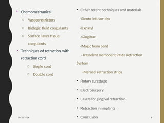
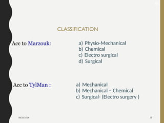
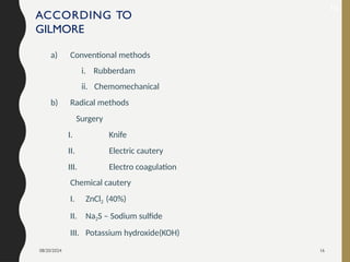
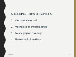
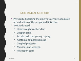
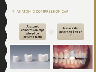
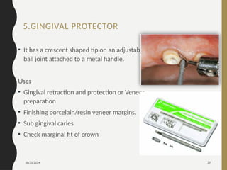
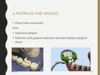
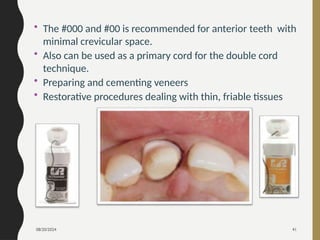
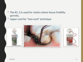
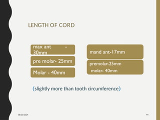
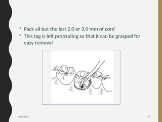
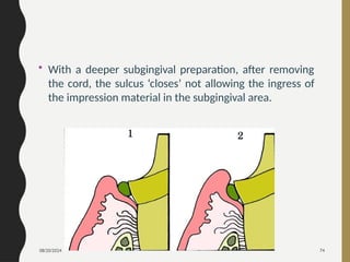
08/20/2024 1
08/20/2024 2
08/20/2024 3
SOFT TISSUE MANAGEMENT IN FPD
Presented by,
Dr.TESSA KURIACHAN
READER
DEPT OF PROSTHODONTICS
SREE ANJANEYA INSTITUE OF
08/20/2024 5
CONTENTS
• Introduction
• Definition
• Pre-retraction assessment of gingival tissues.
• Indications
• Classification
Mechanical
• Heavy weight rubber dam
• Copper band
• Acrylic resin temporary coping
• Anatomic compression cap
• Gingival protector
• Matrices and wedges
• Retraction cord
2
08/20/2024 6
• Chemomechanical
o Vasoconstrictors
o Biologic fluid coagulants
o Surface layer tissue
coagulants
• Techniques of retraction with
retraction cord
o Single cord
o Double cord
3
• Other recent techniques and materials
-Dento-infusor tips
-Expasyl
-Gingitrac
-Magic foam cord
-Traxodent Hemodent Paste Retraction
System
-Merocel retraction strips
• Rotary curettage
• Electrosurgery
• Lasers for gingival retraction
• Retraction in implants
• Conclusion
08/20/2024 7
INTRODUCTION
• A perfect impression should include finish lines which in turn results in good
marginal fit of the restoration. The gingiva must be displaced to make a complete
impression and some times even to permit completion of the preparation and
cementation of the restoration.
• The aesthetics and longevity of restorations is significantly dependent on
gingival and periodontal factors.
• The intimate interaction between the restorations and the surrounding
soft tissues means that all procedures performed should keep the health
of the gingiva and periodontium under consideration.
08/20/2024 8
DEFINITION
 Gingival tissue displacement is the deflection of the marginal
gingiva away from a tooth- GPT 9
 It is defined as the procedure of
 temporary eversion (widening of gingival sulcus)
 resection of gingiva away from the tooth surface or
 deepening of gingival sulcus to expose the cervical portion of
tooth in order to have proper marginal finish to the restoration
and recording the preparation accurately.
6
08/20/2024 9
PRE-RETRACTION ASSESSMENT OF
GINGIVAL TISSUES
• The gingival tissues intended to be retracted should be normal
in colour and firm.
• The contour, consistency and any pain originating from the
gingiva or supporting tissues should be evaluated
• There should be minimum or no bleeding on probing
• ideally to control the apical extent of the preparation so as not
to encroach on the epithelial and connective tissue attachment
08/20/2024 10
# 1
# 2
# 3
# 1 – Sulcular Depth
0.69mm
# 2 – Junctional Epithelial
Attachment = 0.97mm
# 3 – Connective Tissue Attachment
1.07mm
Gargiulo et al. reported in 1961
08/20/2024 11
Based on the sulcus depth, the following three rules can be used
to place intracrevicular margins:
1. If the sulcus probes are 1.5 mm or less, the restorative margin
could be placed 0.5 mm below the gingival tissue crest.
2. If the sulcus probes are more than 1.5 mm, the restorative
margin can be placed in half the depth of the sulcus.
3. If the sulcus is greater than 2 mm, gingivectomy could be
performed to lengthen the tooth and create a 1.5 mm sulcus.
The patient can then be treated as per rule1.
08/20/2024 12
The criteria which should be used in evaluating the retraction:
( 1) A trough or space must be created which makes the
subgingivally prepared margins both accessible and visible.
(2) Must be wide enough to accommodate elastic impression
material of sufficient thickness and strength so that it cannot be
torn during the removal of the finished impression.
(3) Must be free of blood and tissue fluids and must remain dry
for a time sufficient for placement and set (or gel) of the elastic
impression materials.
08/20/2024 13
(4) There must be minimum tissue damage resulting from the
gingival displacement procedure, as well as minimum tissue
damage resulting from the preparation of the subgingival
margins.
(5) The tissues must recover within a reasonable period of time.
(6) The resulting tissue contours must be predictable.
(7) The general systemic effect must be minimal and certainly
must be tolerable to the individual patient.
08/20/2024 14
INDICATIONS OF GINGIVAL RETRACTION:
 Esthetic consideration-Subgingival finish lines
 To control haemorrhage and gingival seepage
 Presence of Sub gingival Caries.
 Cervical abrasion or erosion
 Visualize margins and remove excess cement during final
cementation
 Protection of the gingiva during preparation of tooth for
direct or indirect restoration with subgingival margins.
08/20/2024 15
CLASSIFICATION
Acc to Marzouk: a) Physio-Mechanical
b) Chemical
c) Electro surgical
d) Surgical
Acc to TylMan : a) Mechanical
b) Mechanical – Chemical
c) Surgical- (Electro surgery )
18
08/20/2024 16
ACCORDING TO
GILMORE
a) Conventional methods
i. Rubberdam
ii. Chemomechanical
b) Radical methods
Surgery
I. Knife
II. Electric cautery
III. Electro coagulation
Chemical cautery
I. ZnCl2 (40%)
II. Na2S – Sodium sulfide
III. Potassium hydroxide(KOH)
19
08/20/2024 17
20
ACCORDING TO B.W.BENSON ET AL
1. Mechanical method
2. Mechanico-chemical method
3. Rotary gingival curettage
4. Electrosurgical methods.
08/20/2024 18
MECHANICAL METHODS
• Physically displacing the gingiva to ensure adequate
reproduction of the preparaed finish line.
• Methods used:
- Heavy weight rubber dam
- Copper band
- Acrylic resin temporary coping
- Anatomic compression cap
- Gingival protector
- Matrices and wedges.
- Retraction cord
21
08/20/2024 19
• Use of heavy weight rubber dam with proper interceptal
dimensions, has an effect which is immediate.
• According to Gilmore it can be called as
gum compression rather than displacement.
1. HEAVY WEIGHT RUBBER DAM
22
08/20/2024 20
Indication:
• When a limited number of teeth in one quadrant
are being restored.
• When preparations do not have to be extended too
far sub-gingivally
Limitations:
• Full arch impressions are difficult
• Should not be used with PVS because the rubber
inhibits their polymerization
26
08/20/2024 21
2. COPPER BAND
28
• A copper band or tube can serve as a means of
carrying the impression material as well as a
mechanism for displacing the gingiva to ensure that
the gingival finish line is captured in the impression.
• Impression compound or elastomeric impression
materials can be used along with this band.
08/20/2024 22
29
08/20/2024 23
Gingival margin are crimped to adapt to
gingival contour
08/20/2024 24
Advantage
• The gingival tissue retraction is maximal.
• the margins may be established at the maximum subgingival
depth
• the band impression made under pressure with no air bubbles
or voids
• the technique can be used for either sectional or complete-
arch impression, inlays, three-quarter crowns, or full-coverage
crowns;
• it is not necessary to pour the cast as soon as the impressionis
made
32
08/20/2024 25
Disadvantage
• Can cause incisional injuries of gingival tissues.
• More time necessary to fit and adapt the band
• Difficulty in removing the modelling compound filled
band from undercuts.
08/20/2024 26
3. ACRYLIC RESIN TEMPORARY COPING
33
Temporary acrylic resin coping
constructed
Tray adhesive applied
Filled with elastomeric impression
material and reseated
Tissue displacement occurs
Full arch impression made
08/20/2024 27
4. ANATOMIC COMPRESSION CAP
Anatomic
compression caps
placed on
patient’s teeth
Instruct the
patient to bite on
it
08/20/2024 28
Advantages:
• Stops bleeding due to compression
• Opens the sulcus wide
• Ensures clean , dry area with well defined gingival margin
08/20/2024 29
5.GINGIVAL PROTECTOR
• It has a crescent shaped tip on an adjustable
ball joint attached to a metal handle.
Uses
• Gingival retraction and protection or Veneer
preparation
• Finishing porcelain/resin veneer margins.
• Sub gingival caries
• Check marginal fit of crown
08/20/2024 30
6.MATRICES AND WEDGES
• Placed inter proximally
Uses
• Depresses gingiva
• Matrices with gingival extension provides displace gingival
tissue
08/20/2024 31
7. RETRACTION CORDS
• These are ready made cotton or synthetic woven cords,
frequently with metallic or resin wire wrapped around
them to assure their compactness, immobility, and non
shredding.
• They come in different sizes, arbitrarily numbered by
their manufacturers.
• They may be supplied already impregnated with the
chemical, or the chemical may be added before
insertion of the cord or after insertion while the cord is
within the sulcus.
40
08/20/2024 32
MECHANISM OF RETRACTION
• Deformation of gingival
tissues during retraction
and impression
procedures involves four
forces:
• Retraction,
• Relapse
• Displacement
• Collapse.
42
08/20/2024 33
CLASSIFICATION OF RETRACTION CORDS
Depending on
the
configuration
• Twisted
• Knitted
• Braided
Depending on
surface finish
• Wax
• Unwaxed
08/20/2024 34
Depending on
the chemical
treatment
• Plain
• Impregnated
Depending on
number strands
• Single
• Double-string
08/20/2024 35
Depending
on thickness-
color coded
• Black- 000 ( extra small)
• Yellow – 00 ( small)
• Purple- 0
• Blue- 1
• Green – 2
• Red- 3 ( extra large)
47
08/20/2024 36
DESIRABLE QUALITIES OF CORD
• Dark color ---contrast with tissues, tooth and cord
• Absorbent ---wet medicament
• Spontaneously reversible
• Safe locally & systemically
• Haemostasis
• Should not cause chemical injury
45
08/20/2024 37
TWISTED GINGIVAL CORDS
• Allow the dentist to customize the cord as individual
strands can be removed
08/20/2024 38
• Knitted cord afford greater inter-thread space than braided
cord
• Knitted to form an interlocking chain of thousands of tiny
loops, making it easy to pack below the gingival margin and
stays when packed into place.
braided
knitted
48
KNITTED GINGIVAL RETRACTION CORD
08/20/2024 39
• knitted cords compresses upon packing, then expands
for tissue displacement.
• Twisted and braided cords can’t offer ease of packability and
tissue displacement like knitted ones.
49
08/20/2024 40
RECOMMENDED SELECTION OF RETRACTION CORD SIZE
• The size of cord is clinically determined by evaluating the
depth of the sulcus with a periodontal probe and observing
the friability of the particular tissue.
08/20/2024 41
• The #000 and #00 is recommended for anterior teeth with
minimal crevicular space.
• Also can be used as a primary cord for the double cord
technique.
• Preparing and cementing veneers
• Restorative procedures dealing with thin, friable tissues
08/20/2024 42
• The #0 is recommended for bicuspids as the primary
cord for the double cord technique.
• The #1 cord is recommended for the secondary cord
• Tissue control and/or displacement when soaked in
coagulative hemostatic solution prior to and/or after crown
preparations
08/20/2024 43
• The #2 ,3 is used for molars where tissue friability
permits.
• Upper cord for "two-cord" technique
08/20/2024 44
LENGTH OF CORD
max ant -
30mm
pre molar- 25mm
Molar - 40mm
mand ant-17mm
premolar-25mm
molar- 40mm
(slightly more than tooth circumference)
08/20/2024 45
2.CHEMO-MECHANICAL METHOD
• Gingival retraction using chemically impregnated
retraction cord is a chemo-mechanical method of
displacement
Mechanical aspect
Chemical aspect
08/20/2024 46
• Combination of a chemical with pressure packing
(mechanical action) –
▫ Leads to enlargement of the gingival sulcus
▫ Control of fluids seeping from the sulcus.
• Either
 Previously impregnated
 Or saturated with solution prior to insertion
 Or placed dry and solution applied.
08/20/2024 47
3 categories
• Vasoconstrictors
• Biologic fluid coagulants
• Surface layer tissue coagulants
08/20/2024 48
Vasoconstrictors
• Physiologically restrict the blood supply to the area
by decreasing the size of the blood capillaries,
tissue fluid seepage and consequently size of the
free gingiva.
- Epinephrine
- Nor–epinephrine
08/20/2024 49
EPINEPHRINE
• most commonly used is 8% epinephrine.
• Other strengths are 2%, 4%, 16% and 32%.
• Because of the high vascularity of the gingival tissue, the systemic
effects exerted by epinephrine have been a cause for concern,
especially if the gingival tissues have been lacerated.
• The systemic effect of epinephrine has been described as
‘epinephrine reaction’ or ‘epinephrine syndrome’ and is associated
with the use of epinephrine-soaked retraction cords
08/20/2024 50
CONTRAINDICATIONS FOR EPINEPHRINE USE
1. Cardiovascular disease
2. Hypertension
3. Hyperthyroidism
4. Hypersensitivity to epinephrine
5. Patients on tricyclic anti-depressants
6. Diabetic patients
08/20/2024 51
BIOLOGIC FLUID COAGULANTS
• Coagulate blood and tissue fluids locally, creating surface
layer that is efficient sealant against blood and crevicular
fluid seepage.
• Safe, with no systemic effects.
• Examples
- 100% alum
- 15-25% aluminium chloride
- 10% aluminium potassium sulphate
- 15% tannic acid
- Ferric subsulfate
08/20/2024 52
ViscoStat Clear
• 25% aluminum chloride gel in a
viscous, aqueous vehicle causes the
collagen in the capillary ends to swell,
thereby closing off the capillaries.
• No coagulum is formed, nor does
hemostatic residue adhere to the
preparation
• Does not stain the hard and/or soft
tissues.
08/20/2024 53
C) SURFACE LAYER TISSUE COAGULANTS
• coagulates surface layer and free gingival epithelium as well
as seeped fluids, this creating temporarily impermeable film
for underlying fluids.
• Examples
-8% zinc chloride
-Silver nitrate
08/20/2024 54
• Local hazards
- Ulceration
- Local necrosis
- changes in location and dimension of free gingiva
• These can happen as a result of an excessive amount or
concentration or excessive time in application of agents
08/20/2024 55
PRODUCTS
BRAIDED
• Hemodent Retraction Cord (Premier Products
Company)
 Nor epinephrine
• Gingibraid+ Van R
 Epinephrine/Alum 87
 or Aluminum Potassium Sulfate
 or Non-Impregnated
• Gingi-Pak Z-Twist
 epinephrine HCI
 aluminum sulfate
 Non- Impregnated
08/20/2024 56
• Unibraid Van R
 Epinephrine/Alum or
 Aluminum Potassium Sulfate
• Sil-Trax® Plus (Pascal Company)
 with reduced Racemic Epinephrine HCl
and Zinc Phenosulfonate
08/20/2024 57
KNITTED
• GingiKnit Knitted Retraction Yarn
• Ultrapak
08/20/2024 58
NON-IMPREGNATED
• Ultrapak Ultradent Products, Inc.
• Gingiplain Original GingiPak
• Gingiplain Soft GingiPak
• Gingiplain Z-Twist GingiPak
• Knit Trax Pascal Company, Inc
• Retrax Pascal Company, Inc.
• Sil-Trax® Plain Pascal Company, Inc.
08/20/2024 59
IMPREGNATED- EPINEPHRINE HCI
• UltraPak E (Ultradent Products, Inc.)
• Crownpak (GingiPak )
• Racord (Pascal Company)
• Racord II (Pascal Company) Reduced Racemic
Epinephrine HCl and Zinc Phenosulfonate
• Sil-Trax® Epi (Pascal Company)
• Sil-Trax® Plus (Pascal Company)
08/20/2024 60
TECHNIQUES FOR CORD PLACEMENT
• Single cord technique
• Double cord technique
08/20/2024 61
ARMAMENTARIUM
1. Saliva ejector
2. Scissors
3. Cotton pliers
4. Mouth mirror
5. Explorer
6. Cord packer
7. Cotton rolls
8. Retraction cord
9. Medicament to be
used
10. Dappen dish
11. Cotton pellets
12. 2×2 gauze sponges
08/20/2024 62
89
08/20/2024 63
SINGLE CORD TECHNIQUE
• The desired length of retraction cord is drawn from the
dispenser bottle with sterile cotton pliers .
08/20/2024 64
• The cord is twisted to make it tight and small.
08/20/2024 65
• The retraction cord should be dipped in the medicament
solution in a dappen dish.
• Hemorrhage can be controlled by using Homeostatic
agents like Hemodent liquid (aluminium chloride)
08/20/2024 66
08/20/2024 67
08/20/2024 68
08/20/2024 69
08/20/2024 70
08/20/2024 71
• Pack all but the last 2.0 or 3.0 mm of cord
• This tag is left protruding so that it can be grasped for
easy removal
08/20/2024 72
• After 10 minutes, remove the cord slowly
toavoid bleeding.
• Inject impression material only if the sulcus
remains clean and dry.
• It may be necessary to gently rinse away any coagulum,
then lightly blow air on it.
08/20/2024 73
DOUBLE CORD TECHNIQUE
• This technique is routinely used while making
impressions when gingival health is compromised and
it is impossible to delay treatment procedures.
• When preparation involves multiple teeth.
08/20/2024 74
• With a deeper subgingival preparation, after removing
the cord, the sulcus ‘closes’ not allowing the ingress of
the impression material in the subgingival area.
08/20/2024 75
08/20/2024 76
ADVANTAGE
• The first cord remains in place within the sulcus thus
reducing the tendency of the gingival cuff to recoil and
displace partially set impression material.
• Helps to control gingival haemorrhage and exudate
• Overcomes the problem of the sulcus impression tearing
because of inadequate bulk.
08/20/2024 77
DISADVANTAGE
• The main disadvantage of the two-cord technique
is difficulty to remove the first cord thereby
inflicting a painful, gingival reaction.
08/20/2024 78
08/20/2024 79
08/20/2024 80
• A specifically designed 1.0cc syringe known as dento-
infusor is used to deposit the hemostatic agent i.e. 20%
ferric sulfate.
• infuse hemostatic agents into bleeding capillaries.
DENTO-INFUSOR TIPS
08/20/2024 81
• The infusor is used with a burnishing motion in the sulcus & is
carried circumferentially around the sulcus.
• The medicament is extruded from the syringe/infusor as the
instrument is manipulated around the gingival sulcus.
• The tip infuses the hemostatic agent into capillaries, forming
a cork-like “plug,” then wipes coagulum away.
08/20/2024 82
• When hemostasis is achieved, a retraction cord soaked in
ferric sulfate solution is packed into the sulcus and left in
place for 1 to 3 minutes.
• This is an effective additional technique for control of
bleeding when using the single cord technique.
08/20/2024 83
• chemo-mechanical technique for sulcus opening and
hemotasis.
• Contains
▫ white clay to ensure the consistency of the paste and its
mechanical action,
▫ aluminium chloride enhances the haemostatic action.
EXPASYL
08/20/2024 84
EQUIPMENT CONSISTS OF:
• capsules;
• injection canulas; and
• applicator
• The Expasyl paste is injected into the sulcus, exerting a
stable, non-damaging pressure of 0.1 N/mm.
08/20/2024 85
The canula is pressed against the tooth and angled until it comes into
contact with the sulcus lining of the gingival edge.
08/20/2024 86
The product enters the sulcus.
08/20/2024 87
The marginal gingiva blanches.
08/20/2024 88
The angle of the canula tip is increased and maintains contact on
the sulcus lining of the gingival edge.
08/20/2024 89
The product is injected into the
interproximal space.
Support points: the thumb guides the
tip of the applicator.
08/20/2024 90
Removal of product by an air and water spray.
08/20/2024 91
Sulcus opening and the absence of oozing provide good access.
08/20/2024 92
08/20/2024 93
Advantages
• Reduced chair time- patient comfort
• Hemostasis
• Safe, no danger of rupturing the epithelial attachments
and causing recession as little or no pressure as
compared to cord.
• Easy access and placement
• Easily removed
• No contamination
08/20/2024 94
GINGITRAC
• GingiTrac delivers the perfect combination of built-in
astringency with fast and gentle retraction assuring the
accurate impressions
08/20/2024 95
08/20/2024 96
156
08/20/2024 97
ADVANTAGES
• Single crown or multiple crown retraction, all without
packing cord
• Works in less than 5 minutes, regardless of number of
preps
• Get more accurate impressions
• Works gently, no tissue trauma or ligament damage
• Contains aluminum sulfate astringent to control
bleeding and oozing
• Easy-to-use 1:150ml automix gun system mixes and
delivers GingiTrac
• Removes in a clean manner, in one piece, without
rinsing
08/20/2024 98
MAGIC FOAM CORD
162
08/20/2024 99
• Magic FoamCord is the first expanding PVS material
designed for easy and fast retraction of the sulcus
without the time consuming packing of retraction
cord
08/20/2024 100
Comprecap anatomic technique
164
08/20/2024 101
0% expansion
immediately
after
application
60%
expansion
after 3 min.
after
application
165
MECHANISM OF ACTION
08/20/2024 102
Advantages
• Non-traumatic method of temporary gingival retraction
• Easy and fast application directly to the sulcus without
pressure or packing
• Comfortable to the patient
• No haemostatic chemicals to contaminate the impression
site – no need for extensive rinsing
• Outstanding retraction for perfect impressions
167
08/20/2024 103
MEROCEL RETRACTION STRIPS
• It is a synthetic material obtained from
biocompatible
polymer, i.e. hydroxylate polyvinyl acetate
• This material is placed in the sulcus, the material absorbs any
secretions present in the sulcus and expands to bring about
gingival displacement.
08/20/2024 104
Advantages
• Chemically pure
• Easily shaped
• Remarkably effective for absorption of intraoral fluids
such as blood, saliva, and crevicular fluid
• Soft and adaptable to the surrounding tissues
• Free of fragments, without debris
08/20/2024 105
SURGICAL METHODS
• Some methods utilized to improve the visualization of the
preparation margins of the tooth are not true retraction
techniques.
• This is because they actually remove some part or all of the
overlying gingival tissue in order to expose the finish line of the
preparation and/or control haemorrhage.
• These techniques are more invasive and should only be used in
cases where there is adequate amounts of attached gingiva.
08/20/2024 106
ROTARY CURETTAGE
• Also known as gingetage, Concept described by Amsterdam in
1954.
• Technique described by Hansing and subsequently enlarged
upon by Ingraham in 1975
• Troughing technique, wherein a portion of the epithelium
within the sulcus is removed to expose the finish line.
• It should be done only on healthy gingival tissue.
08/20/2024 107
THE FOLLOWING CRITERIA SHOULD BE FULFILLED FOR
GINGETTAGE.
• Absence of bleeding upon probing from the gingiva.
• The depth of the sulcus is less than 3mm.
• Presence of adequate keratinized gingiva.
08/20/2024 108
TECHNIQUE
• It is usually done simultaneously along with finish line
preparation.
• Torpedo-tipped diamond is used to remove the epithelial
lining of the sulcus.
08/20/2024 109
• Abundant water should be sprayed during the
procedure.
• A retraction cord impregnated with AlCl3, can be
used to control bleeding.
08/20/2024 110
DISADVANTAGES
• Poor tactile sensation when using diamonds on sulcular
walls can produce deepening of sulcus.
• It can potentially damage the periodontium if used
incorrectly.
08/20/2024 111
ELECTROSURGERY
Definition:
It is defined as the use of specially designed
electronic equipment that produces a limited variety of
high frequency wave forms for the purposes of cutting or
removing soft tissue.
08/20/2024 112
• Electrocautery refers to direct current
• whereas electrosurgery uses alternating current.
08/20/2024 113
• Electrosurgery denotes surgical reduction of sulcular
epithelium using an electrode to produce gingival
retraction
08/20/2024 114
Cutting edge designs :
a) Coagulating probe
b) Diamond loop
c) Round loop
d) Small straight probe
e) Small loop
08/20/2024 115
• Controlled tissue destruction.
• Current flows through a small cutting electrode a vacuum
tube or a transistor to deliver a high frequency electrical
current of at least 1.0 MHz
• The procedure is also called as “Surgical Diathermy”
• It uses radio currents in the range of 1.5 to 7.5 million
cycles/sec.
MECHANISM OF ACTION
08/20/2024 116
• Electrosurgical current flows from the unit to the active
(cutting) electrode to the ground and back to the unit.
08/20/2024 117
• Using the Straight Knife electrode to stop bleeding in
gingival sulcus
08/20/2024 118
• Using the Straight Knife electrode to restore gingival
symmetry
08/20/2024 119
• Using the Straight Knife electrode – best for sulcular
enlargement
08/20/2024 120
• Using the Long Loop electrode to remove occluding
gingiva
08/20/2024 121
• Clinical applications:
▫ Widening gingival sulcus
▫ Crown lengthening
▫ Exposing impacted teeth
▫ Incising abcesses
▫ Removing hyperplastic gingiva
▫ Frenectomy
08/20/2024 122
ADVANTAGES
• Excellent vision of the margins
• Removes unwanted tissue with ease
• Reduces chair time by simplifying operative procedures and
maintaining hemostasis
• Provides outstanding cutting precision for superior
clinical and aesthetic results
• Allows uneventful healing
08/20/2024 123
DISADVANTAGES
• Very technique sensitive
• Application of excessive pressure may produce severe
tissue damage
• Difficult to control lateral dissipation of heat
• It cannot be done in a dry field. The operatory area should
be very moist during the procedure. This leads to
compromised access and visibility
08/20/2024 124
CONTRAINDICATIONS
• Patients with cardiac pacemakers because the frequency of
the electrical current in the electrode can interfere with the
functioning of the pacemaker.
•Not suitable where thin attached gingiva is present.
(labial of maxillary canines)
08/20/2024 125
LASERS
• Lasers helps in exposure of subgingival finish lines,
controls the hemorrhage, and removes just enough
epithelial attachment to facilitate the placement of
retraction cord.
• Minimum gingival recession.
• Laser tips 400-600 micron in diameter.
08/20/2024 126
• Useful for cutting, vaporizing, coagulating, haemostasis on
gingiva and mucosa
• Some clinicians use laser only for haemostasis and follow it
with cord which needs to stay in place only 3-4 mins before
impressions.
08/20/2024 127
• The most commonly used lasers for gingival
displacement are the
▫ 980-nm Diode lasers,
▫ 1064-nm Nd-YAG laser
▫ Erbium class of dental lasers.(2940nm)
08/20/2024 128
ADVANTAGES
• Minimum pain, inconvenience
discomfort
• Less fear anxiety, stress
• Minimum or no anaesthesia
• No drill sounds
• Less chair time
• Reduced post operative complications
• Minimum or no bleeding
08/20/2024 129
DISADVANTAGES
• Cost factor is a drawback and technique sensitive
08/20/2024 130
GINGIVAL RETRACTION AROUND
IMPLANTS
• Indicated only in rare situations
• Fabrication of custom abutment
• The use of chemicals, such as 15% aluminum chloride in an
injectable kaolin matrix, is a better option
08/20/2024 131
08/20/2024 132
08/20/2024 133
BennaniV, Schwass D, Chandler N. Gingival retraction techniques for implants
versus teeth: current status. J Am Dent Assoc 2008; 139: 1354 1363.
−
08/20/2024 134
CONCLUSION
• Perfect tooth preparation are worth less without perfect impressions,
and perfect impressions can easily be achieved by using various gingival
retraction techniques as mentioned above
• Since gingival retraction is an integral part of clinical practice, the clinician
should make an effort to utilize different methods and products available
for retraction of gingival tissues in various clinical scenarios.
• Sometimes a combination of methods may be needed, and some things
may work for one clinician and not for another.
• The effort put into the appropriate retraction of gingival tissues pays off
in terms of longevity of restorations, better margins and aesthetics.
08/20/2024 135
REFERENCES
• Shillingburg HT; Fundamentals of Fixed Prosthodontics; 2012; 4th
edition ; Quintessence publications; USA; pp: 257-279
• Rosenstiel SF; Contemporary Fixed Prosthodontics; 2014; 4th edition;
India; pp: 431- 465
• Livaditis et al, Comparison of the new matrix system with traditional
fixed prosthodontic impression procedures, J Prosthet Dent
1998;79:200-7
• Shah M J et al; Gingival retraction methods in fixed prosthodontics –A
systematic review, Journal of dental sciences;2008, Vol 3(1):4-10
• Thomas MS et al, Nonsurgical gingival displacement in restorative
dentistry, June 2011, Vol32(5),27-39
08/20/2024 136
• Bennani V, Schwass D, Chandler N. Gingival retraction
techniques for implants versus teeth: current status. J Am Dent
Assoc 2008; 139: 1354−1363.
• Evaluation of Gingival Displacement Using Foam Cord and
Retraction Cord: An In Vivo Study
• Shivashakthy M, Comparative study on the efficacy of gingival
retraction using polyvinyl acetate strips and conventional
retraction cord - An in vivo study , Journal of clinical and
diagnostic research, 2013 Oct Vol- 7(10):8-11
• Reiman et al.Exposure of subgingival margins by nonsurgical
gingival displacement. J Prosthet. Dent. Dec 1976
THANK YOU…

SOFT TISSUE MANAGEMENT IN FIXED PARTIAL DENTURE

  • 1.
  • 2.
  • 3.
  • 4.
    SOFT TISSUE MANAGEMENTIN FPD Presented by, Dr.TESSA KURIACHAN READER DEPT OF PROSTHODONTICS SREE ANJANEYA INSTITUE OF
  • 5.
    08/20/2024 5 CONTENTS • Introduction •Definition • Pre-retraction assessment of gingival tissues. • Indications • Classification Mechanical • Heavy weight rubber dam • Copper band • Acrylic resin temporary coping • Anatomic compression cap • Gingival protector • Matrices and wedges • Retraction cord 2
  • 6.
    08/20/2024 6 • Chemomechanical oVasoconstrictors o Biologic fluid coagulants o Surface layer tissue coagulants • Techniques of retraction with retraction cord o Single cord o Double cord 3 • Other recent techniques and materials -Dento-infusor tips -Expasyl -Gingitrac -Magic foam cord -Traxodent Hemodent Paste Retraction System -Merocel retraction strips • Rotary curettage • Electrosurgery • Lasers for gingival retraction • Retraction in implants • Conclusion
  • 7.
    08/20/2024 7 INTRODUCTION • Aperfect impression should include finish lines which in turn results in good marginal fit of the restoration. The gingiva must be displaced to make a complete impression and some times even to permit completion of the preparation and cementation of the restoration. • The aesthetics and longevity of restorations is significantly dependent on gingival and periodontal factors. • The intimate interaction between the restorations and the surrounding soft tissues means that all procedures performed should keep the health of the gingiva and periodontium under consideration.
  • 8.
    08/20/2024 8 DEFINITION  Gingivaltissue displacement is the deflection of the marginal gingiva away from a tooth- GPT 9  It is defined as the procedure of  temporary eversion (widening of gingival sulcus)  resection of gingiva away from the tooth surface or  deepening of gingival sulcus to expose the cervical portion of tooth in order to have proper marginal finish to the restoration and recording the preparation accurately. 6
  • 9.
    08/20/2024 9 PRE-RETRACTION ASSESSMENTOF GINGIVAL TISSUES • The gingival tissues intended to be retracted should be normal in colour and firm. • The contour, consistency and any pain originating from the gingiva or supporting tissues should be evaluated • There should be minimum or no bleeding on probing • ideally to control the apical extent of the preparation so as not to encroach on the epithelial and connective tissue attachment
  • 10.
    08/20/2024 10 # 1 #2 # 3 # 1 – Sulcular Depth 0.69mm # 2 – Junctional Epithelial Attachment = 0.97mm # 3 – Connective Tissue Attachment 1.07mm Gargiulo et al. reported in 1961
  • 11.
    08/20/2024 11 Based onthe sulcus depth, the following three rules can be used to place intracrevicular margins: 1. If the sulcus probes are 1.5 mm or less, the restorative margin could be placed 0.5 mm below the gingival tissue crest. 2. If the sulcus probes are more than 1.5 mm, the restorative margin can be placed in half the depth of the sulcus. 3. If the sulcus is greater than 2 mm, gingivectomy could be performed to lengthen the tooth and create a 1.5 mm sulcus. The patient can then be treated as per rule1.
  • 12.
    08/20/2024 12 The criteriawhich should be used in evaluating the retraction: ( 1) A trough or space must be created which makes the subgingivally prepared margins both accessible and visible. (2) Must be wide enough to accommodate elastic impression material of sufficient thickness and strength so that it cannot be torn during the removal of the finished impression. (3) Must be free of blood and tissue fluids and must remain dry for a time sufficient for placement and set (or gel) of the elastic impression materials.
  • 13.
    08/20/2024 13 (4) Theremust be minimum tissue damage resulting from the gingival displacement procedure, as well as minimum tissue damage resulting from the preparation of the subgingival margins. (5) The tissues must recover within a reasonable period of time. (6) The resulting tissue contours must be predictable. (7) The general systemic effect must be minimal and certainly must be tolerable to the individual patient.
  • 14.
    08/20/2024 14 INDICATIONS OFGINGIVAL RETRACTION:  Esthetic consideration-Subgingival finish lines  To control haemorrhage and gingival seepage  Presence of Sub gingival Caries.  Cervical abrasion or erosion  Visualize margins and remove excess cement during final cementation  Protection of the gingiva during preparation of tooth for direct or indirect restoration with subgingival margins.
  • 15.
    08/20/2024 15 CLASSIFICATION Acc toMarzouk: a) Physio-Mechanical b) Chemical c) Electro surgical d) Surgical Acc to TylMan : a) Mechanical b) Mechanical – Chemical c) Surgical- (Electro surgery ) 18
  • 16.
    08/20/2024 16 ACCORDING TO GILMORE a)Conventional methods i. Rubberdam ii. Chemomechanical b) Radical methods Surgery I. Knife II. Electric cautery III. Electro coagulation Chemical cautery I. ZnCl2 (40%) II. Na2S – Sodium sulfide III. Potassium hydroxide(KOH) 19
  • 17.
    08/20/2024 17 20 ACCORDING TOB.W.BENSON ET AL 1. Mechanical method 2. Mechanico-chemical method 3. Rotary gingival curettage 4. Electrosurgical methods.
  • 18.
    08/20/2024 18 MECHANICAL METHODS •Physically displacing the gingiva to ensure adequate reproduction of the preparaed finish line. • Methods used: - Heavy weight rubber dam - Copper band - Acrylic resin temporary coping - Anatomic compression cap - Gingival protector - Matrices and wedges. - Retraction cord 21
  • 19.
    08/20/2024 19 • Useof heavy weight rubber dam with proper interceptal dimensions, has an effect which is immediate. • According to Gilmore it can be called as gum compression rather than displacement. 1. HEAVY WEIGHT RUBBER DAM 22
  • 20.
    08/20/2024 20 Indication: • Whena limited number of teeth in one quadrant are being restored. • When preparations do not have to be extended too far sub-gingivally Limitations: • Full arch impressions are difficult • Should not be used with PVS because the rubber inhibits their polymerization 26
  • 21.
    08/20/2024 21 2. COPPERBAND 28 • A copper band or tube can serve as a means of carrying the impression material as well as a mechanism for displacing the gingiva to ensure that the gingival finish line is captured in the impression. • Impression compound or elastomeric impression materials can be used along with this band.
  • 22.
  • 23.
    08/20/2024 23 Gingival marginare crimped to adapt to gingival contour
  • 24.
    08/20/2024 24 Advantage • Thegingival tissue retraction is maximal. • the margins may be established at the maximum subgingival depth • the band impression made under pressure with no air bubbles or voids • the technique can be used for either sectional or complete- arch impression, inlays, three-quarter crowns, or full-coverage crowns; • it is not necessary to pour the cast as soon as the impressionis made 32
  • 25.
    08/20/2024 25 Disadvantage • Cancause incisional injuries of gingival tissues. • More time necessary to fit and adapt the band • Difficulty in removing the modelling compound filled band from undercuts.
  • 26.
    08/20/2024 26 3. ACRYLICRESIN TEMPORARY COPING 33 Temporary acrylic resin coping constructed Tray adhesive applied Filled with elastomeric impression material and reseated Tissue displacement occurs Full arch impression made
  • 27.
    08/20/2024 27 4. ANATOMICCOMPRESSION CAP Anatomic compression caps placed on patient’s teeth Instruct the patient to bite on it
  • 28.
    08/20/2024 28 Advantages: • Stopsbleeding due to compression • Opens the sulcus wide • Ensures clean , dry area with well defined gingival margin
  • 29.
    08/20/2024 29 5.GINGIVAL PROTECTOR •It has a crescent shaped tip on an adjustable ball joint attached to a metal handle. Uses • Gingival retraction and protection or Veneer preparation • Finishing porcelain/resin veneer margins. • Sub gingival caries • Check marginal fit of crown
  • 30.
    08/20/2024 30 6.MATRICES ANDWEDGES • Placed inter proximally Uses • Depresses gingiva • Matrices with gingival extension provides displace gingival tissue
  • 31.
    08/20/2024 31 7. RETRACTIONCORDS • These are ready made cotton or synthetic woven cords, frequently with metallic or resin wire wrapped around them to assure their compactness, immobility, and non shredding. • They come in different sizes, arbitrarily numbered by their manufacturers. • They may be supplied already impregnated with the chemical, or the chemical may be added before insertion of the cord or after insertion while the cord is within the sulcus. 40
  • 32.
    08/20/2024 32 MECHANISM OFRETRACTION • Deformation of gingival tissues during retraction and impression procedures involves four forces: • Retraction, • Relapse • Displacement • Collapse. 42
  • 33.
    08/20/2024 33 CLASSIFICATION OFRETRACTION CORDS Depending on the configuration • Twisted • Knitted • Braided Depending on surface finish • Wax • Unwaxed
  • 34.
    08/20/2024 34 Depending on thechemical treatment • Plain • Impregnated Depending on number strands • Single • Double-string
  • 35.
    08/20/2024 35 Depending on thickness- colorcoded • Black- 000 ( extra small) • Yellow – 00 ( small) • Purple- 0 • Blue- 1 • Green – 2 • Red- 3 ( extra large) 47
  • 36.
    08/20/2024 36 DESIRABLE QUALITIESOF CORD • Dark color ---contrast with tissues, tooth and cord • Absorbent ---wet medicament • Spontaneously reversible • Safe locally & systemically • Haemostasis • Should not cause chemical injury 45
  • 37.
    08/20/2024 37 TWISTED GINGIVALCORDS • Allow the dentist to customize the cord as individual strands can be removed
  • 38.
    08/20/2024 38 • Knittedcord afford greater inter-thread space than braided cord • Knitted to form an interlocking chain of thousands of tiny loops, making it easy to pack below the gingival margin and stays when packed into place. braided knitted 48 KNITTED GINGIVAL RETRACTION CORD
  • 39.
    08/20/2024 39 • knittedcords compresses upon packing, then expands for tissue displacement. • Twisted and braided cords can’t offer ease of packability and tissue displacement like knitted ones. 49
  • 40.
    08/20/2024 40 RECOMMENDED SELECTIONOF RETRACTION CORD SIZE • The size of cord is clinically determined by evaluating the depth of the sulcus with a periodontal probe and observing the friability of the particular tissue.
  • 41.
    08/20/2024 41 • The#000 and #00 is recommended for anterior teeth with minimal crevicular space. • Also can be used as a primary cord for the double cord technique. • Preparing and cementing veneers • Restorative procedures dealing with thin, friable tissues
  • 42.
    08/20/2024 42 • The#0 is recommended for bicuspids as the primary cord for the double cord technique. • The #1 cord is recommended for the secondary cord • Tissue control and/or displacement when soaked in coagulative hemostatic solution prior to and/or after crown preparations
  • 43.
    08/20/2024 43 • The#2 ,3 is used for molars where tissue friability permits. • Upper cord for "two-cord" technique
  • 44.
    08/20/2024 44 LENGTH OFCORD max ant - 30mm pre molar- 25mm Molar - 40mm mand ant-17mm premolar-25mm molar- 40mm (slightly more than tooth circumference)
  • 45.
    08/20/2024 45 2.CHEMO-MECHANICAL METHOD •Gingival retraction using chemically impregnated retraction cord is a chemo-mechanical method of displacement Mechanical aspect Chemical aspect
  • 46.
    08/20/2024 46 • Combinationof a chemical with pressure packing (mechanical action) – ▫ Leads to enlargement of the gingival sulcus ▫ Control of fluids seeping from the sulcus. • Either  Previously impregnated  Or saturated with solution prior to insertion  Or placed dry and solution applied.
  • 47.
    08/20/2024 47 3 categories •Vasoconstrictors • Biologic fluid coagulants • Surface layer tissue coagulants
  • 48.
    08/20/2024 48 Vasoconstrictors • Physiologicallyrestrict the blood supply to the area by decreasing the size of the blood capillaries, tissue fluid seepage and consequently size of the free gingiva. - Epinephrine - Nor–epinephrine
  • 49.
    08/20/2024 49 EPINEPHRINE • mostcommonly used is 8% epinephrine. • Other strengths are 2%, 4%, 16% and 32%. • Because of the high vascularity of the gingival tissue, the systemic effects exerted by epinephrine have been a cause for concern, especially if the gingival tissues have been lacerated. • The systemic effect of epinephrine has been described as ‘epinephrine reaction’ or ‘epinephrine syndrome’ and is associated with the use of epinephrine-soaked retraction cords
  • 50.
    08/20/2024 50 CONTRAINDICATIONS FOREPINEPHRINE USE 1. Cardiovascular disease 2. Hypertension 3. Hyperthyroidism 4. Hypersensitivity to epinephrine 5. Patients on tricyclic anti-depressants 6. Diabetic patients
  • 51.
    08/20/2024 51 BIOLOGIC FLUIDCOAGULANTS • Coagulate blood and tissue fluids locally, creating surface layer that is efficient sealant against blood and crevicular fluid seepage. • Safe, with no systemic effects. • Examples - 100% alum - 15-25% aluminium chloride - 10% aluminium potassium sulphate - 15% tannic acid - Ferric subsulfate
  • 52.
    08/20/2024 52 ViscoStat Clear •25% aluminum chloride gel in a viscous, aqueous vehicle causes the collagen in the capillary ends to swell, thereby closing off the capillaries. • No coagulum is formed, nor does hemostatic residue adhere to the preparation • Does not stain the hard and/or soft tissues.
  • 53.
    08/20/2024 53 C) SURFACELAYER TISSUE COAGULANTS • coagulates surface layer and free gingival epithelium as well as seeped fluids, this creating temporarily impermeable film for underlying fluids. • Examples -8% zinc chloride -Silver nitrate
  • 54.
    08/20/2024 54 • Localhazards - Ulceration - Local necrosis - changes in location and dimension of free gingiva • These can happen as a result of an excessive amount or concentration or excessive time in application of agents
  • 55.
    08/20/2024 55 PRODUCTS BRAIDED • HemodentRetraction Cord (Premier Products Company)  Nor epinephrine • Gingibraid+ Van R  Epinephrine/Alum 87  or Aluminum Potassium Sulfate  or Non-Impregnated • Gingi-Pak Z-Twist  epinephrine HCI  aluminum sulfate  Non- Impregnated
  • 56.
    08/20/2024 56 • UnibraidVan R  Epinephrine/Alum or  Aluminum Potassium Sulfate • Sil-Trax® Plus (Pascal Company)  with reduced Racemic Epinephrine HCl and Zinc Phenosulfonate
  • 57.
    08/20/2024 57 KNITTED • GingiKnitKnitted Retraction Yarn • Ultrapak
  • 58.
    08/20/2024 58 NON-IMPREGNATED • UltrapakUltradent Products, Inc. • Gingiplain Original GingiPak • Gingiplain Soft GingiPak • Gingiplain Z-Twist GingiPak • Knit Trax Pascal Company, Inc • Retrax Pascal Company, Inc. • Sil-Trax® Plain Pascal Company, Inc.
  • 59.
    08/20/2024 59 IMPREGNATED- EPINEPHRINEHCI • UltraPak E (Ultradent Products, Inc.) • Crownpak (GingiPak ) • Racord (Pascal Company) • Racord II (Pascal Company) Reduced Racemic Epinephrine HCl and Zinc Phenosulfonate • Sil-Trax® Epi (Pascal Company) • Sil-Trax® Plus (Pascal Company)
  • 60.
    08/20/2024 60 TECHNIQUES FORCORD PLACEMENT • Single cord technique • Double cord technique
  • 61.
    08/20/2024 61 ARMAMENTARIUM 1. Salivaejector 2. Scissors 3. Cotton pliers 4. Mouth mirror 5. Explorer 6. Cord packer 7. Cotton rolls 8. Retraction cord 9. Medicament to be used 10. Dappen dish 11. Cotton pellets 12. 2×2 gauze sponges
  • 62.
  • 63.
    08/20/2024 63 SINGLE CORDTECHNIQUE • The desired length of retraction cord is drawn from the dispenser bottle with sterile cotton pliers .
  • 64.
    08/20/2024 64 • Thecord is twisted to make it tight and small.
  • 65.
    08/20/2024 65 • Theretraction cord should be dipped in the medicament solution in a dappen dish. • Hemorrhage can be controlled by using Homeostatic agents like Hemodent liquid (aluminium chloride)
  • 66.
  • 67.
  • 68.
  • 69.
  • 70.
  • 71.
    08/20/2024 71 • Packall but the last 2.0 or 3.0 mm of cord • This tag is left protruding so that it can be grasped for easy removal
  • 72.
    08/20/2024 72 • After10 minutes, remove the cord slowly toavoid bleeding. • Inject impression material only if the sulcus remains clean and dry. • It may be necessary to gently rinse away any coagulum, then lightly blow air on it.
  • 73.
    08/20/2024 73 DOUBLE CORDTECHNIQUE • This technique is routinely used while making impressions when gingival health is compromised and it is impossible to delay treatment procedures. • When preparation involves multiple teeth.
  • 74.
    08/20/2024 74 • Witha deeper subgingival preparation, after removing the cord, the sulcus ‘closes’ not allowing the ingress of the impression material in the subgingival area.
  • 75.
  • 76.
    08/20/2024 76 ADVANTAGE • Thefirst cord remains in place within the sulcus thus reducing the tendency of the gingival cuff to recoil and displace partially set impression material. • Helps to control gingival haemorrhage and exudate • Overcomes the problem of the sulcus impression tearing because of inadequate bulk.
  • 77.
    08/20/2024 77 DISADVANTAGE • Themain disadvantage of the two-cord technique is difficulty to remove the first cord thereby inflicting a painful, gingival reaction.
  • 78.
  • 79.
  • 80.
    08/20/2024 80 • Aspecifically designed 1.0cc syringe known as dento- infusor is used to deposit the hemostatic agent i.e. 20% ferric sulfate. • infuse hemostatic agents into bleeding capillaries. DENTO-INFUSOR TIPS
  • 81.
    08/20/2024 81 • Theinfusor is used with a burnishing motion in the sulcus & is carried circumferentially around the sulcus. • The medicament is extruded from the syringe/infusor as the instrument is manipulated around the gingival sulcus. • The tip infuses the hemostatic agent into capillaries, forming a cork-like “plug,” then wipes coagulum away.
  • 82.
    08/20/2024 82 • Whenhemostasis is achieved, a retraction cord soaked in ferric sulfate solution is packed into the sulcus and left in place for 1 to 3 minutes. • This is an effective additional technique for control of bleeding when using the single cord technique.
  • 83.
    08/20/2024 83 • chemo-mechanicaltechnique for sulcus opening and hemotasis. • Contains ▫ white clay to ensure the consistency of the paste and its mechanical action, ▫ aluminium chloride enhances the haemostatic action. EXPASYL
  • 84.
    08/20/2024 84 EQUIPMENT CONSISTSOF: • capsules; • injection canulas; and • applicator • The Expasyl paste is injected into the sulcus, exerting a stable, non-damaging pressure of 0.1 N/mm.
  • 85.
    08/20/2024 85 The canulais pressed against the tooth and angled until it comes into contact with the sulcus lining of the gingival edge.
  • 86.
    08/20/2024 86 The productenters the sulcus.
  • 87.
    08/20/2024 87 The marginalgingiva blanches.
  • 88.
    08/20/2024 88 The angleof the canula tip is increased and maintains contact on the sulcus lining of the gingival edge.
  • 89.
    08/20/2024 89 The productis injected into the interproximal space. Support points: the thumb guides the tip of the applicator.
  • 90.
    08/20/2024 90 Removal ofproduct by an air and water spray.
  • 91.
    08/20/2024 91 Sulcus openingand the absence of oozing provide good access.
  • 92.
  • 93.
    08/20/2024 93 Advantages • Reducedchair time- patient comfort • Hemostasis • Safe, no danger of rupturing the epithelial attachments and causing recession as little or no pressure as compared to cord. • Easy access and placement • Easily removed • No contamination
  • 94.
    08/20/2024 94 GINGITRAC • GingiTracdelivers the perfect combination of built-in astringency with fast and gentle retraction assuring the accurate impressions
  • 95.
  • 96.
  • 97.
    08/20/2024 97 ADVANTAGES • Singlecrown or multiple crown retraction, all without packing cord • Works in less than 5 minutes, regardless of number of preps • Get more accurate impressions • Works gently, no tissue trauma or ligament damage • Contains aluminum sulfate astringent to control bleeding and oozing • Easy-to-use 1:150ml automix gun system mixes and delivers GingiTrac • Removes in a clean manner, in one piece, without rinsing
  • 98.
  • 99.
    08/20/2024 99 • MagicFoamCord is the first expanding PVS material designed for easy and fast retraction of the sulcus without the time consuming packing of retraction cord
  • 100.
  • 101.
  • 102.
    08/20/2024 102 Advantages • Non-traumaticmethod of temporary gingival retraction • Easy and fast application directly to the sulcus without pressure or packing • Comfortable to the patient • No haemostatic chemicals to contaminate the impression site – no need for extensive rinsing • Outstanding retraction for perfect impressions 167
  • 103.
    08/20/2024 103 MEROCEL RETRACTIONSTRIPS • It is a synthetic material obtained from biocompatible polymer, i.e. hydroxylate polyvinyl acetate • This material is placed in the sulcus, the material absorbs any secretions present in the sulcus and expands to bring about gingival displacement.
  • 104.
    08/20/2024 104 Advantages • Chemicallypure • Easily shaped • Remarkably effective for absorption of intraoral fluids such as blood, saliva, and crevicular fluid • Soft and adaptable to the surrounding tissues • Free of fragments, without debris
  • 105.
    08/20/2024 105 SURGICAL METHODS •Some methods utilized to improve the visualization of the preparation margins of the tooth are not true retraction techniques. • This is because they actually remove some part or all of the overlying gingival tissue in order to expose the finish line of the preparation and/or control haemorrhage. • These techniques are more invasive and should only be used in cases where there is adequate amounts of attached gingiva.
  • 106.
    08/20/2024 106 ROTARY CURETTAGE •Also known as gingetage, Concept described by Amsterdam in 1954. • Technique described by Hansing and subsequently enlarged upon by Ingraham in 1975 • Troughing technique, wherein a portion of the epithelium within the sulcus is removed to expose the finish line. • It should be done only on healthy gingival tissue.
  • 107.
    08/20/2024 107 THE FOLLOWINGCRITERIA SHOULD BE FULFILLED FOR GINGETTAGE. • Absence of bleeding upon probing from the gingiva. • The depth of the sulcus is less than 3mm. • Presence of adequate keratinized gingiva.
  • 108.
    08/20/2024 108 TECHNIQUE • Itis usually done simultaneously along with finish line preparation. • Torpedo-tipped diamond is used to remove the epithelial lining of the sulcus.
  • 109.
    08/20/2024 109 • Abundantwater should be sprayed during the procedure. • A retraction cord impregnated with AlCl3, can be used to control bleeding.
  • 110.
    08/20/2024 110 DISADVANTAGES • Poortactile sensation when using diamonds on sulcular walls can produce deepening of sulcus. • It can potentially damage the periodontium if used incorrectly.
  • 111.
    08/20/2024 111 ELECTROSURGERY Definition: It isdefined as the use of specially designed electronic equipment that produces a limited variety of high frequency wave forms for the purposes of cutting or removing soft tissue.
  • 112.
    08/20/2024 112 • Electrocauteryrefers to direct current • whereas electrosurgery uses alternating current.
  • 113.
    08/20/2024 113 • Electrosurgerydenotes surgical reduction of sulcular epithelium using an electrode to produce gingival retraction
  • 114.
    08/20/2024 114 Cutting edgedesigns : a) Coagulating probe b) Diamond loop c) Round loop d) Small straight probe e) Small loop
  • 115.
    08/20/2024 115 • Controlledtissue destruction. • Current flows through a small cutting electrode a vacuum tube or a transistor to deliver a high frequency electrical current of at least 1.0 MHz • The procedure is also called as “Surgical Diathermy” • It uses radio currents in the range of 1.5 to 7.5 million cycles/sec. MECHANISM OF ACTION
  • 116.
    08/20/2024 116 • Electrosurgicalcurrent flows from the unit to the active (cutting) electrode to the ground and back to the unit.
  • 117.
    08/20/2024 117 • Usingthe Straight Knife electrode to stop bleeding in gingival sulcus
  • 118.
    08/20/2024 118 • Usingthe Straight Knife electrode to restore gingival symmetry
  • 119.
    08/20/2024 119 • Usingthe Straight Knife electrode – best for sulcular enlargement
  • 120.
    08/20/2024 120 • Usingthe Long Loop electrode to remove occluding gingiva
  • 121.
    08/20/2024 121 • Clinicalapplications: ▫ Widening gingival sulcus ▫ Crown lengthening ▫ Exposing impacted teeth ▫ Incising abcesses ▫ Removing hyperplastic gingiva ▫ Frenectomy
  • 122.
    08/20/2024 122 ADVANTAGES • Excellentvision of the margins • Removes unwanted tissue with ease • Reduces chair time by simplifying operative procedures and maintaining hemostasis • Provides outstanding cutting precision for superior clinical and aesthetic results • Allows uneventful healing
  • 123.
    08/20/2024 123 DISADVANTAGES • Verytechnique sensitive • Application of excessive pressure may produce severe tissue damage • Difficult to control lateral dissipation of heat • It cannot be done in a dry field. The operatory area should be very moist during the procedure. This leads to compromised access and visibility
  • 124.
    08/20/2024 124 CONTRAINDICATIONS • Patientswith cardiac pacemakers because the frequency of the electrical current in the electrode can interfere with the functioning of the pacemaker. •Not suitable where thin attached gingiva is present. (labial of maxillary canines)
  • 125.
    08/20/2024 125 LASERS • Lasershelps in exposure of subgingival finish lines, controls the hemorrhage, and removes just enough epithelial attachment to facilitate the placement of retraction cord. • Minimum gingival recession. • Laser tips 400-600 micron in diameter.
  • 126.
    08/20/2024 126 • Usefulfor cutting, vaporizing, coagulating, haemostasis on gingiva and mucosa • Some clinicians use laser only for haemostasis and follow it with cord which needs to stay in place only 3-4 mins before impressions.
  • 127.
    08/20/2024 127 • Themost commonly used lasers for gingival displacement are the ▫ 980-nm Diode lasers, ▫ 1064-nm Nd-YAG laser ▫ Erbium class of dental lasers.(2940nm)
  • 128.
    08/20/2024 128 ADVANTAGES • Minimumpain, inconvenience discomfort • Less fear anxiety, stress • Minimum or no anaesthesia • No drill sounds • Less chair time • Reduced post operative complications • Minimum or no bleeding
  • 129.
    08/20/2024 129 DISADVANTAGES • Costfactor is a drawback and technique sensitive
  • 130.
    08/20/2024 130 GINGIVAL RETRACTIONAROUND IMPLANTS • Indicated only in rare situations • Fabrication of custom abutment • The use of chemicals, such as 15% aluminum chloride in an injectable kaolin matrix, is a better option
  • 131.
  • 132.
  • 133.
    08/20/2024 133 BennaniV, SchwassD, Chandler N. Gingival retraction techniques for implants versus teeth: current status. J Am Dent Assoc 2008; 139: 1354 1363. −
  • 134.
    08/20/2024 134 CONCLUSION • Perfecttooth preparation are worth less without perfect impressions, and perfect impressions can easily be achieved by using various gingival retraction techniques as mentioned above • Since gingival retraction is an integral part of clinical practice, the clinician should make an effort to utilize different methods and products available for retraction of gingival tissues in various clinical scenarios. • Sometimes a combination of methods may be needed, and some things may work for one clinician and not for another. • The effort put into the appropriate retraction of gingival tissues pays off in terms of longevity of restorations, better margins and aesthetics.
  • 135.
    08/20/2024 135 REFERENCES • ShillingburgHT; Fundamentals of Fixed Prosthodontics; 2012; 4th edition ; Quintessence publications; USA; pp: 257-279 • Rosenstiel SF; Contemporary Fixed Prosthodontics; 2014; 4th edition; India; pp: 431- 465 • Livaditis et al, Comparison of the new matrix system with traditional fixed prosthodontic impression procedures, J Prosthet Dent 1998;79:200-7 • Shah M J et al; Gingival retraction methods in fixed prosthodontics –A systematic review, Journal of dental sciences;2008, Vol 3(1):4-10 • Thomas MS et al, Nonsurgical gingival displacement in restorative dentistry, June 2011, Vol32(5),27-39
  • 136.
    08/20/2024 136 • BennaniV, Schwass D, Chandler N. Gingival retraction techniques for implants versus teeth: current status. J Am Dent Assoc 2008; 139: 1354−1363. • Evaluation of Gingival Displacement Using Foam Cord and Retraction Cord: An In Vivo Study • Shivashakthy M, Comparative study on the efficacy of gingival retraction using polyvinyl acetate strips and conventional retraction cord - An in vivo study , Journal of clinical and diagnostic research, 2013 Oct Vol- 7(10):8-11 • Reiman et al.Exposure of subgingival margins by nonsurgical gingival displacement. J Prosthet. Dent. Dec 1976
  • 137.

Editor's Notes

  • #9 it is important to assess the gingival tissues and adjacent supporting structures thoroughly. This is essential because the placement of subgingival margins and the procedures undertaken to record these margins can damage the delicate gingiva. indicates inflamed and damaged gingiva, which is difficult to isolate and is more likely to get damaged during the retraction and displacement process
  • #11 Wagenberg concluded that at least 5 ‑ 5.25 mm of hard tooth substance above the bone margins is necessary for a correctly prepared restoration placement
  • #12 The sulcular width should be at least 0.2 mm so that the impression material does not tear or distort when removed from the sulcus Reiman et al.Exposure of subgingival margins by nonsurgical gingival displacement. J Prosthet. Dent. Dec 1976
  • #19 Retainers are also used to retract gingival tissue. The prongs of some retainers are gingivally directed (inverted) and are helpful when additional soft tissue retraction is indicated
  • #20 , Single tooth or quadrant impressions are only feasible with this technique.
  • #27 Provides safe, effective temporary gingival retraction and fluid absorption during impressioning. Much more effective than biting on sideways cotton rolls. Creates a dry, wide-open sulcus that allows hemostatic agents like Parkell's Dryz or Dryz Blu to penetrate deep into the pocket. Made of pure, naturally-absorbent starched cotton to maintain its shape during hard biting. Available in two styles, Anatomic and Non-Anatomic, and 3 sizes, Anterior, Premolar and Molar.
  • #32 Retraction is the downward and outward movement of the free gingival margin that is caused by the retraction material and the technique used. Relapse is the tendency of the gingival cuff to go back to its original position.
  • #38 Braided: Firm ,Flexible, Multistrand
  • #39 Absorption capability of knitted and braided retraction cords Independently of the soaking time and cord thickness, the braided cords absorbed significantly more hemostatic solution when compared to the knitted cords
  • #42 When luting near gingival and subgingival veneers, Class III, IV and V restorations
  • #48 Ex: epinephrine and norepinephrine.
  • #49 Sulcular epithelium is a semi-permeable membrane & it will allow the passage of molecules. It was hypothesized that epinephrine could enter the vascular bed by osmosis under these circumstances. The literature on the absorption and effects of epinephrine from gingival retraction cords is somewhat contradictory. Dent Clin N Am 48 (2004) 433–444 Current concepts in gingival displacement
  • #50 Through the direct action of epinephrine-greater probability of acute hypertensive crisis (dangerously high blood pressure), angina pectoris and myocardial infarction, as well as cardiac arrthymias. Brought about by the interaction of epinephrine and some antihypertensive medications-acute hypertensive or hypotensive crisis.
  • #62 Smooth, nonserrated circular heads can be used to place and compress twisted cord with a sliding motion. Serrated circular heads for use with braided cords. The thin edges of these serrated circular heads sink into the braided cord, and the fine serrations keep it from slipping off and cutting the gingival attachment.
  • #63 The operating area should be dry. Fluid control should be done with an evacuating device and the quadrant containing the prepared tooth is isolate with cotton rolls.
  • #66 Form the cord into a "U" and loop it around the prepared tooth Hold the cord between the thumb and forefinger, and apply slight tension in an apical direction.
  • #68 In some instances where there is a shallow sulcus or a finish line with drastically changing contours, it may be necessary to hold the cord already placed in position with a instrument held in the left hand.
  • #69 Instrument must be angled towards the root
  • #70 Continue on around to the mesial, firmly securing the cord where it was lightly packed before. Cut off the length of cord protruding from the mesial sulcus as closely as possible to the interdental papilla. Placement of distal end till it s overlapping the mesial part of cord
  • #75 First step involved is placement of a small- diameter retraction cord in the sulcus.A second cord- the largest diameter that can be placed in the sulcus is soaked in the hemostatic agent and placed in the sulcus above the first cord.
  • #92 2.33 mins
  • #94 15% aluminium sulfate + vinyl polysiloxane.
  • #111 harris
  • #114 An electrosurgical probe comprises of a shank and a cutting edge.
  • #115 In electrosurgery, the patient is included in the circuit, and current enters the patient's body. Cutting with the current During electrocautery, current does not enter the patient's body. Only the heated wire comes in contact with tissue.
  • #121 This technique is frequently employed in conjunction with retraction cords,especially in cases of gingival hyperplasia, excessive haemorrhage, deep subgingival preparation margins and to widen the gingival sulcus
  • #124  cardio-verter defibrillators, as the electromagnetic interference created by the electro-surgical units can detrimentally affect the working of the cardiac defibrillators
  • #125 gingival tissue displacement with lasers is less painful and can even be used without anaesthesia in selected cases.5 They result in minimal post-operative pain, haemorrhage and gingival recession