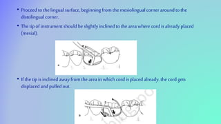
SOFT TISSUE MANAGEMENT
IN FPDS
DELLA S INDRAN
II MDS
LIST OF CONTENTS
• INTRODUCTION
• FINISH LINE EXPOSURE
• GINGIVAL SULCUS ENLARGEMENT
• REMOVAL OF EDENTULOUS CUFF
• CROWN LENGTHENING
• RECENT ADVANCES
• CONCLUSION
• REVIEW OF LITERATURE
• REFERENCES
INTRODUCTION
• Complete control of the environment of the operative site is essential during restorative
dental procedures.
• Control of oral environment extends to the gingiva surrounding the teeth to be restored.
• The gingiva must be displaced to make a complete impression and even sometimes to permit
completion of preparation and cementation of restoration.
• At times its necessary to completely alter the gingival teeth contours to ensure better long-
lastingresult of fixed restoration.
FINISH LINE EXPOSURE(gingival sulcus
enlargement)
• It is complicated to get a complete impression, when finish line lies at or apical to crest to the
free gingiva.
• In such case, there is a need for temporarily expose the finishline.
• The techniques for exposing finishlines are;
Mechanical
Chemicomechanical
surgical
MECHANICAL METHOD
• Using copperband
• A copper band or tube used as a means to carry impression material as well to displace the
gingiva to expose finish line.
• One end of the tube is trimmed or festooned to follow the gingival finish line profile, which
in turn follows the free gingival margin contours.
• Impressiondone: impression compound or elastomeric impression material.
• Indication: several teeth have been prepared.
• Advantage: minimal recession ranging from 0.1 in healthy adolescence to 0.3 mm in general
population.
• Disadvantage: can causeincisionalinjuries of gingival tissues.
Copper band displacing free gingiva
• Rubberdam
Indications: when limited number of teeth are
to be restored .
• when preparations are not extended far subgingivally.
Note: rubber dam not used with polyvinyl siloxane impression material as the sulphur content
in rubber inhibits polymerization.
• Cotton cord
• Plain cotton cords used for sulcus enlargement by physically pushing gingiva away from the
finishline.
• It is ineffective asthe pressure alone willnot control sulcularhemorrhage.
CHEMICOMECHANICAL (RETRACTION
CORD)
• Combination of chemical action and pressure packing.
• Retraction cords can be non impregnated or impregnated with hemostatic agents.
• Eg: 8% racemic epinephrine, aluminum chloride, ferric sulphate
• Criteria for selection of gingival retraction material:
 effectiveness in gingival displacement andhemostasis
 absence of irreversible damage to gingiva
 minimal systemic effects
• EPINEPHRINE
• Concentrations of 0.001%, 0.01% ,0.1% w/v used.
Mechanism of hemostatic action
• Induce local vasoconstriction by binding to alpha 1 adregenic receptors
• Mechanicalcompression during placement
Indications
• Gingival sulcusenlargement
• For hemostasis
• Contraindications
• Not used in patients using beta blockers and anti hypertensive drugs
• Epinephrine cause elevation of blood pressure and increased heart rate, so contra
indicated in those with cardiac, diabetic, hypersensitive patients.
• Epinephrine syndrome: tachycardia, rapid respiration, anxiety, elevated blood
pressure and postoperative depression.
• ALUMINIUM CHLORIDE 5 to 25%
• 25% approx. doubles the hemostatic effect of other chemicals.
Advantages
• No known contraindications andminimal side effects.
• Effective in controlling bleeding andtissue displacement with minimal damage.
Disadvantage
• Less than10% causelocal tissue destruction.
• FERRIC SULPHATE
• It doesn’t traumatize the tissuesand healingis more rapid than aluminum chloride
• It is compatible with aluminum chloride not with epinephrine
• When used with epinephrine ,it develops massive blue precipitate.
• Coagulates blood quickly
• Time of use is 1to 3 min and 10 to 20 min max
• Tissuedisplacement maintained for 30 min
• Corrosive effect absent, unpleasant taste and tissue discoloration.
ARMAMENTARIUM
• Evacuator.
• Scissors.
• Cotton pliers.
• Dappen dish
• Explorer.
• Cotton pellets
• Fischer ultrapakpacker.
• Gauze sponges (2*2 inch)
• Double ended plastic filling instrument
• Cotton rolls
• Retraction cord
• Hemodont liquid
RETRACTIONCORDS
a) Braided or woven cords
• Have tight and consistent weave
• Easierto placein sulcuswith serrated or non serrated instruments
• Twisting is not required before placing
b) Knitted or twisted cord
• They unravel and fray less when cut during placement
• They expand when wet, so willopen the sulcus greater than the original diameter of the cord.
• Twisting required to produce a tight cord of small diameter.
• TECHNIQUE
• Retraction cords available in packets as well in bulk dispenser
bottles(2 inch cut off).
2 inch piece of retraction cord
cut off
Cord twisted to make it small and tight.
If woven or braided cord used , twisting not needed
• The retraction cord must be moistened with 25 % aluminum chloride (hemostatic agent) in a
dappen dish.
• Removing dry cord cancause injury to epithelial lining.
• The cord is formed into aU andlooped around the prepared tooth.
• The cord held between thumb andfore finger, a slight tension applied in apicaldirection.
• The cord gently slipped between tooth and gingiva in the mesial interproximal area with a
Fischer packinginstrument.
• Proceed to the lingualsurface, beginning from the mesiolingual corner around to the
distolingual corner.
• The tip of instrument should be slightly inclined to the area where cord is already placed
(mesial).
• If the tip is inclined away from the area in which cord is placed already, the cord gets
displaced and pulledout.
• The cord gently pressed apically with the instrument, directing the tip slightly towards the
root.
• The cord slid gingivally along the preparation until the finish line is felt, then cord pushed
into the crevice.
• If the instrument directed totally in apical direction, the cord will rebound off the gingiva and
roll out of sulcus.
• Work continues around to the mesial, firmly securing where it was lightly tacked before.
• Excesscord is cut off from the mesial interproximal area.
• Placement of distal end continued until it overlaps the mesial end .
• A bulk of gauze placed in patients mouth to close on to make patient comfortable and will
keep the area dry.
• After 10 minutes cord removed slowlyto avoid bleeding.
• In case of bleeding, electrocoagulation and ferric sulfate canbe used
• Impression material injected to the dry and cleansulcus
Application of hemostatic agent
SURGICAL METHODS
• Rotary curettage
• Troughing technique, the purpose is to produce limited removal of epithelial tissues in the
sulcuswhile a chamfer finish line is created in tooth structure.
• Also called asgingetage, its used with the subgingival placement of restoration margins.
• Suitability of gingiva for this method determined by factors;
1. Absence of bleeding on probing
2. Sulcus depth less than 3mm
3. Presence of adequate keratinized gingiva.
• Procedure
• A finish line is prepared at the level of the gingival crest with a flatend tapered diamond.
• A torpedo nosed diamond of 150 to 180 grit used to extend the finish line apically,one half
to two thirds the depth of the sulcus,converting the finish line to chamfer.
Prior to rotary curettage, a
shoulder formed at the level
of gingival crest.
Finish line formed
simultaneously, removing
epithelial lining of sulcus.
• Agenerouswater sprayisusedwhilepreparingthefinishlineandcurettingtheadjacentgingiva.
• Cordimpregnatedwithaluminumchlorideor alum, tocontrolhemorrhage.
• Thecordisremovedafter 4to8minutesandthesulcusthoroughlyirrigatedwithwater.
Disadvantage
• Becauseofpoortactilesensation,cancauseoverdeepeningofsulcus.
ELECTROSURGEY
• Electrosurgery done in case of inflammation and granulation tissues around the teeth, where
retraction is not possible along with retraction cord.
• An electrosurgery unit is a high frequency oscillator or radio transmitter that uses a vacuum
tube or a transistor to deliver high frequency electrical current of 1 MHz , hence its called
surgicaldiathermy.
Electrosurgical
electrode enlarging
gingival sulcus.
A: grounding electrode
B: active electrode
• Mechanism of electrosurgery
• Electro surgery produces a controlled tissue destruction to achieve surgicalresult.
• Current flows from a small cutting electrode that produces high current density and rapid
temperature rise at the point of tissue contact.
• The cellsdirectly in contact with electrode is destroyed by temperature increase.
• Cutting electrodes are designed to take advantage of this property.
SURGICAL ELECTRODES
A: coagulating
B: diamond loop
C: round loop
D: small straight
E: small loop
• Types of current
1.unrectifies,damped current
• Characterized by recurrent peaks of power thatrapidly diminish.
• It is generated by old hyfurcator or spark gap generator, and gives intense dehydration and
necrosis.
• Healing is slow and painful.
• Referred as oudin or teslacurrent
• Partially rectified, damped (half wave modulated)
• Thiscurrent produces awave form with adamping in the second halfof each cycle.
• There is lateralpenetration of heat, with slow heat occurring in deep tissues.
• The damping effect produces good coagulation and hemostasis, but healingis slow.
• Fully rectified (full wave modulated)
• Continuous flow of energy
• Better cutting characteristics
• Haemostasiasobtained
• Fully rectified (filtered)
• Continuous wave that have excellent cutting efficiency.
• Less tissuedamage and healing of tissue is better.
Grounding
• For the safety of patient, its important to complete the circuit using ground
electrodes.
• The ground electrodes also known as ground plate, indifferent plate,
indifferent electrodes, passive electrodes.
• There are bipolar units , that eliminates the need of grounding plate. They
have dual electrodes , among one acts as passive electrodes and other as
an active electrodes.
G: grounding
unit
Bipolar unit
Contra indications
• Not used in patient with cardiac pacemaker.
• Not used in flammable environment, as it can lead to fire.
Armamentarium
• Electrosurgical unit.
• Set of electrodes.
• Cotton pliers.
• Mouth mirror
• Fischer ultraPak packer
• DE plastic fillinginstrument
• High volume vacuum with plastictip
• retraction cord
• alcoholsponges
• dappen dish
• hydrogen peroxide
• Aromatic oil
• Cotton tip applicator
• Cotton rolls
Technique
• With a cotton tipped applicator, place a drop of aromatic oil such as peppermint, at the upper lip
border to mask the unpleasant order of flesh burning.
• Make surethe connections are solid. cutting electrodes should be seated properly on handpiece.
• Cutting electrode be applied with very light pressure andquick deft strokes.
• A high volume vacuum tip placed adjacentto electrode to draw the unpleasantodors
• Cutting should be stopped frequently to clean any fragments of tissue from the electrode by
wipping with an alchoholsoaked 4x4 inch sponge.
• Proper technique canbe summed up:
Proper power setting
Quick passes with the electrode
Adequate time intervals between strokes
Electrode should pass from
facial, mesial, lingual and distal
surfaces.
GINGIVAL SULCUS ENLARGEMENT
• For gingival sulcusenlargement, a smallstraight or jshaped electrode is selected.
• It is used with a wire parallel to the long axis of tooth so the tissue is removed from the inner
wallof sulcus(0.1 mm of gingival height).
• With the electro surgery unit off, the electrode is held over the teeth to be treated, and the
cutting strokes are traced over the tissue.
• The foot switch is depressed before contact is made with the tissues, and then electrode is
moved through the first pass.
• Its takenfrom facial,mesial,lingualand distalat a speed of less than7 mm per second.
• In case second pass required in any one area, 8 to 10 sec waited for the second stroke to
pass.(minimize lateral heat).
• After each stroke, debris is cleaned off.
• A cotton pellet dipped in hydrogen peroxide used to remove the debris.
REMOVAL OF AN EDENTULOUS
CUFF
• Interdental papilla adjacent to edentulous space rolls up and makes it difficult to fabricate a
pontic with cleansableembrasures and strong connectors.
• A large loop electrode with high power setting ,required to remove thistissues.
LOOP removing
cuff
CROWN LENGTHENING
• There are circumstances where its desirable to have long clinicalcrowns thannormal.
• Wide band of attached gingiva around the tooth is removed by gingivectomy using diamond
electrode.
• A second series of cut done, to form a bevel which gives a better tissue contour without a
hard to clean edge near the tooth.
• In case of extensive wound, a periodontal dressing provided which has to be replaced in
every 7 days.
LENGTHENED TOOTH AFTER
GINGIVECTOMY.
LASERS
• LASER stands for light amplificationby stimulated emission of radiation.
• It’s a non ionizing radiation with low term risk effects.
• Hazards include: eye damage, skindamage and fire risks.
• Lasers are high powered focused beam which causes tissue vaporization at 100oC to 150oC.
CORDLESS LASER WITH STYLUS a. stylus b.
foot control c. charger.
• Choice of laser depends on: wave length, pulse characteristics, maximum wattage.
• Shorter the wave length, greater the hemostasis. Longer the wavelength, more cleaner the
incision.
• Lasers provide continuous wave and pulse modes( allows less tissue damage and thermal
damage).
• Most soft tissue procedure done with dental diode of 1 to 2 watts of power.
• Eg: diode laser
Wave length near infrared
Notissue recession
Minimal or no patient discomfort
Better hemostasis than conventional method
• The other examples include co2 laser system, Nd:YAG, Er:YAG, Er,Cr:YSGG.
RECENT ADVANCES
• Magic foam
• Magic Foam Cord is a flowable and expanding polyvinyl siloxane (PVS) material designed for easy
and fast retraction of the sulcus.
• This technique is atraumatic and is easy to use even for a beginner who has limited experience with
gingival retraction.
• The material is a 1:1 combination of the base and catalyst that should be dispensed via a syringe
with a fine tip, into the sulcus.
• After setting (setting time-2 mins), the material is washed off and animpression is made.
• A com- pre cap is used to encircle the tooth Gingitrac impression material, Gingitrac- Centrix. This
enables the material to be pushed downward, thereby causing displacement of the gingiva.
Alternatively a cotton roll may alsobe used.
• Expasylretractionsystem
• It’s achemicomechanicalcordlessretractionsystemthatdisplacesgingivawellas produces
hemostasis.
• was introducedbyPierre Rolandin 1988
• COMPOSITION:
• Kaolin
Aluminiumchloride
• Water
• Oiloflemon
• Colorant
• Thesystemconsistsofaluminiumchloridethatprovideshomeostasisas wellas displacementofthe
gingivaltissue.
•
Itisavailableingunandcartridgesystemwithcurvedfinetips,thathelpindispensingthematerial
intothegingivalsulcus.
Once the cartridge is loaded into the gun the material should be injected into the of the
prepared tooth surface
The paste is injected into the sulcusat astable pressure of 0.1 N/mm
• The material should be allowed to set for about 2 mins following which it should be washed
anddried obtain asulcus opening of 0.5mm for 2 mins
• advantages:increased hemostasis
• Easierto dispense and save chairside time
• Decreased incidence of gingival recession
• Disadvantages:intialcost of kitis expensive
• Aquasil ultra cordless tissue retraction system
• Dentsply® International launched a signature cordless tissue retraction that uses air pressure
to inject material into the sulcus for gingival retraction
The components of this system include
• Digit dispenser with intra sulculartips
• Preimpression surface optimizer—mainly used to decrease the surface tension over the
prepared tooth surface.
• Tissuewash material
• Tray material
• The digit dispenser has anintegrated air channelwith mixing unit that simultaneously mixes
and dispenses the material into the sulcularcavity.
• The company claims the following advantages:
• Higher tear strength of the tissue washmaterial
• Decreased chair side time takenper impression.
• Better patient compliance
• Disadvantage
• Cost is higher
• Merocel
• Marco Ferrari et al in 1996 found merocele,a synthetic material ie hydroxylate polyvinyl
acetate.
CONCLUSION
• Fixed dental prosthesis success requires appropriate impression making of the prepared finish line.
As the finish line is adjacent to the gingival sulcus, gingival retraction should be used to decrease
the marginal discrepancy among the restoration and the prepared abutment which is one of the
factors required for the success of the restoration.
REVIEW OF LITERATURE
Efficacy of two gingival retraction systems on lateral
gingival displacement: A prospective clinical study.
• Anupam, Vibha. Journal of oral biology and craniofacial research. 2013 May 1;3(2):68-72.
• Aim: This study aimed to compare the efficacy of a new retraction cord (Stay-Put) and a
conventional retraction cord (Ultra pak) on lateral gingival displacement in continuation with
the treatment protocol of the subjects for fixed dental prosthesis.
• Method: Thirty subjects were selected who needed bilateral fixed dental restoration. In
selected subjects both gingival retraction cords were placed bilaterally buccolingually by
simple randomization method. After removing the cords, impressions were made and
undamaged definitive casts were retrieved. The abutment teeth were sectioned
buccolingually at the buccal ridge followed by decimal measurement of the width (mm) of
the retracted gingival sulcus,under a traveling microscope.
• Result : mean gingival retraction in Stay-Put system (0.528 mm) was higher as compared
to that in Ultrapak(0.487 mm)
Laser Gingival Retraction: A Quantitative Assessment
• Ch VK, Gupta , Reddy KM. Journal of clinical and diagnostic research. 2013 Aug;7(8):1787.
• Aims: The present study was intended to assess the amount of lateral gingival retraction
achieved quantitatively by using diode lasers.
• Settings and Design: Study was carried on 20 patients attended to a dental
institutionthat underwent root canaltreatment and indicated for fabrication of crowns.
• Material and Methods: Gingival retraction was carried out on 20 teeth and
elastomeric impressions were obtained. Models retrieved from the impressions were
sectioned and the lateral distance between finish line and the marginal gingival was
measured using tool makers microscope. Retraction was measured in mid buccal, mesio
buccal anddisto buccal regions.
• Results: Mean retraction values of 399.5 µm, 445.5 µm and 422.5µm were obtained in
mid buccal, mesio buccal and disto buccal regions respectively. Conclusions: Gingival
Retraction achieved was closer to the thickness of sulcular epithelium and greater than the
minimum required retraction of 200um.
Comparison of gingival retraction produced by retraction cord and
expasyl retraction systems - An in vivo study
Nallaswamy D. Drug Invention Today. 2018 Jan 1;10(1).
• Aim: The aim of this study is to evaluate the amount of gingival retraction produced by
expasylretraction paste andplain retraction cord.
• Methods and Materials: This study included 39 subjects. After abutment, teeth were
prepared for fixed partial denture, plain retraction cord or expasyl retraction paste was
placed into the sulcus of the prepared teeth, and time taken for application was recorded and
bleeding was noted after removal of retraction material. Gingival sulcus width was measured
by travelling microscope. The gingival recession was measured using digital caliper.
• Results: The mean gingival width of retracted sulcus in both the groups showed no
statisticallysignificantdifference between the two.
• Conclusion: From the study results, amount of gingival retraction with the use of expasyl
retraction paste is almost similar in comparison to plain retraction cord; expasyl retraction
system appears to produce less hemorrhage andneeds less clinicaltime for application.
A: plain retraction
B: expasyl retraction
Gingival displacement using diode laser or
retraction cords: A comparative clinical study. Melilli D,
Mauceri R, Albanese A, Matranga D, Pizzo G. American journal of dentistry. 2018 Jun 1;31(3):131-4.
• Purpose
• To compare two systems used for conditioning the gingival sulcus and
exposing the finish line before the final impression for a fixed
denture: retraction cords and diode laser.
• Methods
• All subjects participating in the study had healthy gingival and
periodontal status before intervention for fixed prosthesis. 74
abutments for complete crown restoration were randomly divided
into two groups for displacing the gingival sulcus before the final
impression: gingival retraction cords (RC) and diode laser (DL).
• The height of the clinical crowns was measured by a blinded examiner
in three points of the buccal surface (mesial, midline and distal) at
four different times: after tooth preparation (T0), 15 days after tooth
preparation, before exposing the finish line with RC or with DL (T1),
10 minutes after exposing the finish line (T2), and 15 days after the
final impression was taken (T3). The amount of gingival retraction
produced (ΔT2-T1) and restoration to baseline (ΔT3-T1) were
calculated. Ease of technique and patient comfort were evaluated
through the Visual Analog Scale. The time required to carry out the
technique and bleeding during and after the conditioning procedure
were also evaluated.
• Results
• There was no difference between the two techniques with regard to the height differences.
Comparative Evaluation of the Amount of Gingival
Displacement Using Three Recent Gingival Retraction
Systems – In vivo Study Qureshi SM, Anasane NS, Kakade D. Contemporary
Clinical Dentistry. 2020 Jan;11(1):28.
• AIM: study was conducted to compare the efficacy of three recent
gingival displacement materials in achieving gingival tissue
displacement.
• Materials and Methods:
• A total of 10 subjects was selected and 40 samples were made for the
study. Samples were divided into four groups depending on the
materials used for gingival displacement.
• On day 1, baseline impression was made without gingival displacement. On day 2, day 22,
and day 42 impressions were made after gingival displacement on intact maxillary right
central incisor with any one of the three agents. The amount of gingival displacement was
then measured as a distance from the tooth to the crest of the gingiva in a horizontal plane
using stereomicroscope.
Gingival retraction with stay-put, expasyl, and astringent
retraction material
• RESULT: Astringent gingival retraction paste showed the highest value for gingival
displacement (0.50 mm) followed by the stay-put retraction cord (0.48 mm), whereas
expasyl(0.34 mm) showed the least value.
• Conclusion:
• Within the limitations of this invivostudy, astringent gingival retraction paste showed the
highest value for gingival displacement followed by stay-put retraction cord whereas, expasyl
showed the leastvalue.
Effect of different retraction and impression techniques on
the marginal fit of crowns
Peter Rehmanna , Dieter Trost. journalof dentistry 36(2008)508–512
• Objective: objective of this study to compare the marginal fit in fixed restorations using
two modes of gingivalretraction and two different impression techniques in ananimal
model.
• Methods: To simulate clinical conditions, 6 teeth in each of 10 lower jaws of freshly slaughtered
cows were prepared with subgingival finish lines. Two different retraction techniques were used to
expose the finish line: retraction cords containing epinephrine (Surgident) and electro-surgery were
applied contra-laterally at 3 teeth per quadrant. Two impressions per jaw were taken in a two-step
putty-washtechnique(TPW)andaone-stepputty-washtechnique(OPW),respectively.
• On the casts, measurement copings were fabricated and seated on the extracted original
tooth. In each coping the marginal discrepancy was assessed at 8 reference marks. Since the
data was normally distributed, results were subjected to parametric statistics (T-test; p =
0.05). Results: Overall marginal discrepancies ranged between 0 and 200 mm. There was a
small but not significantdifference between electro-surgery and the retraction cords.
• Conclusions: Within the limits of the study it can be concluded that the use of gingival
retraction cords as wellas electro-surgery lead to acceptable results.
Comparative Study on the Efficacy of Gingival Retraction using
Polyvinyl Acetate Strips and Conventional Retraction Cord – An in
Vivo Study
Shivasakthy M, Ali SA. Journal of clinical and diagnostic research. 2013 Oct;7(10):2368.
• Purpose of the Study: This study aimed to determine whether the polyvinyl acetate strips
are able to effectively displace the gingival tissues in comparison with the conventional
retraction cord.
• Material and Methods: Complete metal ceramic preparation with supra-gingival margin
was performed in fourteen maxillary incisors and gingival retraction was done using Merocel
strips and conventional retraction cords alternatively in 2 weeks time interval. The amount of
displacement was compared using a digital vernier caliper of 0.01mm accuracy. Results were
analyzed statisticallyusing Paired students t-test.
• Results: The statistical analysis of the data revealed that both the
conventional retraction cord and the Merocel strip produce significant
retraction. Among both the materials, Merocel proved to be significantly
more effective.
• Conclusion: Merocel strip produces more gingival displacement than the
conventional retraction cord.
Polyvinyl acetate strips (Merocel, Mystic,Conn)
A Comparative Evaluation of Efficacy of Gingival Retraction Using
Polyvinyl Siloxane Foam Retraction System, Vinyl Polysiloxane Paste
Retraction System, and Copper Wire Reinforced Retraction Cord in
Endodontically Treated Teeth: An in vivo Study
Mehta S, Virani H, Memon S, Nirmal N. Contemporary clinical dentistry. 2019 Jul;10(3):428.
• The purpose of the study is to evaluate the efficacy of three gingival
retraction systems such as polyvinyl siloxane foam retraction system
(magic foam cord; Coltene/WhaledentInc), polysiloxane paste
retraction system (GingiTrac; Centrix), and aluminum chloride
impregnated twisted retraction cord (Stay-Put; Roeko) in
endodontically treated teeth.
• Materials and Methods:
• Patients who were endodontically treated for molars and requiring crown for the same, were
selected for the present study with sample size of 45. The 45 participants were divided into
three groups. Group 1 was treated with Stay-Put, Group 2 with Magic Foam, and Group 3
with GingiTrac. About 90 elastomeric impressions of the participants were taken—45
impressions before retraction and 45 impressions after retraction. The sulcus width was
measured on the die obtained from the elastomeric impressions by placing the dies under
OVI-200 optical microscope in combination with X soft imaging system software attached to
a computer.
•
• Results:
• The study indicated 0.46 mm of gingival retraction for aluminum chloride impregnated
retraction cord, 0.21 mm of gingival retraction for GingiTrac paste, and 0.29 mm of gingival
retraction for magic foam cord.
REFERENCES
• Luthardt, R.G.; Stossel, M.; Hinz, M.; Vollandt, R. Clinical performance and periodontal outcome of
temporary crowns and fixed partial dentures: A randomized clinical trial. J. Prosthet. Dent. 2000, 83, 32–39.
[CrossRef]
• Donaldson, M.; Goodchild, J.H. Local and systemic effects of mechanico-chemical retraction. Compend.
Contin. Educ. Dent. 2013, 34, 1–7, quiz p8.
• Loe, H. The Gingival Index, the Plaque Index and the Retention Index Systems. J. Periodontol. 1967, 38, 610–
616. [CrossRef]
• Bennani, V.; Aarts, J.M.; Schumayer, D. Correlation of pressure and displacement during gingival
displacement: An in vitro study.
• J. Prosthet. Dent. 2016, 115, 296–300. [CrossRef]
• Laufer, B.Z.; Baharav, H.; Ganor, Y.; Cardash, H.S. The effect of marginal thickness on the distortion of
different impression
• materials. J. Prosthet. Dent. 1996, 76, 466–471, Erratum in: J. Prosthet. Dent. 1997, 4, 452. [CrossRef]
• Gupta, A.; Prithviraj, D.R.; Gupta, D.; Shruti, D.P. Clinical evaluation of three new gingival retraction systems:
A research report.
• J. Indian Prosthodont. Soc. 2013, 13, 36–42. [CrossRef]
• Phatale, S.; Marawar, P.P.; Byakod, G.; Lagdive, S.B.; Kalburge, J.V. Effect of retraction materials on gingival
health: A histopatho-
• logical study. J. Indian Soc. Periodontol. 2010, 14, 35–39. [CrossRef]
• Sachdev, P.A.; Arora, A.; Nanda, S. A Comparative Evaluation of Different Gingival
Retraction Methods-an In Vivo Study. Oral
• Health Case Rep. 2018, 4, 142. [CrossRef]
• Song, J.-E.; Um, Y.-J.; Kim, C.-S.; Choi, S.-H.; Cho, K.S.; Kim, C.-K.; Chai, J.-K.; Jung, U.-W.
Thickness of posterior palatal
• masticatory mucosa: The use of computerized tomography. J. Periodontol. 2008, 79, 406–
412. [CrossRef] [PubMed]
• Mythri, S.; Arunkumar, S.M.; Hegde, S.; Rajesh, S.K.; Munaz, M.; Ashwin, D. Etiology and
occurrence of gingival recession—An
• epidemiological study. J. Indian Soc. Periodontol. 2015, 19, 671–675. [CrossRef]
[PubMed]
• van Palenstein Helderman, W.H.; Lembariti, B.S.; van der Weijden, G.A.; van’t Hof, M.A.
Gingival recession and its association
• with calculus in subjects deprived of prophylactic dental care. J. Clin. Periodontol. 1998,
25, 106–111. [CrossRef] [PubMed]
• Rayyan, M.M.; Hussien, A.N.M.; Sayed, N.M.; Abdallah, R.; Osman, E.; El Saad, N.A.;
Ramadan, S. Comparison of four cordless
• gingival displacement systems: A clinical study. J. Prosthet. Dent. 2019, 121, 265–270.
[CrossRef] [PubMed]
THANKYOU

Soft tissue management

  • 1.
    SOFT TISSUE MANAGEMENT INFPDS DELLA S INDRAN II MDS
  • 2.
    LIST OF CONTENTS •INTRODUCTION • FINISH LINE EXPOSURE • GINGIVAL SULCUS ENLARGEMENT • REMOVAL OF EDENTULOUS CUFF • CROWN LENGTHENING • RECENT ADVANCES • CONCLUSION • REVIEW OF LITERATURE • REFERENCES
  • 3.
    INTRODUCTION • Complete controlof the environment of the operative site is essential during restorative dental procedures. • Control of oral environment extends to the gingiva surrounding the teeth to be restored. • The gingiva must be displaced to make a complete impression and even sometimes to permit completion of preparation and cementation of restoration. • At times its necessary to completely alter the gingival teeth contours to ensure better long- lastingresult of fixed restoration.
  • 4.
    FINISH LINE EXPOSURE(gingivalsulcus enlargement) • It is complicated to get a complete impression, when finish line lies at or apical to crest to the free gingiva. • In such case, there is a need for temporarily expose the finishline. • The techniques for exposing finishlines are; Mechanical Chemicomechanical surgical
  • 5.
    MECHANICAL METHOD • Usingcopperband • A copper band or tube used as a means to carry impression material as well to displace the gingiva to expose finish line. • One end of the tube is trimmed or festooned to follow the gingival finish line profile, which in turn follows the free gingival margin contours. • Impressiondone: impression compound or elastomeric impression material. • Indication: several teeth have been prepared.
  • 6.
    • Advantage: minimalrecession ranging from 0.1 in healthy adolescence to 0.3 mm in general population. • Disadvantage: can causeincisionalinjuries of gingival tissues. Copper band displacing free gingiva
  • 7.
    • Rubberdam Indications: whenlimited number of teeth are to be restored . • when preparations are not extended far subgingivally. Note: rubber dam not used with polyvinyl siloxane impression material as the sulphur content in rubber inhibits polymerization. • Cotton cord • Plain cotton cords used for sulcus enlargement by physically pushing gingiva away from the finishline. • It is ineffective asthe pressure alone willnot control sulcularhemorrhage.
  • 8.
    CHEMICOMECHANICAL (RETRACTION CORD) • Combinationof chemical action and pressure packing. • Retraction cords can be non impregnated or impregnated with hemostatic agents. • Eg: 8% racemic epinephrine, aluminum chloride, ferric sulphate • Criteria for selection of gingival retraction material:  effectiveness in gingival displacement andhemostasis  absence of irreversible damage to gingiva  minimal systemic effects
  • 9.
    • EPINEPHRINE • Concentrationsof 0.001%, 0.01% ,0.1% w/v used. Mechanism of hemostatic action • Induce local vasoconstriction by binding to alpha 1 adregenic receptors • Mechanicalcompression during placement Indications • Gingival sulcusenlargement • For hemostasis
  • 10.
    • Contraindications • Notused in patients using beta blockers and anti hypertensive drugs • Epinephrine cause elevation of blood pressure and increased heart rate, so contra indicated in those with cardiac, diabetic, hypersensitive patients. • Epinephrine syndrome: tachycardia, rapid respiration, anxiety, elevated blood pressure and postoperative depression.
  • 11.
    • ALUMINIUM CHLORIDE5 to 25% • 25% approx. doubles the hemostatic effect of other chemicals. Advantages • No known contraindications andminimal side effects. • Effective in controlling bleeding andtissue displacement with minimal damage. Disadvantage • Less than10% causelocal tissue destruction. • FERRIC SULPHATE • It doesn’t traumatize the tissuesand healingis more rapid than aluminum chloride • It is compatible with aluminum chloride not with epinephrine
  • 12.
    • When usedwith epinephrine ,it develops massive blue precipitate. • Coagulates blood quickly • Time of use is 1to 3 min and 10 to 20 min max • Tissuedisplacement maintained for 30 min • Corrosive effect absent, unpleasant taste and tissue discoloration.
  • 13.
    ARMAMENTARIUM • Evacuator. • Scissors. •Cotton pliers. • Dappen dish • Explorer. • Cotton pellets • Fischer ultrapakpacker. • Gauze sponges (2*2 inch) • Double ended plastic filling instrument • Cotton rolls • Retraction cord • Hemodont liquid
  • 14.
    RETRACTIONCORDS a) Braided orwoven cords • Have tight and consistent weave • Easierto placein sulcuswith serrated or non serrated instruments • Twisting is not required before placing b) Knitted or twisted cord • They unravel and fray less when cut during placement • They expand when wet, so willopen the sulcus greater than the original diameter of the cord. • Twisting required to produce a tight cord of small diameter.
  • 15.
    • TECHNIQUE • Retractioncords available in packets as well in bulk dispenser bottles(2 inch cut off). 2 inch piece of retraction cord cut off Cord twisted to make it small and tight. If woven or braided cord used , twisting not needed
  • 16.
    • The retractioncord must be moistened with 25 % aluminum chloride (hemostatic agent) in a dappen dish. • Removing dry cord cancause injury to epithelial lining. • The cord is formed into aU andlooped around the prepared tooth. • The cord held between thumb andfore finger, a slight tension applied in apicaldirection. • The cord gently slipped between tooth and gingiva in the mesial interproximal area with a Fischer packinginstrument.
  • 17.
    • Proceed tothe lingualsurface, beginning from the mesiolingual corner around to the distolingual corner. • The tip of instrument should be slightly inclined to the area where cord is already placed (mesial). • If the tip is inclined away from the area in which cord is placed already, the cord gets displaced and pulledout.
  • 18.
    • The cordgently pressed apically with the instrument, directing the tip slightly towards the root. • The cord slid gingivally along the preparation until the finish line is felt, then cord pushed into the crevice. • If the instrument directed totally in apical direction, the cord will rebound off the gingiva and roll out of sulcus.
  • 19.
    • Work continuesaround to the mesial, firmly securing where it was lightly tacked before. • Excesscord is cut off from the mesial interproximal area. • Placement of distal end continued until it overlaps the mesial end .
  • 20.
    • A bulkof gauze placed in patients mouth to close on to make patient comfortable and will keep the area dry. • After 10 minutes cord removed slowlyto avoid bleeding. • In case of bleeding, electrocoagulation and ferric sulfate canbe used • Impression material injected to the dry and cleansulcus Application of hemostatic agent
  • 22.
    SURGICAL METHODS • Rotarycurettage • Troughing technique, the purpose is to produce limited removal of epithelial tissues in the sulcuswhile a chamfer finish line is created in tooth structure. • Also called asgingetage, its used with the subgingival placement of restoration margins. • Suitability of gingiva for this method determined by factors; 1. Absence of bleeding on probing 2. Sulcus depth less than 3mm 3. Presence of adequate keratinized gingiva.
  • 23.
    • Procedure • Afinish line is prepared at the level of the gingival crest with a flatend tapered diamond. • A torpedo nosed diamond of 150 to 180 grit used to extend the finish line apically,one half to two thirds the depth of the sulcus,converting the finish line to chamfer. Prior to rotary curettage, a shoulder formed at the level of gingival crest. Finish line formed simultaneously, removing epithelial lining of sulcus.
  • 24.
    • Agenerouswater sprayisusedwhilepreparingthefinishlineandcurettingtheadjacentgingiva. •Cordimpregnatedwithaluminumchlorideor alum, tocontrolhemorrhage. • Thecordisremovedafter 4to8minutesandthesulcusthoroughlyirrigatedwithwater. Disadvantage • Becauseofpoortactilesensation,cancauseoverdeepeningofsulcus.
  • 25.
    ELECTROSURGEY • Electrosurgery donein case of inflammation and granulation tissues around the teeth, where retraction is not possible along with retraction cord. • An electrosurgery unit is a high frequency oscillator or radio transmitter that uses a vacuum tube or a transistor to deliver high frequency electrical current of 1 MHz , hence its called surgicaldiathermy. Electrosurgical electrode enlarging gingival sulcus. A: grounding electrode B: active electrode
  • 26.
    • Mechanism ofelectrosurgery • Electro surgery produces a controlled tissue destruction to achieve surgicalresult. • Current flows from a small cutting electrode that produces high current density and rapid temperature rise at the point of tissue contact. • The cellsdirectly in contact with electrode is destroyed by temperature increase. • Cutting electrodes are designed to take advantage of this property. SURGICAL ELECTRODES A: coagulating B: diamond loop C: round loop D: small straight E: small loop
  • 27.
    • Types ofcurrent 1.unrectifies,damped current • Characterized by recurrent peaks of power thatrapidly diminish. • It is generated by old hyfurcator or spark gap generator, and gives intense dehydration and necrosis. • Healing is slow and painful. • Referred as oudin or teslacurrent
  • 28.
    • Partially rectified,damped (half wave modulated) • Thiscurrent produces awave form with adamping in the second halfof each cycle. • There is lateralpenetration of heat, with slow heat occurring in deep tissues. • The damping effect produces good coagulation and hemostasis, but healingis slow.
  • 29.
    • Fully rectified(full wave modulated) • Continuous flow of energy • Better cutting characteristics • Haemostasiasobtained • Fully rectified (filtered) • Continuous wave that have excellent cutting efficiency. • Less tissuedamage and healing of tissue is better.
  • 30.
    Grounding • For thesafety of patient, its important to complete the circuit using ground electrodes. • The ground electrodes also known as ground plate, indifferent plate, indifferent electrodes, passive electrodes. • There are bipolar units , that eliminates the need of grounding plate. They have dual electrodes , among one acts as passive electrodes and other as an active electrodes. G: grounding unit Bipolar unit
  • 31.
    Contra indications • Notused in patient with cardiac pacemaker. • Not used in flammable environment, as it can lead to fire. Armamentarium • Electrosurgical unit. • Set of electrodes. • Cotton pliers. • Mouth mirror • Fischer ultraPak packer • DE plastic fillinginstrument • High volume vacuum with plastictip
  • 32.
    • retraction cord •alcoholsponges • dappen dish • hydrogen peroxide • Aromatic oil • Cotton tip applicator • Cotton rolls
  • 33.
    Technique • With acotton tipped applicator, place a drop of aromatic oil such as peppermint, at the upper lip border to mask the unpleasant order of flesh burning. • Make surethe connections are solid. cutting electrodes should be seated properly on handpiece.
  • 34.
    • Cutting electrodebe applied with very light pressure andquick deft strokes. • A high volume vacuum tip placed adjacentto electrode to draw the unpleasantodors
  • 35.
    • Cutting shouldbe stopped frequently to clean any fragments of tissue from the electrode by wipping with an alchoholsoaked 4x4 inch sponge. • Proper technique canbe summed up: Proper power setting Quick passes with the electrode Adequate time intervals between strokes
  • 36.
    Electrode should passfrom facial, mesial, lingual and distal surfaces.
  • 38.
    GINGIVAL SULCUS ENLARGEMENT •For gingival sulcusenlargement, a smallstraight or jshaped electrode is selected. • It is used with a wire parallel to the long axis of tooth so the tissue is removed from the inner wallof sulcus(0.1 mm of gingival height). • With the electro surgery unit off, the electrode is held over the teeth to be treated, and the cutting strokes are traced over the tissue. • The foot switch is depressed before contact is made with the tissues, and then electrode is moved through the first pass. • Its takenfrom facial,mesial,lingualand distalat a speed of less than7 mm per second. • In case second pass required in any one area, 8 to 10 sec waited for the second stroke to pass.(minimize lateral heat).
  • 39.
    • After eachstroke, debris is cleaned off. • A cotton pellet dipped in hydrogen peroxide used to remove the debris.
  • 40.
    REMOVAL OF ANEDENTULOUS CUFF • Interdental papilla adjacent to edentulous space rolls up and makes it difficult to fabricate a pontic with cleansableembrasures and strong connectors. • A large loop electrode with high power setting ,required to remove thistissues. LOOP removing cuff
  • 41.
    CROWN LENGTHENING • Thereare circumstances where its desirable to have long clinicalcrowns thannormal. • Wide band of attached gingiva around the tooth is removed by gingivectomy using diamond electrode.
  • 42.
    • A secondseries of cut done, to form a bevel which gives a better tissue contour without a hard to clean edge near the tooth. • In case of extensive wound, a periodontal dressing provided which has to be replaced in every 7 days. LENGTHENED TOOTH AFTER GINGIVECTOMY.
  • 43.
    LASERS • LASER standsfor light amplificationby stimulated emission of radiation. • It’s a non ionizing radiation with low term risk effects. • Hazards include: eye damage, skindamage and fire risks. • Lasers are high powered focused beam which causes tissue vaporization at 100oC to 150oC. CORDLESS LASER WITH STYLUS a. stylus b. foot control c. charger.
  • 44.
    • Choice oflaser depends on: wave length, pulse characteristics, maximum wattage. • Shorter the wave length, greater the hemostasis. Longer the wavelength, more cleaner the incision. • Lasers provide continuous wave and pulse modes( allows less tissue damage and thermal damage). • Most soft tissue procedure done with dental diode of 1 to 2 watts of power.
  • 45.
    • Eg: diodelaser Wave length near infrared Notissue recession Minimal or no patient discomfort Better hemostasis than conventional method • The other examples include co2 laser system, Nd:YAG, Er:YAG, Er,Cr:YSGG.
  • 47.
    RECENT ADVANCES • Magicfoam • Magic Foam Cord is a flowable and expanding polyvinyl siloxane (PVS) material designed for easy and fast retraction of the sulcus. • This technique is atraumatic and is easy to use even for a beginner who has limited experience with gingival retraction. • The material is a 1:1 combination of the base and catalyst that should be dispensed via a syringe with a fine tip, into the sulcus. • After setting (setting time-2 mins), the material is washed off and animpression is made.
  • 48.
    • A com-pre cap is used to encircle the tooth Gingitrac impression material, Gingitrac- Centrix. This enables the material to be pushed downward, thereby causing displacement of the gingiva. Alternatively a cotton roll may alsobe used.
  • 50.
    • Expasylretractionsystem • It’sachemicomechanicalcordlessretractionsystemthatdisplacesgingivawellas produces hemostasis. • was introducedbyPierre Rolandin 1988 • COMPOSITION: • Kaolin Aluminiumchloride • Water • Oiloflemon • Colorant • Thesystemconsistsofaluminiumchloridethatprovideshomeostasisas wellas displacementofthe gingivaltissue. • Itisavailableingunandcartridgesystemwithcurvedfinetips,thathelpindispensingthematerial intothegingivalsulcus.
  • 51.
    Once the cartridgeis loaded into the gun the material should be injected into the of the prepared tooth surface The paste is injected into the sulcusat astable pressure of 0.1 N/mm • The material should be allowed to set for about 2 mins following which it should be washed anddried obtain asulcus opening of 0.5mm for 2 mins • advantages:increased hemostasis • Easierto dispense and save chairside time • Decreased incidence of gingival recession • Disadvantages:intialcost of kitis expensive
  • 54.
    • Aquasil ultracordless tissue retraction system • Dentsply® International launched a signature cordless tissue retraction that uses air pressure to inject material into the sulcus for gingival retraction The components of this system include • Digit dispenser with intra sulculartips • Preimpression surface optimizer—mainly used to decrease the surface tension over the prepared tooth surface. • Tissuewash material • Tray material • The digit dispenser has anintegrated air channelwith mixing unit that simultaneously mixes and dispenses the material into the sulcularcavity.
  • 55.
    • The companyclaims the following advantages: • Higher tear strength of the tissue washmaterial • Decreased chair side time takenper impression. • Better patient compliance • Disadvantage • Cost is higher
  • 57.
    • Merocel • MarcoFerrari et al in 1996 found merocele,a synthetic material ie hydroxylate polyvinyl acetate.
  • 58.
    CONCLUSION • Fixed dentalprosthesis success requires appropriate impression making of the prepared finish line. As the finish line is adjacent to the gingival sulcus, gingival retraction should be used to decrease the marginal discrepancy among the restoration and the prepared abutment which is one of the factors required for the success of the restoration.
  • 59.
  • 60.
    Efficacy of twogingival retraction systems on lateral gingival displacement: A prospective clinical study. • Anupam, Vibha. Journal of oral biology and craniofacial research. 2013 May 1;3(2):68-72. • Aim: This study aimed to compare the efficacy of a new retraction cord (Stay-Put) and a conventional retraction cord (Ultra pak) on lateral gingival displacement in continuation with the treatment protocol of the subjects for fixed dental prosthesis.
  • 61.
    • Method: Thirtysubjects were selected who needed bilateral fixed dental restoration. In selected subjects both gingival retraction cords were placed bilaterally buccolingually by simple randomization method. After removing the cords, impressions were made and undamaged definitive casts were retrieved. The abutment teeth were sectioned buccolingually at the buccal ridge followed by decimal measurement of the width (mm) of the retracted gingival sulcus,under a traveling microscope. • Result : mean gingival retraction in Stay-Put system (0.528 mm) was higher as compared to that in Ultrapak(0.487 mm)
  • 62.
    Laser Gingival Retraction:A Quantitative Assessment • Ch VK, Gupta , Reddy KM. Journal of clinical and diagnostic research. 2013 Aug;7(8):1787. • Aims: The present study was intended to assess the amount of lateral gingival retraction achieved quantitatively by using diode lasers. • Settings and Design: Study was carried on 20 patients attended to a dental institutionthat underwent root canaltreatment and indicated for fabrication of crowns.
  • 63.
    • Material andMethods: Gingival retraction was carried out on 20 teeth and elastomeric impressions were obtained. Models retrieved from the impressions were sectioned and the lateral distance between finish line and the marginal gingival was measured using tool makers microscope. Retraction was measured in mid buccal, mesio buccal anddisto buccal regions.
  • 64.
    • Results: Meanretraction values of 399.5 µm, 445.5 µm and 422.5µm were obtained in mid buccal, mesio buccal and disto buccal regions respectively. Conclusions: Gingival Retraction achieved was closer to the thickness of sulcular epithelium and greater than the minimum required retraction of 200um.
  • 65.
    Comparison of gingivalretraction produced by retraction cord and expasyl retraction systems - An in vivo study Nallaswamy D. Drug Invention Today. 2018 Jan 1;10(1). • Aim: The aim of this study is to evaluate the amount of gingival retraction produced by expasylretraction paste andplain retraction cord. • Methods and Materials: This study included 39 subjects. After abutment, teeth were prepared for fixed partial denture, plain retraction cord or expasyl retraction paste was placed into the sulcus of the prepared teeth, and time taken for application was recorded and bleeding was noted after removal of retraction material. Gingival sulcus width was measured by travelling microscope. The gingival recession was measured using digital caliper.
  • 66.
    • Results: Themean gingival width of retracted sulcus in both the groups showed no statisticallysignificantdifference between the two. • Conclusion: From the study results, amount of gingival retraction with the use of expasyl retraction paste is almost similar in comparison to plain retraction cord; expasyl retraction system appears to produce less hemorrhage andneeds less clinicaltime for application. A: plain retraction B: expasyl retraction
  • 67.
    Gingival displacement usingdiode laser or retraction cords: A comparative clinical study. Melilli D, Mauceri R, Albanese A, Matranga D, Pizzo G. American journal of dentistry. 2018 Jun 1;31(3):131-4. • Purpose • To compare two systems used for conditioning the gingival sulcus and exposing the finish line before the final impression for a fixed denture: retraction cords and diode laser. • Methods • All subjects participating in the study had healthy gingival and periodontal status before intervention for fixed prosthesis. 74 abutments for complete crown restoration were randomly divided into two groups for displacing the gingival sulcus before the final impression: gingival retraction cords (RC) and diode laser (DL).
  • 68.
    • The heightof the clinical crowns was measured by a blinded examiner in three points of the buccal surface (mesial, midline and distal) at four different times: after tooth preparation (T0), 15 days after tooth preparation, before exposing the finish line with RC or with DL (T1), 10 minutes after exposing the finish line (T2), and 15 days after the final impression was taken (T3). The amount of gingival retraction produced (ΔT2-T1) and restoration to baseline (ΔT3-T1) were calculated. Ease of technique and patient comfort were evaluated through the Visual Analog Scale. The time required to carry out the technique and bleeding during and after the conditioning procedure were also evaluated.
  • 69.
    • Results • Therewas no difference between the two techniques with regard to the height differences.
  • 70.
    Comparative Evaluation ofthe Amount of Gingival Displacement Using Three Recent Gingival Retraction Systems – In vivo Study Qureshi SM, Anasane NS, Kakade D. Contemporary Clinical Dentistry. 2020 Jan;11(1):28. • AIM: study was conducted to compare the efficacy of three recent gingival displacement materials in achieving gingival tissue displacement. • Materials and Methods: • A total of 10 subjects was selected and 40 samples were made for the study. Samples were divided into four groups depending on the materials used for gingival displacement.
  • 71.
    • On day1, baseline impression was made without gingival displacement. On day 2, day 22, and day 42 impressions were made after gingival displacement on intact maxillary right central incisor with any one of the three agents. The amount of gingival displacement was then measured as a distance from the tooth to the crest of the gingiva in a horizontal plane using stereomicroscope. Gingival retraction with stay-put, expasyl, and astringent retraction material
  • 72.
    • RESULT: Astringentgingival retraction paste showed the highest value for gingival displacement (0.50 mm) followed by the stay-put retraction cord (0.48 mm), whereas expasyl(0.34 mm) showed the least value. • Conclusion: • Within the limitations of this invivostudy, astringent gingival retraction paste showed the highest value for gingival displacement followed by stay-put retraction cord whereas, expasyl showed the leastvalue.
  • 73.
    Effect of differentretraction and impression techniques on the marginal fit of crowns Peter Rehmanna , Dieter Trost. journalof dentistry 36(2008)508–512 • Objective: objective of this study to compare the marginal fit in fixed restorations using two modes of gingivalretraction and two different impression techniques in ananimal model. • Methods: To simulate clinical conditions, 6 teeth in each of 10 lower jaws of freshly slaughtered cows were prepared with subgingival finish lines. Two different retraction techniques were used to expose the finish line: retraction cords containing epinephrine (Surgident) and electro-surgery were applied contra-laterally at 3 teeth per quadrant. Two impressions per jaw were taken in a two-step putty-washtechnique(TPW)andaone-stepputty-washtechnique(OPW),respectively.
  • 74.
    • On thecasts, measurement copings were fabricated and seated on the extracted original tooth. In each coping the marginal discrepancy was assessed at 8 reference marks. Since the data was normally distributed, results were subjected to parametric statistics (T-test; p = 0.05). Results: Overall marginal discrepancies ranged between 0 and 200 mm. There was a small but not significantdifference between electro-surgery and the retraction cords. • Conclusions: Within the limits of the study it can be concluded that the use of gingival retraction cords as wellas electro-surgery lead to acceptable results.
  • 75.
    Comparative Study onthe Efficacy of Gingival Retraction using Polyvinyl Acetate Strips and Conventional Retraction Cord – An in Vivo Study Shivasakthy M, Ali SA. Journal of clinical and diagnostic research. 2013 Oct;7(10):2368. • Purpose of the Study: This study aimed to determine whether the polyvinyl acetate strips are able to effectively displace the gingival tissues in comparison with the conventional retraction cord. • Material and Methods: Complete metal ceramic preparation with supra-gingival margin was performed in fourteen maxillary incisors and gingival retraction was done using Merocel strips and conventional retraction cords alternatively in 2 weeks time interval. The amount of displacement was compared using a digital vernier caliper of 0.01mm accuracy. Results were analyzed statisticallyusing Paired students t-test.
  • 76.
    • Results: Thestatistical analysis of the data revealed that both the conventional retraction cord and the Merocel strip produce significant retraction. Among both the materials, Merocel proved to be significantly more effective. • Conclusion: Merocel strip produces more gingival displacement than the conventional retraction cord. Polyvinyl acetate strips (Merocel, Mystic,Conn)
  • 77.
    A Comparative Evaluationof Efficacy of Gingival Retraction Using Polyvinyl Siloxane Foam Retraction System, Vinyl Polysiloxane Paste Retraction System, and Copper Wire Reinforced Retraction Cord in Endodontically Treated Teeth: An in vivo Study Mehta S, Virani H, Memon S, Nirmal N. Contemporary clinical dentistry. 2019 Jul;10(3):428. • The purpose of the study is to evaluate the efficacy of three gingival retraction systems such as polyvinyl siloxane foam retraction system (magic foam cord; Coltene/WhaledentInc), polysiloxane paste retraction system (GingiTrac; Centrix), and aluminum chloride impregnated twisted retraction cord (Stay-Put; Roeko) in endodontically treated teeth.
  • 78.
    • Materials andMethods: • Patients who were endodontically treated for molars and requiring crown for the same, were selected for the present study with sample size of 45. The 45 participants were divided into three groups. Group 1 was treated with Stay-Put, Group 2 with Magic Foam, and Group 3 with GingiTrac. About 90 elastomeric impressions of the participants were taken—45 impressions before retraction and 45 impressions after retraction. The sulcus width was measured on the die obtained from the elastomeric impressions by placing the dies under OVI-200 optical microscope in combination with X soft imaging system software attached to a computer. •
  • 80.
    • Results: • Thestudy indicated 0.46 mm of gingival retraction for aluminum chloride impregnated retraction cord, 0.21 mm of gingival retraction for GingiTrac paste, and 0.29 mm of gingival retraction for magic foam cord.
  • 81.
    REFERENCES • Luthardt, R.G.;Stossel, M.; Hinz, M.; Vollandt, R. Clinical performance and periodontal outcome of temporary crowns and fixed partial dentures: A randomized clinical trial. J. Prosthet. Dent. 2000, 83, 32–39. [CrossRef] • Donaldson, M.; Goodchild, J.H. Local and systemic effects of mechanico-chemical retraction. Compend. Contin. Educ. Dent. 2013, 34, 1–7, quiz p8. • Loe, H. The Gingival Index, the Plaque Index and the Retention Index Systems. J. Periodontol. 1967, 38, 610– 616. [CrossRef] • Bennani, V.; Aarts, J.M.; Schumayer, D. Correlation of pressure and displacement during gingival displacement: An in vitro study. • J. Prosthet. Dent. 2016, 115, 296–300. [CrossRef] • Laufer, B.Z.; Baharav, H.; Ganor, Y.; Cardash, H.S. The effect of marginal thickness on the distortion of different impression • materials. J. Prosthet. Dent. 1996, 76, 466–471, Erratum in: J. Prosthet. Dent. 1997, 4, 452. [CrossRef] • Gupta, A.; Prithviraj, D.R.; Gupta, D.; Shruti, D.P. Clinical evaluation of three new gingival retraction systems: A research report. • J. Indian Prosthodont. Soc. 2013, 13, 36–42. [CrossRef] • Phatale, S.; Marawar, P.P.; Byakod, G.; Lagdive, S.B.; Kalburge, J.V. Effect of retraction materials on gingival health: A histopatho- • logical study. J. Indian Soc. Periodontol. 2010, 14, 35–39. [CrossRef]
  • 82.
    • Sachdev, P.A.;Arora, A.; Nanda, S. A Comparative Evaluation of Different Gingival Retraction Methods-an In Vivo Study. Oral • Health Case Rep. 2018, 4, 142. [CrossRef] • Song, J.-E.; Um, Y.-J.; Kim, C.-S.; Choi, S.-H.; Cho, K.S.; Kim, C.-K.; Chai, J.-K.; Jung, U.-W. Thickness of posterior palatal • masticatory mucosa: The use of computerized tomography. J. Periodontol. 2008, 79, 406– 412. [CrossRef] [PubMed] • Mythri, S.; Arunkumar, S.M.; Hegde, S.; Rajesh, S.K.; Munaz, M.; Ashwin, D. Etiology and occurrence of gingival recession—An • epidemiological study. J. Indian Soc. Periodontol. 2015, 19, 671–675. [CrossRef] [PubMed] • van Palenstein Helderman, W.H.; Lembariti, B.S.; van der Weijden, G.A.; van’t Hof, M.A. Gingival recession and its association • with calculus in subjects deprived of prophylactic dental care. J. Clin. Periodontol. 1998, 25, 106–111. [CrossRef] [PubMed] • Rayyan, M.M.; Hussien, A.N.M.; Sayed, N.M.; Abdallah, R.; Osman, E.; El Saad, N.A.; Ramadan, S. Comparison of four cordless • gingival displacement systems: A clinical study. J. Prosthet. Dent. 2019, 121, 265–270. [CrossRef] [PubMed]
  • 83.