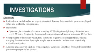
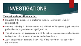
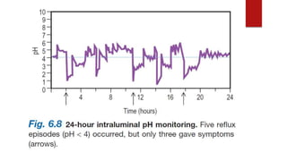
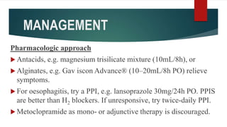
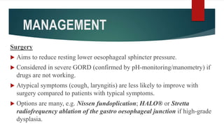
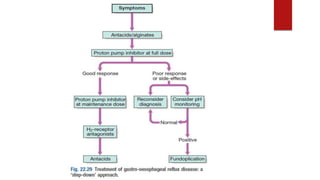
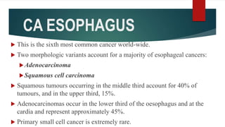
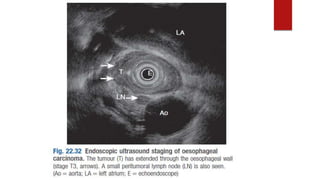
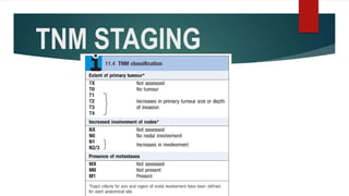
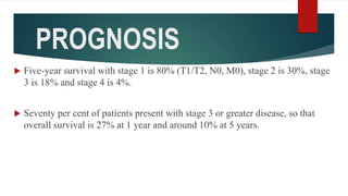
GERD, CA ESOPHAGUS
Harrison Mbohe, MBChB Level 4 JKUAT
GERD
 Occurs when reflux of stomach contents (acid ± bile) causes
troublesome symptoms (≥2 heartburn episodes/week) and/or
complications.
 Commonest cause of esophagitis.
 Normally, small amounts of gastro-oesophageal reflux occur
owing to transient relaxations of the lower esophageal sphincter.
AETIOPATHOGENESIS
 Esophagitis results from excessive reflux, often accompanied by impaired
clearance of the refluxed gastric juice.
 Three dominant mechanisms of esophagogastric junction incompetence are
recognized:
Transient LES relaxations (a vagovagal reflex in which lower esophageal
sphincter (LES) relaxation is elicited by gastric distention)
LES hypotension, or
Anatomic distortion of the esophagogastric junction inclusive of hiatus
hernia.
AETIOPATHOGENESIS
 Transient LES relaxations account for at least 90% of reflux in normal
subjects or GERD patients without hiatus hernia.
 In oesophagitis, the apical junctional complexes between the squamous
cells are damaged leading to increased H+ diffusion and cellular damage.
 Hydrogen ions also impair the growth and replication of damaged cells.
 Acid stimulates primary sensory neurones in the oesophagus by activating
the vanilloid receptor-1 (VR1). This initiates inflammation and release of
proinflammatory substances from the tissue and produce pain. Pain can
also be due to contraction of longitudinal oesophageal muscle.
AETIOPATHOGENESIS
Factors tending to exacerbate reflux regardless of
mechanism are
Abdominal obesity and pregnancy which increase
intraabdominal pressure
Gastric hypersecretory states
Delayed gastric emptying,
Disruption of esophageal peristalsis
AETIOPATHOGENESIS
Gluttony
 Delayed oesophageal clearance
Abnormalities of the lower esophageal sphincter e.g. achalasia
Dietary fat, chocolate, alcohol and coffee relax the lower
esophageal sphincter tone.
Hiatus hernia
CLINICAL FEATURES
 Heartburn (burning, retrosternal discomfort after meals, lying, stooping or straining, relieved
by antacids).
 Belching.
 Acid brash (acid or bile regurgitation).
 Water brash (increased salivation: “My mouth fills with saliva”).
 Odynophagia (painful swallowing, e.g. from oesophagitis or ulceration).
 Nocturnal asthma.
 Chronic cough.
 Laryngitis (hoarseness, throat clearing).
 Sinusitis.
COMPLICATIONS
Chronic esophagitis.
Barrett's esophagus.
Iron deficiency anaemia.
Benign oesophageal stricture.
Gastric volvulus.
Esophageal adenocarcinoma
INVESTIGATIONS
Endoscopy.
 Rationale - to exclude other upper gastrointestinal diseases that can mimic gastro-oesophageal
reflux and to identify complications.
 Indications:
 Symptoms for >4weeks; Persistent vomiting, GI bleeding/iron deficiency; Palpable mass;
Age >55 years; Dysphagia; Symptoms despite treatment; Relapsing symptoms; Weight loss.
 Young patients who present with typical symptoms of gastro-oesophageal reflux, without
worrying features such as dysphagia, weight loss or anaemia, can be treated empirically
without investigation.
 A normal endoscopy in a patient with compatible symptoms should not preclude treatment for
gastro-oesophageal reflux disease.
INVESTIGATIONS
Twenty-four-hour pH monitoring
 Indicated if the diagnosis is unclear or surgical intervention is under
consideration.
 Involves tethering a slim catheter with a terminal radio telemetry pH-sensitive
probe above the gastro-oesophageal junction.
 The intraluminal pH is recorded whilst the patient undergoes normal activities,
and episodes of symptoms are noted and related to pH.
 A pH of less than 4 for more than 6–7% of the study time is diagnostic of
reflux disease
INVESTIGATIONS
Barium Meal
Is the initial procedure for evaluation of dysphagia to
exclude subtle rings or strictures and assess for achalasia.
May also show hiatus hernia
LOS ANGELES CLASSIFICATION
OF GERD
 ≥1 mucosal break(s) <5mm long not extending beyond 2 mucosal
fold tops.
 Mucosal break >5mm long limited to the space between 2 mucosal
fold tops.
 Mucosal break continuous between the tops of 2 or more mucosal
folds but which involves less than 75% of the oesophageal
circumference.
 Mucosal break involving ≥75% of the oesophageal circumference.
DIFFERENTIAL DIAGNOSIS
Infectious, pill, or eosinophilic esophagitis,
Peptic ulcer disease
Dyspepsia
Biliary colic
Coronary artery disease
Esophageal motility disorders.
MANAGEMENT
Lifestyle modifications
 Avoidance of foods that reduce lower esophageal sphincter
pressure, making them “refluxogenic” (these commonly include
fatty foods, alcohol, spearmint, peppermint, tomato-based foods,
possibly coffee and tea)
 Avoidance of acidic foods that are inherently irritating
 Adoption of behaviors to minimize reflux and/or heartburn.
MANAGEMENT
Pharmacologic approach
 Antacids, e.g. magnesium trisilicate mixture (10mL/8h), or
 Alginates, e.g. Gav iscon Advance® (10–20mL/8h PO) relieve
symptoms.
 For oesophagitis, try a PPI, e.g. lansoprazole 30mg/24h PO. PPIS
are better than H2 blockers. If unresponsive, try twice-daily PPI.
 Metoclopramide as mono- or adjunctive therapy is discouraged.
MANAGEMENT
Surgery
 Aims to reduce resting lower oesophageal sphincter pressure.
 Considered in severe GORD (confirmed by pH-monitoring/manometry) if
drugs are not working.
 Atypical symptoms (cough, laryngitis) are less likely to improve with
surgery compared to patients with typical symptoms.
 Options are many, e.g. Nissen fundoplication; HALO® or Stretta
radiofrequency ablation of the gastro oesophageal junction if high-grade
dysplasia.
THANK YOU
CA ESOPHAGUS
 This is the sixth most common cancer world-wide.
 Two morphologic variants account for a majority of esophageal cancers:
Adenocarcinoma
Squamous cell carcinoma
 Squamous tumours occurring in the middle third account for 40% of
tumours, and in the upper third, 15%.
 Adenocarcinomas occur in the lower third of the oesophagus and at the
cardia and represent approximately 45%.
 Primary small cell cancer is extremely rare.
CA ESOPHAGUS
 Worldwide, squamous cell carcinoma is more common, but in the
United States and other Western countries adenocarcinoma is on
the rise.
 Squamous cell carcinoma is common in Ethiopia, China, South and
east Africa and in the Caspian regions of Iran. By contrast, North,
Middle and West Africa have low rates.
 The incidence of adenocarcinomas is increasing in western
industrialized countries – by three- to fivefold over the last 40 years
in Europe and the USA.
AETIOLOGY: SCC
AETIOPATHOGENESIS: SCC
 A majority of esophageal squamous cell carcinomas are at least partially
attributable to the use of alcohol and tobacco, the effects of which synergize to
increase risk.
 Other factors such as, nutritional deficiencies, as well as polycyclic
hydrocarbons, nitrosamines, and other mutagenic compounds, such as those
found in fungus-contaminated foods, have been considered as possible risk
factors.
 HPV infection also has been implicated in esophageal squamous cell
carcinoma in high-risk but not in low-risk regions.
AETIOPATHOGENESIS: AC
 Progression of Barrett esophagus to adenocarcinoma occurs over an extended
period through the stepwise acquisition of genetic and epigenetic changes.
 Epithelial clones in non-dysplastic Barrett metaplasia persist and accumulate
mutations during progression to dysplasia and invasive carcinoma.
 Chromosomal abnormalities and TP53 mutation are often present at early
stages of esophageal adenocarcinoma.
 Additional genetic changes and inflammation also are thought to contribute
to neoplastic progression.
CLINICAL FEATURES
 Progressive painless solid food dysphagia.
 Weight loss and anorexia.
 Odynophagia
 Iron deficiency
 Hoarseness from left recurrent laryngeal nerve injury with mid-esophageal
tumours.
 Coughing and aspiration into the lungs is common due esophageal obstruction.
 Cervical lymphadenopathy or other evidence of metastatic spread is common.
INVESTIGATIONS
 Endoscopy with biopsy provides histological or cytological proof of the
carcinoma; 90% of oesophageal carcinomas can be confirmed with this
technique.
 Barium swallow - useful where the differential diagnosis of dysphagia
includes a motility disorder such as achalasia.
 CT scan of the thorax and upper abdomen shows the volume of tumour,
local invasion, peritumoral and coeliac lymph node involvement and
metastases in the lung and elsewhere. MRI is equivalent to CT in local
staging but not as good for pulmonary metastases.
Barium swallow, showing
an irregular narrowed area
(arrows) at the lower end
of the oesophagus.
INVESTIGATIONS
 Endoscopic ultrasound has an accuracy rate of nearly 90% for assessing depth
of tumour and infiltration and 80% for staging lymph node involvement. It is
useful if CT does not show a lesion already too advanced for surgery.
 A fine needle aspiration (FNA) of lymph nodes improves staging accuracy.
 Laparoscopy is useful if the tumour is at the cardia, to look for peritoneal and
node metastases.
 Positron emission tomography (PET) after fluorodeoxyglucose is used
principally to confirm distant metastases suspected on CT.
TNM STAGING
TREATMENT
 Depends on the age and performance status of the patient and the stage of the
disease.
 Treatment modalities include;
 Surgery provides the best chance of a cure and indicated only when imaging
has shown that the tumour has not infiltrated outside the oesophageal wall
(Stage 1). Surgery in this group shows an 80% survival rate if the postoperative
pathology confirms the staging.
 Chemoradiation. Pre-operative (‘neo-adjuvant’) chemoradiation therapy may
benefit patients with stage 2b and 3 disease.
TREATMENT
 Palliative therapy is often the only realistic possibility.
Dilatation which involves insertion of an expanding metal stent allows
liquids and soft foods to be eaten but is only of short term benefit.
Brachytherapy prolongs luminal patency after stenting.
Endoscopic laser destruction, or alcohol injections can be useful in
proliferative tumours for relieving the dysphagia but repeated treatments
are necessary.
 Nutritional support, as well as support for the patient and their family, is
vital in this distressing condition and improving the quality of life.
PROGNOSIS
 Five-year survival with stage 1 is 80% (T1/T2, N0, M0), stage 2 is 30%, stage
3 is 18% and stage 4 is 4%.
 Seventy per cent of patients present with stage 3 or greater disease, so that
overall survival is 27% at 1 year and around 10% at 5 years.

Gerd, ca esophagus by Harrison Mbohe

  • 1.
    GERD, CA ESOPHAGUS HarrisonMbohe, MBChB Level 4 JKUAT
  • 2.
    GERD  Occurs whenreflux of stomach contents (acid ± bile) causes troublesome symptoms (≥2 heartburn episodes/week) and/or complications.  Commonest cause of esophagitis.  Normally, small amounts of gastro-oesophageal reflux occur owing to transient relaxations of the lower esophageal sphincter.
  • 3.
    AETIOPATHOGENESIS  Esophagitis resultsfrom excessive reflux, often accompanied by impaired clearance of the refluxed gastric juice.  Three dominant mechanisms of esophagogastric junction incompetence are recognized: Transient LES relaxations (a vagovagal reflex in which lower esophageal sphincter (LES) relaxation is elicited by gastric distention) LES hypotension, or Anatomic distortion of the esophagogastric junction inclusive of hiatus hernia.
  • 4.
    AETIOPATHOGENESIS  Transient LESrelaxations account for at least 90% of reflux in normal subjects or GERD patients without hiatus hernia.  In oesophagitis, the apical junctional complexes between the squamous cells are damaged leading to increased H+ diffusion and cellular damage.  Hydrogen ions also impair the growth and replication of damaged cells.  Acid stimulates primary sensory neurones in the oesophagus by activating the vanilloid receptor-1 (VR1). This initiates inflammation and release of proinflammatory substances from the tissue and produce pain. Pain can also be due to contraction of longitudinal oesophageal muscle.
  • 5.
    AETIOPATHOGENESIS Factors tending toexacerbate reflux regardless of mechanism are Abdominal obesity and pregnancy which increase intraabdominal pressure Gastric hypersecretory states Delayed gastric emptying, Disruption of esophageal peristalsis
  • 6.
    AETIOPATHOGENESIS Gluttony  Delayed oesophagealclearance Abnormalities of the lower esophageal sphincter e.g. achalasia Dietary fat, chocolate, alcohol and coffee relax the lower esophageal sphincter tone. Hiatus hernia
  • 8.
    CLINICAL FEATURES  Heartburn(burning, retrosternal discomfort after meals, lying, stooping or straining, relieved by antacids).  Belching.  Acid brash (acid or bile regurgitation).  Water brash (increased salivation: “My mouth fills with saliva”).  Odynophagia (painful swallowing, e.g. from oesophagitis or ulceration).  Nocturnal asthma.  Chronic cough.  Laryngitis (hoarseness, throat clearing).  Sinusitis.
  • 9.
    COMPLICATIONS Chronic esophagitis. Barrett's esophagus. Irondeficiency anaemia. Benign oesophageal stricture. Gastric volvulus. Esophageal adenocarcinoma
  • 10.
    INVESTIGATIONS Endoscopy.  Rationale -to exclude other upper gastrointestinal diseases that can mimic gastro-oesophageal reflux and to identify complications.  Indications:  Symptoms for >4weeks; Persistent vomiting, GI bleeding/iron deficiency; Palpable mass; Age >55 years; Dysphagia; Symptoms despite treatment; Relapsing symptoms; Weight loss.  Young patients who present with typical symptoms of gastro-oesophageal reflux, without worrying features such as dysphagia, weight loss or anaemia, can be treated empirically without investigation.  A normal endoscopy in a patient with compatible symptoms should not preclude treatment for gastro-oesophageal reflux disease.
  • 11.
    INVESTIGATIONS Twenty-four-hour pH monitoring Indicated if the diagnosis is unclear or surgical intervention is under consideration.  Involves tethering a slim catheter with a terminal radio telemetry pH-sensitive probe above the gastro-oesophageal junction.  The intraluminal pH is recorded whilst the patient undergoes normal activities, and episodes of symptoms are noted and related to pH.  A pH of less than 4 for more than 6–7% of the study time is diagnostic of reflux disease
  • 13.
    INVESTIGATIONS Barium Meal Is theinitial procedure for evaluation of dysphagia to exclude subtle rings or strictures and assess for achalasia. May also show hiatus hernia
  • 14.
    LOS ANGELES CLASSIFICATION OFGERD  ≥1 mucosal break(s) <5mm long not extending beyond 2 mucosal fold tops.  Mucosal break >5mm long limited to the space between 2 mucosal fold tops.  Mucosal break continuous between the tops of 2 or more mucosal folds but which involves less than 75% of the oesophageal circumference.  Mucosal break involving ≥75% of the oesophageal circumference.
  • 15.
    DIFFERENTIAL DIAGNOSIS Infectious, pill,or eosinophilic esophagitis, Peptic ulcer disease Dyspepsia Biliary colic Coronary artery disease Esophageal motility disorders.
  • 16.
    MANAGEMENT Lifestyle modifications  Avoidanceof foods that reduce lower esophageal sphincter pressure, making them “refluxogenic” (these commonly include fatty foods, alcohol, spearmint, peppermint, tomato-based foods, possibly coffee and tea)  Avoidance of acidic foods that are inherently irritating  Adoption of behaviors to minimize reflux and/or heartburn.
  • 17.
    MANAGEMENT Pharmacologic approach  Antacids,e.g. magnesium trisilicate mixture (10mL/8h), or  Alginates, e.g. Gav iscon Advance® (10–20mL/8h PO) relieve symptoms.  For oesophagitis, try a PPI, e.g. lansoprazole 30mg/24h PO. PPIS are better than H2 blockers. If unresponsive, try twice-daily PPI.  Metoclopramide as mono- or adjunctive therapy is discouraged.
  • 18.
    MANAGEMENT Surgery  Aims toreduce resting lower oesophageal sphincter pressure.  Considered in severe GORD (confirmed by pH-monitoring/manometry) if drugs are not working.  Atypical symptoms (cough, laryngitis) are less likely to improve with surgery compared to patients with typical symptoms.  Options are many, e.g. Nissen fundoplication; HALO® or Stretta radiofrequency ablation of the gastro oesophageal junction if high-grade dysplasia.
  • 20.
  • 21.
    CA ESOPHAGUS  Thisis the sixth most common cancer world-wide.  Two morphologic variants account for a majority of esophageal cancers: Adenocarcinoma Squamous cell carcinoma  Squamous tumours occurring in the middle third account for 40% of tumours, and in the upper third, 15%.  Adenocarcinomas occur in the lower third of the oesophagus and at the cardia and represent approximately 45%.  Primary small cell cancer is extremely rare.
  • 22.
    CA ESOPHAGUS  Worldwide,squamous cell carcinoma is more common, but in the United States and other Western countries adenocarcinoma is on the rise.  Squamous cell carcinoma is common in Ethiopia, China, South and east Africa and in the Caspian regions of Iran. By contrast, North, Middle and West Africa have low rates.  The incidence of adenocarcinomas is increasing in western industrialized countries – by three- to fivefold over the last 40 years in Europe and the USA.
  • 23.
  • 24.
    AETIOPATHOGENESIS: SCC  Amajority of esophageal squamous cell carcinomas are at least partially attributable to the use of alcohol and tobacco, the effects of which synergize to increase risk.  Other factors such as, nutritional deficiencies, as well as polycyclic hydrocarbons, nitrosamines, and other mutagenic compounds, such as those found in fungus-contaminated foods, have been considered as possible risk factors.  HPV infection also has been implicated in esophageal squamous cell carcinoma in high-risk but not in low-risk regions.
  • 25.
    AETIOPATHOGENESIS: AC  Progressionof Barrett esophagus to adenocarcinoma occurs over an extended period through the stepwise acquisition of genetic and epigenetic changes.  Epithelial clones in non-dysplastic Barrett metaplasia persist and accumulate mutations during progression to dysplasia and invasive carcinoma.  Chromosomal abnormalities and TP53 mutation are often present at early stages of esophageal adenocarcinoma.  Additional genetic changes and inflammation also are thought to contribute to neoplastic progression.
  • 26.
    CLINICAL FEATURES  Progressivepainless solid food dysphagia.  Weight loss and anorexia.  Odynophagia  Iron deficiency  Hoarseness from left recurrent laryngeal nerve injury with mid-esophageal tumours.  Coughing and aspiration into the lungs is common due esophageal obstruction.  Cervical lymphadenopathy or other evidence of metastatic spread is common.
  • 27.
    INVESTIGATIONS  Endoscopy withbiopsy provides histological or cytological proof of the carcinoma; 90% of oesophageal carcinomas can be confirmed with this technique.  Barium swallow - useful where the differential diagnosis of dysphagia includes a motility disorder such as achalasia.  CT scan of the thorax and upper abdomen shows the volume of tumour, local invasion, peritumoral and coeliac lymph node involvement and metastases in the lung and elsewhere. MRI is equivalent to CT in local staging but not as good for pulmonary metastases.
  • 28.
    Barium swallow, showing anirregular narrowed area (arrows) at the lower end of the oesophagus.
  • 29.
    INVESTIGATIONS  Endoscopic ultrasoundhas an accuracy rate of nearly 90% for assessing depth of tumour and infiltration and 80% for staging lymph node involvement. It is useful if CT does not show a lesion already too advanced for surgery.  A fine needle aspiration (FNA) of lymph nodes improves staging accuracy.  Laparoscopy is useful if the tumour is at the cardia, to look for peritoneal and node metastases.  Positron emission tomography (PET) after fluorodeoxyglucose is used principally to confirm distant metastases suspected on CT.
  • 31.
  • 32.
    TREATMENT  Depends onthe age and performance status of the patient and the stage of the disease.  Treatment modalities include;  Surgery provides the best chance of a cure and indicated only when imaging has shown that the tumour has not infiltrated outside the oesophageal wall (Stage 1). Surgery in this group shows an 80% survival rate if the postoperative pathology confirms the staging.  Chemoradiation. Pre-operative (‘neo-adjuvant’) chemoradiation therapy may benefit patients with stage 2b and 3 disease.
  • 33.
    TREATMENT  Palliative therapyis often the only realistic possibility. Dilatation which involves insertion of an expanding metal stent allows liquids and soft foods to be eaten but is only of short term benefit. Brachytherapy prolongs luminal patency after stenting. Endoscopic laser destruction, or alcohol injections can be useful in proliferative tumours for relieving the dysphagia but repeated treatments are necessary.  Nutritional support, as well as support for the patient and their family, is vital in this distressing condition and improving the quality of life.
  • 34.
    PROGNOSIS  Five-year survivalwith stage 1 is 80% (T1/T2, N0, M0), stage 2 is 30%, stage 3 is 18% and stage 4 is 4%.  Seventy per cent of patients present with stage 3 or greater disease, so that overall survival is 27% at 1 year and around 10% at 5 years.