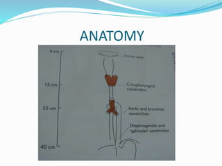
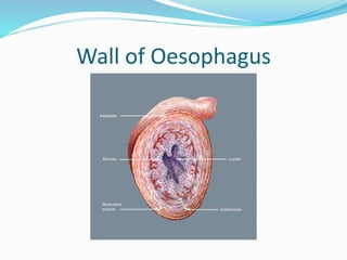
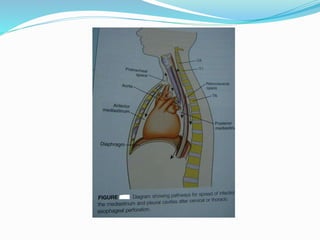
Professor
Abdulsalam Y Taha
School of Medicine
University of Sulaimani
Iraq
https://sulaimaniu.academia.edu/AbdulsalamTaha
LEARNING OBJECTIVES
To understand:
The anatomy and physiology of the
oesophagus and their relationship to
disease.
The clinical features, investigations, and
treatment of benign and malignant disease
with particular reference to the common
adult disorders.
TOPICS
 Surgical anatomy
 Physiology
 Symptoms
 Investigations
 Congenital lesions:
TOF and Atresia
Benign tumours.
Cancer of oesophagus
Others.
 Foreign bodies.
 Oesophageal
perforation.
 Gastro-oesophageal
reflux diease.
 Hiatal hernia.
 Oesophageal motility
disorders: achalasia and
diffuse spasm.
 Oesophgeal diverticula.
SURGICAL ANATOMY
 The oesophagus is a fibromuscular tube 25 cm long.
 Occupying the posterior mediastinum.
 Extending from the cricopharyngeal sphincter to the
cardia of the stomach.
 4 cm of this tube lies below the diaphragm.
 The musculature of the upper one third is mainly
striated, giving way to smooth muscle below.
 It is lined by squamous epithelium except the lower 3
cms which are lined by specialized mucosa.
ANATOMY
Wall of Oesophagus
PHYSIOLOGY
 To transfer food from the mouth to the stomach.
 Sequential contraction of oropharyngeal musculature
+ simultaneous closure of nasal and respiratory
passages+ opening of the cricopharyngeal sphincter.
 Involuntary peristaltic wave in the body of oesophagus
then sweeps food bolus downwards.
 Through a relaxed gastro-oesophageal sphincter zone
into the stomach.
 The upper sphincter is normally closed at rest to
prevent regurgitation. Failure of it to relax on
swallowing may cause propulsion diverticulum.
PHYSIOLOGY
 At the lower end of the oesophagus there is a
physiological sphincter which together with other
anatomical mechanisms prevent reflux of gastric acid
and bile.
 The tone of this sphincter is influenced by
gastrointestinal hormones, anti-cholinergic drugs and
smoking.
 The displaced sphincter loses its tone and permits
reflux to occur.
 The normal GOJ is 3-4 cm long and has a pressure of
30 cm H2O.
SYMPTOMS
 Difficulty in swallowing described as food or fluid
sticking ( oesophageal dysphagia). Must rule out
malignancy.
 Pain on swallowing ( odynophagia). Suggest
inflamation and ulceration.
 Regurgitation or reflux ( heartburn). Common in
gastro-oesophageal reflux disease ( GORD).
 Chest pain; difficult to distinguish from cardiac pain.
 Loss of weight, anaemia, cachexia and change of voice
are other important symptoms.
INVESTIGATIONS
 Radiography.
plain CXR, contrast oesophagography ( barium or
gastrographin swallow) and CT scan of chest.
 Endoscopy: rigid and flexible oesophagoscopy.
 Endosonography: endoscopic ultrasonography.
 Oesophageal manametry: to diagnose oesophageal
motility disorders.
 24-hour pH monitoring: the most accurate method for
the diagnosis of gastro-oesophageal reflux.
BARIUM SWALLOW
BARIUM SWALLOW
ENDOSONOGRAPHY
RIGID OESOPHAGOSCOPY
CONGENITAL ABNORMALITIES
Atresia with or without tracheo-
oesophageal fistula.
Stenosis-rare.
Short oesophagus with hiatus hernia.
Dysphagia lusoria ( compression by an
abnormal artery).
TYPES OF TOF
IT SHOULD BE SUSPECTED IN ALL CASES OF HYDRAMNIOS,
A CONDITION WHICH IS PRESENT IN 50% OF CASES OF ATRESIA.
RECOGNITION WITHIN FORTY-EIGHT HOURS OF BIRTH,
AND SUBSEQUENT SURGICAL CORRECTION, IS THE
ONLY HOPE OF SURVIVAL.
CONGENITAL TRACHEO-OESOPHAGEAL
FISTULA
FOREIGN BODIES IN OESOPHAGUS
 Adults as well as children are prone to ingest FBs.
 Varieties of FBs have been encountered. The most
common impacted material is food.
 Dysphagia, odynophagia and drooling of saliva.
 Plain X- ray +_ contrast study to confirm diagnosis.
 Removal should be done as early as possible.
 Complications: perforation of oesophagus, aspiration,
fistula formation with aorta.
 Removal is by rigid or flexible oesophagoscopy.
Surgery may be needed for sharp or impacted FBs
which fail to be extracted by endoscopy.
SWALLOWED FOREIGN BODIES
PERFORATION OF OESOPHAGUS
 Potentially lethal complication due to mediastinitis
and septic shock.
 Numerous causes, but may be iatrogenic.
 Surgical emphysema is virtually pathognomonic.
 Treatment is urgent; it may be conservative or surgical,
but requires specialized care.
 May be spontaneous ( due to barotrauma): Borehaave
syndrome; is the most serious form of perforation
because of large volume of material that is released
under pressure into the mediastinum and pleura. It is
caused by vomiting against a closed glottis, sometimes
following labour or weight lifting. The tear is in the
weakest point in the lower third.
PERFORATION OF OESOPHAGUS
 Instrumentation is by far the most common cause of
perforation.
 Diagnostic upper GI endoscopy has a rate of 1: 4000
perforation rate.
 Therapeutic endoscopy has a rate of 1: 400 perforation
rate.
 Diagnosis is based on clinical features, plain x-ray,
contrast study and CT scan.
 Prompt and thorough investigations is the key to
management.
PERFORATION OF CERVICAL OESOPHAGUS
Management Options in Perforation of the Oesophagus
Factors that favour non-
operative management
Factors that favour operative
Repair
 Small septic load.
 Minimal cardiovascular
upset.
 Perforation confined to
mediastinum.
 Perforation by flexible
endoscope.
 Perforation of cervical
oesophagus.
 Large septic load.
 Septic shock.
 Pleura breached.
 Boerhave,s synrome.
 Perforation of abdominal
oesophagus.
INGESTION OF CORROSIVE AGENTS
 Corrosives such as
sodium hydroxide (
caustic soda) or
sulphuric acid may be
ingested accidentally or
intentionally causing
chemical burn of
oesophagus.
 Severe strictures may
develop.
MANAGEMENT
 The management in the acute stage is
controversial.
 Nothing by mouth, steroids to reduce fibrosis and
parenteral nutrition. Followed by careful
oesophagoscopy.
 Dilatation may be helpful for short strictures.
 Long strictures are better managed surgically.
 Surgical options include: replacement of
oesophagus by stomach, colon or jujenum.
ACHALASIA CARDIA
MANAGEMENT
HELLER,S OP DILATATION
TOPICS
Oesophageal Diverticulae.
Gastro-oesophageal reflux
diease.
Hiatal hernia.
Benign tumours.
Cancer of oesophagus
BENIGN TUMOURS
 Leiomyomas.
 Benign intraluminal tumours:
Mucosal polyps
Lipomas
Fibrolipomas
Myxofibromas
Leiomyomas
 Account for two thirds of all benign tumours of the
oesophagus.
 Symptoms: dysphagia occurs when leiyomyomas
exceed a diameter of 5 cm as they grow within the
muscular wall, leaving the overlying mucosa intact.
 Diagnosis: Dysphagia, barium swallow and
oesophagoscopy.
 Biopsy is contraindicated.
Leiyomyoma
The characteristic radiographic finding of an esophageal leiyomyoma
on barium esophagogram, a smooth concave filling defect, created
by a well- defined lesion, with sharp, intact mucosal shadow with
abrupt angle where the tumour meets the normal esophageal wall.
Surgical treatment
 Enucleation: in symptomatic patients, the
tumour is enucleated from the oesophageal wall
without violating the mucosa.
 A limited oesophageal resection is indicated if the
tumour lies in the lower oesophagus and can not
be enucleated.
Benign intraluminal tumours
 Oesophagoscopy is performed to confirm the
diagnosis and to rule out malignancy.
 Surgical treatment:
Oesophagotomy, removal of the tumour, and repair of
the oesophagomyotomy.
Endoscopy should not be used to remove these tumours
because of the possibility of oesophageal perforation.
Malignant Tumours
 Incidence:
 in the US, the incidence of oesophageal
carcinoma ranges from 3.5 in 1 million for whites
to 13.5 in 100,000 for blacks.
 The highest incidence of oesophageal carcinoma
is noted in the Hunan Chinese population with as
many as 130 in 100,000 inviduals affected.
Aetiology
 The exact cause is unknown.
 Associated factors are:
tobacco use, excessive alcohol ingestion,
nitrosamines, poor dental hygiene, and hot
beverages.
 Premalignant conditions:
Achalasia
Barrett,s oesophagus.
Pathology
 Squamous cell carcinoma is the most common form.
 Adenocarcinoma, the next commonest, is the type
that occurs in patients with Barrett,s oesophagus.
 Rare tumours include mucoepidermoid carcinoma
and adenoid cystic carcinoma.
 Tumour spread: direct invasion, lymphatic and
haematogenic spread.
DIAGNOSIS
 History: dysphagia and weight loss.
 Contrast study.
 CT: depth of invasion, lymphatic spread and distant
metastases.
 Oesophagoscopy: for tissue diagnosis.
 Endoscopic ultrasonography: depth of invasion and
staging.
 Bronchoscopy: for proximal lesions to exclude invasion
of the bronchial tree.
TREATMENT
 Surgery provides the only cure.
 Operative mortality is less than 5%.
 Types: Ivor-Lewis op.
Transhiatal oesophagectomy.
Left thoraco-abdominal approach.
Radiotherapy and chemotherapy: either as adjuvant
to surgery or as a primary treatment option.
Paliative Treatment for inoperable cases: stenting.
GASTRO-OESOPHAGEAL
REFLUX
 This is a common condition affecting 80% of
population.
 LES is a physiological sphincter normally has
an intra-abdominal position. Loss of LES
pressure results in gastric reflux.
 Oesophageal motility causes refluxed
secretions to be cleared by oesophageal
peristalsis.
 Gastric secretions, gastric acid, pepsin and bile
reflux produce severe oesophagitis.
DIAGNOSIS
 Symptoms: substernal pain, heartburn and
regurgitation.
 Manometry: decreased LES pressure.
 Oesophagoscopy: oesophagitis.
 24-hr pH monitoring: increased acidity.
 Cineradiography: correlates the amount of reflux via
motion pictures.
TREATMENT
 MEDICAL:
PPI, H2- receptor antagonists, cisapride and
metoclapramide increasing rate of gastric
emptying, antacids, weight reduction, abstinence
from smoking and alcohol and elevation of the
head of bed at night.
SURGERY:
Antireflux operations: Nissen fundoplication, Belsey
Mark IV op and Hill repair.
diseasesofoesophagus-141014153402-conversion-gate01.pdf

diseasesofoesophagus-141014153402-conversion-gate01.pdf

  • 1.
    Professor Abdulsalam Y Taha Schoolof Medicine University of Sulaimani Iraq https://sulaimaniu.academia.edu/AbdulsalamTaha
  • 2.
    LEARNING OBJECTIVES To understand: Theanatomy and physiology of the oesophagus and their relationship to disease. The clinical features, investigations, and treatment of benign and malignant disease with particular reference to the common adult disorders.
  • 3.
    TOPICS  Surgical anatomy Physiology  Symptoms  Investigations  Congenital lesions: TOF and Atresia Benign tumours. Cancer of oesophagus Others.  Foreign bodies.  Oesophageal perforation.  Gastro-oesophageal reflux diease.  Hiatal hernia.  Oesophageal motility disorders: achalasia and diffuse spasm.  Oesophgeal diverticula.
  • 4.
    SURGICAL ANATOMY  Theoesophagus is a fibromuscular tube 25 cm long.  Occupying the posterior mediastinum.  Extending from the cricopharyngeal sphincter to the cardia of the stomach.  4 cm of this tube lies below the diaphragm.  The musculature of the upper one third is mainly striated, giving way to smooth muscle below.  It is lined by squamous epithelium except the lower 3 cms which are lined by specialized mucosa.
  • 5.
  • 6.
  • 8.
    PHYSIOLOGY  To transferfood from the mouth to the stomach.  Sequential contraction of oropharyngeal musculature + simultaneous closure of nasal and respiratory passages+ opening of the cricopharyngeal sphincter.  Involuntary peristaltic wave in the body of oesophagus then sweeps food bolus downwards.  Through a relaxed gastro-oesophageal sphincter zone into the stomach.  The upper sphincter is normally closed at rest to prevent regurgitation. Failure of it to relax on swallowing may cause propulsion diverticulum.
  • 9.
    PHYSIOLOGY  At thelower end of the oesophagus there is a physiological sphincter which together with other anatomical mechanisms prevent reflux of gastric acid and bile.  The tone of this sphincter is influenced by gastrointestinal hormones, anti-cholinergic drugs and smoking.  The displaced sphincter loses its tone and permits reflux to occur.  The normal GOJ is 3-4 cm long and has a pressure of 30 cm H2O.
  • 10.
    SYMPTOMS  Difficulty inswallowing described as food or fluid sticking ( oesophageal dysphagia). Must rule out malignancy.  Pain on swallowing ( odynophagia). Suggest inflamation and ulceration.  Regurgitation or reflux ( heartburn). Common in gastro-oesophageal reflux disease ( GORD).  Chest pain; difficult to distinguish from cardiac pain.  Loss of weight, anaemia, cachexia and change of voice are other important symptoms.
  • 11.
    INVESTIGATIONS  Radiography. plain CXR,contrast oesophagography ( barium or gastrographin swallow) and CT scan of chest.  Endoscopy: rigid and flexible oesophagoscopy.  Endosonography: endoscopic ultrasonography.  Oesophageal manametry: to diagnose oesophageal motility disorders.  24-hour pH monitoring: the most accurate method for the diagnosis of gastro-oesophageal reflux.
  • 12.
  • 13.
  • 14.
  • 16.
  • 17.
    CONGENITAL ABNORMALITIES Atresia withor without tracheo- oesophageal fistula. Stenosis-rare. Short oesophagus with hiatus hernia. Dysphagia lusoria ( compression by an abnormal artery).
  • 18.
    TYPES OF TOF ITSHOULD BE SUSPECTED IN ALL CASES OF HYDRAMNIOS, A CONDITION WHICH IS PRESENT IN 50% OF CASES OF ATRESIA. RECOGNITION WITHIN FORTY-EIGHT HOURS OF BIRTH, AND SUBSEQUENT SURGICAL CORRECTION, IS THE ONLY HOPE OF SURVIVAL.
  • 19.
  • 20.
    FOREIGN BODIES INOESOPHAGUS  Adults as well as children are prone to ingest FBs.  Varieties of FBs have been encountered. The most common impacted material is food.  Dysphagia, odynophagia and drooling of saliva.  Plain X- ray +_ contrast study to confirm diagnosis.  Removal should be done as early as possible.  Complications: perforation of oesophagus, aspiration, fistula formation with aorta.  Removal is by rigid or flexible oesophagoscopy. Surgery may be needed for sharp or impacted FBs which fail to be extracted by endoscopy.
  • 21.
  • 22.
    PERFORATION OF OESOPHAGUS Potentially lethal complication due to mediastinitis and septic shock.  Numerous causes, but may be iatrogenic.  Surgical emphysema is virtually pathognomonic.  Treatment is urgent; it may be conservative or surgical, but requires specialized care.  May be spontaneous ( due to barotrauma): Borehaave syndrome; is the most serious form of perforation because of large volume of material that is released under pressure into the mediastinum and pleura. It is caused by vomiting against a closed glottis, sometimes following labour or weight lifting. The tear is in the weakest point in the lower third.
  • 23.
    PERFORATION OF OESOPHAGUS Instrumentation is by far the most common cause of perforation.  Diagnostic upper GI endoscopy has a rate of 1: 4000 perforation rate.  Therapeutic endoscopy has a rate of 1: 400 perforation rate.  Diagnosis is based on clinical features, plain x-ray, contrast study and CT scan.  Prompt and thorough investigations is the key to management.
  • 24.
  • 25.
    Management Options inPerforation of the Oesophagus Factors that favour non- operative management Factors that favour operative Repair  Small septic load.  Minimal cardiovascular upset.  Perforation confined to mediastinum.  Perforation by flexible endoscope.  Perforation of cervical oesophagus.  Large septic load.  Septic shock.  Pleura breached.  Boerhave,s synrome.  Perforation of abdominal oesophagus.
  • 26.
    INGESTION OF CORROSIVEAGENTS  Corrosives such as sodium hydroxide ( caustic soda) or sulphuric acid may be ingested accidentally or intentionally causing chemical burn of oesophagus.  Severe strictures may develop.
  • 27.
    MANAGEMENT  The managementin the acute stage is controversial.  Nothing by mouth, steroids to reduce fibrosis and parenteral nutrition. Followed by careful oesophagoscopy.  Dilatation may be helpful for short strictures.  Long strictures are better managed surgically.  Surgical options include: replacement of oesophagus by stomach, colon or jujenum.
  • 28.
  • 29.
  • 30.
  • 31.
    BENIGN TUMOURS  Leiomyomas. Benign intraluminal tumours: Mucosal polyps Lipomas Fibrolipomas Myxofibromas
  • 32.
    Leiomyomas  Account fortwo thirds of all benign tumours of the oesophagus.  Symptoms: dysphagia occurs when leiyomyomas exceed a diameter of 5 cm as they grow within the muscular wall, leaving the overlying mucosa intact.  Diagnosis: Dysphagia, barium swallow and oesophagoscopy.  Biopsy is contraindicated.
  • 33.
    Leiyomyoma The characteristic radiographicfinding of an esophageal leiyomyoma on barium esophagogram, a smooth concave filling defect, created by a well- defined lesion, with sharp, intact mucosal shadow with abrupt angle where the tumour meets the normal esophageal wall.
  • 34.
    Surgical treatment  Enucleation:in symptomatic patients, the tumour is enucleated from the oesophageal wall without violating the mucosa.  A limited oesophageal resection is indicated if the tumour lies in the lower oesophagus and can not be enucleated.
  • 35.
    Benign intraluminal tumours Oesophagoscopy is performed to confirm the diagnosis and to rule out malignancy.  Surgical treatment: Oesophagotomy, removal of the tumour, and repair of the oesophagomyotomy. Endoscopy should not be used to remove these tumours because of the possibility of oesophageal perforation.
  • 36.
    Malignant Tumours  Incidence: in the US, the incidence of oesophageal carcinoma ranges from 3.5 in 1 million for whites to 13.5 in 100,000 for blacks.  The highest incidence of oesophageal carcinoma is noted in the Hunan Chinese population with as many as 130 in 100,000 inviduals affected.
  • 37.
    Aetiology  The exactcause is unknown.  Associated factors are: tobacco use, excessive alcohol ingestion, nitrosamines, poor dental hygiene, and hot beverages.  Premalignant conditions: Achalasia Barrett,s oesophagus.
  • 38.
    Pathology  Squamous cellcarcinoma is the most common form.  Adenocarcinoma, the next commonest, is the type that occurs in patients with Barrett,s oesophagus.  Rare tumours include mucoepidermoid carcinoma and adenoid cystic carcinoma.  Tumour spread: direct invasion, lymphatic and haematogenic spread.
  • 39.
    DIAGNOSIS  History: dysphagiaand weight loss.  Contrast study.  CT: depth of invasion, lymphatic spread and distant metastases.  Oesophagoscopy: for tissue diagnosis.  Endoscopic ultrasonography: depth of invasion and staging.  Bronchoscopy: for proximal lesions to exclude invasion of the bronchial tree.
  • 40.
    TREATMENT  Surgery providesthe only cure.  Operative mortality is less than 5%.  Types: Ivor-Lewis op. Transhiatal oesophagectomy. Left thoraco-abdominal approach. Radiotherapy and chemotherapy: either as adjuvant to surgery or as a primary treatment option. Paliative Treatment for inoperable cases: stenting.
  • 43.
    GASTRO-OESOPHAGEAL REFLUX  This isa common condition affecting 80% of population.  LES is a physiological sphincter normally has an intra-abdominal position. Loss of LES pressure results in gastric reflux.  Oesophageal motility causes refluxed secretions to be cleared by oesophageal peristalsis.  Gastric secretions, gastric acid, pepsin and bile reflux produce severe oesophagitis.
  • 44.
    DIAGNOSIS  Symptoms: substernalpain, heartburn and regurgitation.  Manometry: decreased LES pressure.  Oesophagoscopy: oesophagitis.  24-hr pH monitoring: increased acidity.  Cineradiography: correlates the amount of reflux via motion pictures.
  • 45.
    TREATMENT  MEDICAL: PPI, H2-receptor antagonists, cisapride and metoclapramide increasing rate of gastric emptying, antacids, weight reduction, abstinence from smoking and alcohol and elevation of the head of bed at night. SURGERY: Antireflux operations: Nissen fundoplication, Belsey Mark IV op and Hill repair.