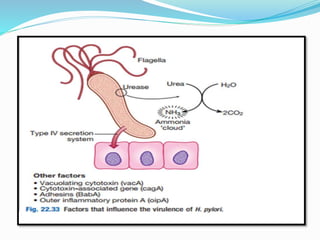
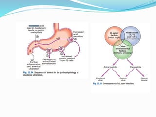
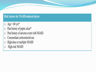
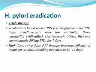
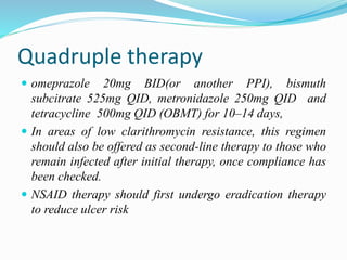
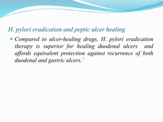
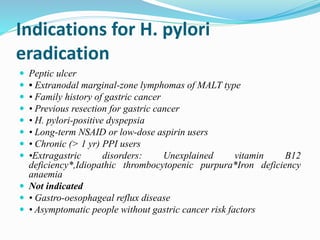
2. ULCERS
Ulcers are defined histologically as a breach in the
mucosa of alimentary tract that extends through
muscularis mucosa into submucosa or deeper.
Ulcers are distinguished from erosion in which there is
epithelial disruption with in mucosa but no breach of the
muscularis mucosa
3. PEPTIC ULCERS:
Peptic ulcers are chronic,most often solitary,lesions that
occur in any portion of the gastrointestinal tract exposed
to aggressive action of acid or peptic juices.
peptic ulcers are usually solitary lesions less than 4cm in
diameter
4. Predilection site of ulcer
Lower oesophagus
Stomach ,usually antrum
Duodenum,first portion
With in margins of gastrojejunostomy
In the ileum adjacent to a meckel’s diverticulum.
In the duodenum,stomach, jejunum of patients with
zollinger-Ellison syndrome
5.
6. Gastric and duodenal ulcer
The prevalence of peptic ulcer (0.1–0.2%) is decreasing in
many Western communities as a result of widespread use
of Helicobacter pylori eradication therapy but it remains
high in developing countries.
The male-to female ratio for duodenal ulcer varies from
5 : 1 to 2 : 1
for gastric ulcer is 2:1 or less.
The prevalence in developed nations rises with age, and in
the UK approximately 50% of people over the age of 50
years are infected while in developing countries adults
suffer 90 %
7. Risk factors for Peptic Ulcer
Disease
H. pylori infection
Cigarette use (synergizes with H. pylori for gastric PUD)
Chronic obstructive pulmonary disease
Illicit drugs, e.g. cocaine, that reduce mucosal blood flow
NSAIDs (potentiated by corticosteroids)
Alcoholic cirrhosis (primarily duodenal PUD)
Psychological stress (can increase gastric acid secretion)
Endocrine cell hyperplasia (can stimulate parietal cell growth
and gastric acid secretion)
Zollinger-Ellison Syndrome (PUD of stomach, duodenum, and
jejunum)
Viral infection (CMV, herpes simplex virus)
8. PATHOPHYSIOLOGY
Peptic ulceration is strongly associated with H. pylori
infection.
H. pylori is Gram-negative and spiral, and has multiple
flagella at one end, which make it motile, allowing it to
burrow and live beneath the mucus layer adherent to the
epithelial surface.
It uses an adhesin molecule (BabA) to bind to the Lewis b
antigen on epithelial cells. Here the surface pH is close to
neutral and any acidity is buffered by the organism’s
production of the enzyme urease.
9. This produces ammonia from urea and raises the pH
around the bacterium and between its two cell membrane
layers. H.
H. pylori exclusively colonises gastric-type epithelium and
is only found in the duodenum in association with patches
of gastric metaplasia.
It causes chronic gastritis by provoking a local
inflammatory response in the underlying epithelium.
10. H. pylori causes localised antral gastritis associated with
depletion of somatostatin (from D cells) and increased
gastrin release from G cells. The subsequent
hypergastrinaemia stimulates increased acid
14. Smoking
Smoking confers an increased risk of gastric ulcer and, to
a lesser extent, duodenal ulcer.
Once the ulcer has formed, it is more likely to cause
complications and less likely to heal if the patient
continues to smoke.
15. CLINICAL FEATURES
It is a chronic condition with spontaneous relapses and
remissions lasting for decades, if not for life.
The most common presentation is with recurrent
abdominal pain which has three notable characteristics:
1. localisation to the epigastrium
2. relationship to food and episodic occurrence
3. Occasional vomiting occurs in about 40% of ulcer
subjects; persistent daily vomiting suggests gastric
outlet obstruction
16. Contd…
IN ELDERLY PEOPLE OR THOSE TAKING NSAIDS
In them, pain may be absent or so slight that it is experienced
only as a vague sense of epigastric unease. Occasionally, the
only symptoms are anorexia and nausea, or early satiety after
meals.
In some patients, the ulcer is completely ‘silent’, presenting for
the first time with anaemia from chronic undetected blood loss,
as an abrupt haematemesis or as acute perforation;
in others, there is recurrent acute bleeding without ulcer pain.
The diagnostic value of individual symptoms for peptic ulcer
disease is poor; the history is therefore a poor predictor of the
presence of an ulcer.
17. INVESTIGATIONS
Endoscopy
Biopsy
Test for H.pylori infection (Some are invasive and some
are non invasive tests)
Breath tests or faecal antigen tests are best because of
accuracy, simplicity and non-invasiveness
18. Methods for the diagnosis of Helicobacter pylori infection
Non-invasive
Serology
13C-urea breath tests
Faecal antigen test
Invasive (antral biopsy)
Histology
Rapid urease test
microbial culture
19. MANAGEMENT
The aims of management are to relieve symptoms, induce
healing and prevent recurrence.
1. H. pylori eradication is the cornerstone of therapy for
peptic ulcers, as this will successfully prevent relapse
and eliminate the need for long-term therapy in the
majority of patients
2. Drugs that inhibit gastric acid secretion
3. Drugs that neutralize gastric acid (Antacids)
4. Ulcer protectives
21. H. pylori eradication
Triple therapy
Treatment is based upon a PPI (i.e omeprazole 20mg BID
taken simultaneously with two antibiotics (from
amoxicillin 1000mgBID, clarithromycin 500mg BID and
metronidazole 500mg BID) for 7 days .
High-dose, twice-daily PPI therapy increases efficacy of
treatment, as does extending treatment to 10–14 days.
22. Quadruple therapy
omeprazole 20mg BID(or another PPI), bismuth
subcitrate 525mg QID, metronidazole 250mg QID and
tetracycline 500mg QID (OBMT) for 10–14 days,
In areas of low clarithromycin resistance, this regimen
should also be offered as second-line therapy to those who
remain infected after initial therapy, once compliance has
been checked.
NSAID therapy should first undergo eradication therapy
to reduce ulcer risk
23. H. pylori eradication and peptic ulcer healing
Compared to ulcer-healing drugs, H. pylori eradication
therapy is superior for healing duodenal ulcers and
affords equivalent protection against recurrence of both
duodenal and gastric ulcers.’
24. Indications for H. pylori
eradication
Peptic ulcer
• Extranodal marginal-zone lymphomas of MALT type
• Family history of gastric cancer
• Previous resection for gastric cancer
• H. pylori-positive dyspepsia
• Long-term NSAID or low-dose aspirin users
• Chronic (> 1 yr) PPI users
•Extragastric disorders: Unexplained vitamin B12
deficiency*,Idiopathic thrombocytopenic purpura*Iron deficiency
anaemia
Not indicated
• Gastro-oesophageal reflux disease
• Asymptomatic people without gastric cancer risk factors
25. General measures
Cigarette smoking, aspirin and NSAIDs should be
avoided. Alcohol in moderation is not harmful and no
special dietary advice is required.
26. Maintenance treatment
Continuous maintenance treatment should not be
necessary after successful H. pylori eradication. For the
minority who do require it, the lowest effective dose of PPI
should be used
27. Common side-effects of H. pylori
eradication therapy
Diarrhoea: 30–50% of patients; usually mild but
Clostridium difficile-associated diarrhoea can occur
• Flushing and vomiting when taken with alcohol
(metronidazole)
• Nausea, vomiting
• Abdominal cramps
• Headache
• Rash
28. SURGICAL TREATMENT
Surgery is now rarely required for peptic ulcer disease
but it is needed in some cases
Indications for surgery in peptic ulcer
Emergency
Perforation and Haemorrhage
Elective
Gastric outflow obstruction
Persistent ulceration despite adequate medical therapy
Recurrent ulcer following gastric surgery
29. Complications of gastric resection
or vagotomy
Dumping.
Chemical (bile reflux) gastropathy
Diarrhoea and maldigestion.
Weight loss
Anaemia.
Metabolic bone disease.
Gastric cancer.