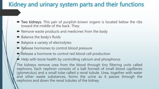
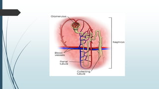
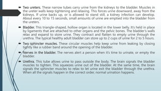
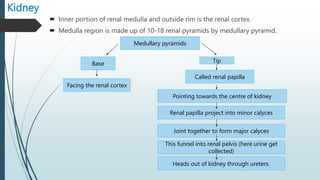
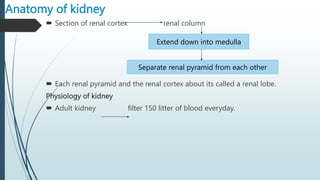
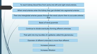
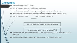
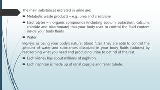
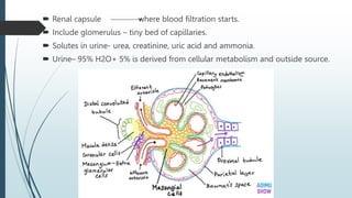
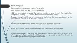
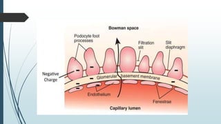
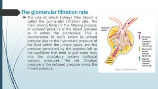
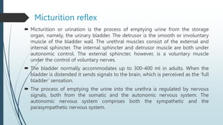
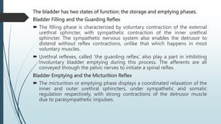
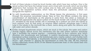
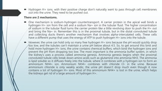
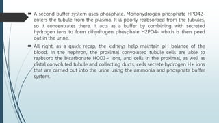
SlideShare a Scribd company logo
URINARY SYSTEM
Presented by:-
Miss. Gayatri K. Bahatkar
Assistant Professor,
P. R. Patil Institute Of Pharmacy, Talegoan(sp)
Physical features and location of kidney
 Weight – 150 g
 Size- 10-12 cm long
 Colour- reddish
 Located between the T12 and T13 vertebra partially protected floating
ribs(11 & 12).
 Sits behind the peritoneum membrane alongside the vertebral column.
 Each kidney contain 1.2 million nephron .
 The right side kidney is pushed down by liver so its site slightly lower the
left kidney
 The middle of each kidney there is an indentation that form renal hilum.
 Hilum is the entry and exist point for the ureter , renal artery , renal veins,
lymphatic and nervous goes into and come out of the kidney.
Why kidney is called as the workhouse of the
urinary system?
 It clears the harmful substances by filtering the blood
 It regulate blood pH, volume, pressure, osmolarity as well as production.
Kidney is surrounded by 3 layer of tissue:-
 Renal fascia – outermost layer- thin layer of dense tissue
 Adipose capsule – middle layer- fatty layer
 Renal capsule – inner most layer- smooth transparent sheet of dense
connective tissue
Kidney and urinary system parts and their functions
 Two kidneys. This pair of purplish-brown organs is located below the ribs
toward the middle of the back. They:
 Remove waste products and medicines from the body
 Balance the body’s fluids
 Balance a variety of electrolytes
 Release hormones to control blood pressure
 Release a hormone to control red blood cell production
 Help with bone health by controlling calcium and phosphorus
The kidneys remove urea from the blood through tiny filtering units called
nephrons. Each nephron consists of a ball formed of small blood capillaries
(glomerulus) and a small tube called a renal tubule. Urea, together with water
and other waste substances, forms the urine as it passes through the
nephrons and down the renal tubules of the kidney.
 Two ureters. These narrow tubes carry urine from the kidneys to the bladder. Muscles in
the ureter walls keep tightening and relaxing. This forces urine downward, away from the
kidneys. If urine backs up, or is allowed to stand still, a kidney infection can develop.
About every 10 to 15 seconds, small amounts of urine are emptied into the bladder from
the ureters.
 Bladder. This triangle-shaped, hollow organ is located in the lower belly. It’s held in place
by ligaments that are attached to other organs and the pelvic bones. The bladder’s walls
relax and expand to store urine. They contract and flatten to empty urine through the
urethra. The typical healthy adult bladder can store up to 2 cups of urine for 2 to 5 hours.
 Two sphincter muscles. These circular muscles help keep urine from leaking by closing
tightly like a rubber band around the opening of the bladder.
 Nerves in the bladder. The nerves alert a person when it’s time to urinate, or empty the
bladder.
 Urethra. This tube allows urine to pass outside the body. The brain signals the bladder
muscles to tighten. This squeezes urine out of the bladder. At the same time, the brain
signals the sphincter muscles to relax to let urine exit the bladder through the urethra.
When all the signals happen in the correct order, normal urination happens.
Kidney
 Inner portion of renal medulla and outside rim is the renal cortex.
 Medulla region is made up of 10-18 renal pyramids by medullary pyramid.
Medullary pyramids
Base Tip
Facing the renal cortex
Called renal papilla
Pointing towards the centre of kidney
Renal papilla project into minor calyces
Joint together to form major calyces
This funnel into renal pelvis (here urine get
collected)
Heads out of kidney through ureters.
Anatomy of kidney
 Section of renal cortex renal column
 Each renal pyramid and the renal cortex about its called a renal lobe.
Physiology of kidney
 Adult kidney filter 150 litter of blood everyday.
Extend down into medulla
Separate renal pyramid from each other
To reach kidney blood flow from aorta into left and right renal arteries.
When renal arteries enter the kidney they get divided into segmental arteries.
Then into Interglobal arteries passes through the renal column then to accurate arteries
that
Bases of renal pyramids
Continue to divide eventually forming afferent arterioles
That split into tiny bundles of capillaries called the glomerulus
Diameter of afferent arterioles is more than efferent.
Increases pressure
Increases filtration
Glomerulus
 Site were blood filtration starts.
 This is 50 time more permeable than capillaries.
 Over the blood leaves from the glomeruli dose not enter into venules.
 These peritubular capillaries then units to become the cortical radiates veins.
 Then the arcuate veins then to interlobular veins
 Flow of vein are equal to or similar to the flow of artery but in reverse (opposite
direction)
 The only difference is there is segmental artery but no segmental vein.
and then finally into the left and right renal veins
Which connect to the inferior vena cava (I.V.C)
The main substances excreted in urine are:
 Metabolic waste products – e.g., urea and creatinine
 Electrolytes – inorganic compounds (including sodium, potassium, calcium,
chloride and bicarbonate) that your body uses to control the fluid content
inside your body fluids
 Water
kidneys as being your body’s natural blood filter. They are able to control the
amount of water and substances dissolved in your body fluids (solutes) by
reabsorbing what you need and producing urine to get rid of the rest.
 Each kidney has about millions of nephron.
 Each nephron is made up of renal capsule and renal tubule.
 Renal capsule where blood filtration starts.
 Include glomerulus – tiny bed of capillaries.
 Solutes in urine- urea, creatinine, uric acid and ammonia.
 Urine– 95% H2O+ 5% is derived from cellular metabolism and outside source.
Bowmans capsule
Surrounded the glomerulus ( made of renal cells)
Now as blood flows into the glomerulus
H2O and some solute in blood like sodium, are able to pass through the endothelium
lining of the capillary across the basement membrane.(6-7 nm)
Through the epithelial lining of nephron and finally into the bowman's (space) of the
nephron itself at which point it is called filtrates.
The epithelium of nephron is made up of specialised cell called podocytes
Which wrap around the basement membrane like the tentacles of an octopus.
Between this tentacles – like projection are tiny gaps called filtration slits that act like sieve
allowing only small particular such as H2O, glucose and ionic salts to pass through while to
to pass trough while blocking large protein and RBC.
The glomerular filtration rate
 The rate at which kidneys filter blood is
called the glomerular filtration rate. The
main driving force for the filtering process,
or outward pressure is the blood pressure
as it enters the glomerulus. This is
counteracted to some extent by inward
pressure due to the hydrostatic pressure of
the fluid within the urinary space, and the
pressure generated by the proteins left in
the capillaries that tend to pull water back
into the circulatory system (colloidal
osmotic pressure). The net filtration
pressure is the outward pressure minus the
inward pressure.
Micturition reflex
 Micturition or urination is the process of emptying urine from the storage
organ, namely, the urinary bladder. The detrusor is the smooth or involuntary
muscle of the bladder wall. The urethral muscles consist of the external and
internal sphincter. The internal sphincter and detrusor muscle are both under
autonomic control. The external sphincter, however, is a voluntary muscle
under the control of voluntary nerves.
 The bladder normally accommodates up to 300-400 ml in adults. When the
bladder is distended it sends signals to the brain, which is perceived as the ‘full
bladder’ sensation.
 The process of emptying the urine into the urethra is regulated by nervous
signals, both from the somatic and the autonomic nervous system. The
autonomic nervous system comprises both the sympathetic and the
parasympathetic nervous system.
The bladder has two states of function; the storage and emptying phases.
Bladder Filling and the Guarding Reflex
 The filling phase is characterized by voluntary contraction of the external
urethral sphincter, with sympathetic contraction of the inner urethral
sphincter. The sympathetic nervous system also enables the detrusor to
distend without reflex contractions, unlike that which happens in most
voluntary muscles.
 Urethral reflexes, called ‘the guarding reflex,’ also play a part in inhibiting
involuntary bladder emptying during this process. The afferents are all
conveyed through the pelvic nerves to initiate a spinal reflex.
Bladder Emptying and the Micturition Reflex
 The micturition or emptying phase displays a coordinated relaxation of the
inner and outer urethral sphincters, under sympathetic and somatic
regulation respectively, with strong contractions of the detrusor muscle
due to parasympathetic impulses.
Micturition Reflex Process
 When the bladder is full, it sends the signal to the brain for the process of emptying.
The bladder emptying phase is called micturition, and it involves the coordinated
reflexes of the outer and inner urethral sphincter under somatic and sympathetic
regulation, respectively.
 At first, the afferent impulses or the sensory impulses from the receptors reach the
spinal cord through the sensory fibers of the pelvic nerve (parasympathetic nerve).
 The motor impulses created in the spinal cord run through the motor fibers of the
pelvic nerve towards the bladder and the internal sphincter.
 These motor impulses (efferent impulses) create contraction of the detrusor muscle
and also the relaxation of the internal sphincter. Thus, the urine enters the urethra
from the urinary bladder.
 Once urine reaches the urethra, the stretch receptors present in the urethra are
stimulated, and they send afferent impulses to the spinal cord through pelvic nerve
fibers.
 Now the impulses created from the spinal centres obstruct the pudendal nerve. This
leads to the relaxation of the external sphincter, and micturition occurs.
 Facilitatory centers for micturition are present in the pons, and some are even
in the cerebral cortex. It facilitates micturition through spinal centers.
 Inhibitory centers for micturition are present in the cerebral cortex and
midbrain. It inhibits the micturition by repressing spinal micturition centers.
 The micturition reflex process is self-regenerative. This is because the initial
contraction of the urinary bladder activates the receptors to create an
increase in sensory impulses from the urethra as well as the bladder. These
impulses, in turn, create a further increase in reflex contraction of the urinary
bladder. This cycle of events repeats until the force of contraction of the
urinary bladder reaches the maximum, leading to the voiding of urine or the
micturition process. During micturition, the urine flow is facilitated by the
increase in abdominal pressure. This is due to the voluntary contraction of the
muscles in the abdomen.
Micturition is thus characterized by:
 Relaxation of the striated sphincter (somatic innervation)
 Relaxation of the smooth muscle sphincter and opening of the bladder neck
(sympathetic innervation)
 Detrusor contraction (parasympathetic innervation)
 The distension of the urinary bladder wall causes wall tension to rise very
slightly. However, when the bladder is almost full, at about 300-400 ml, the
inherent contractility of the detrusor causes reflex contractions to occur,
which are less powerful than the voiding contraction. Afferent firing
frequency increases with filling, but cortical control still overrides the
micturition reflex until voluntary voiding is determined upon.
 During micturition, urinary flow is assisted by additional detrusor
contractions and external sphincter relaxation which further lowers
resistance to the passage of urine. The abdominal wall and pelvic floor
musculature also participates by increasing the force on the bladder to help
achieve complete emptying.
Role of kidneys in acid base balance
 The kidneys have two main ways to maintain acid-base balance – their cells
reabsorb bicarbonate HCO3− from the urine back to the blood and they
secrete hydrogen H+ ions into the urine. By adjusting the amounts
reabsorbed and secreted, they balance the bloodstream’s pH.
 Our kidneys filter blood continuously by distributing the blood that comes
into the kidney to millions of tiny functional units called nephrons. Each
nephron is made up of the glomerulus, or a tiny clump of capillaries, where
blood filtration begins. When blood passes through a glomerulus, about one-
fifth of the plasma leaves the glomerular capillaries and goes into the renal
tubule. « Reabsorption of the good stuff---water and electrolytes---and
leaving behind the bad stuff---waste products and acid--- is the job of the the
renal tubular system. The renal tubule is a structure with several segments:
the proximal convoluted tubule, the U- shaped loop of Henle with a thin
descending and a thick ascending limb, and the distal convoluted tubule,
which winds and twists back up again, before emptying into the collecting
duct, which collects the final urine.
 Each of these tubules is lined by brush border cells which have two surfaces. One is the
apical surface that faces the tubular lumen and is lined with microvilli, which are tiny little
projections that increase the cell’s surface area to help with solute reabsorption. The
other is the basolateral surface, which faces the peritubular capillaries, which run
alongside the nephron.
 So with bicarbonate reabsorption, as the filtrate leaves the glomerulus, it first goes
through the proximal convoluted tubule. Now at first, this filtrate contains the same
concentration of electrolytes as the plasma it came from. But when a molecule of
bicarbonate approaches the apical surface of the brush border cell it binds to hydrogen
H+ secreted by the brush border cell in exchange for a sodium ion from the tubule to
form carbonic acid. At that point, an enzyme called carbonic anhydrase type 4 which
lurks in the tubule in the microvilli like a shark, swims along and splits the carbonic acid
into water and carbon dioxide.
 Unlike charged bicarbonate anions, which are stuck in the tubule, the water and carbon
dioxide happily diffuse across the membrane into the cells where carbonic anhydrase
type 2 facilitates the reverse reaction – combining them to form carbonic acid, which
dissolves into bicarbonate and hydrogen. A sodium bicarbonate cotransporter on the
basolateral surface snatches up the bicarbonate and a nearby sodium, and shuttles both
into the blood. Alternatively, a bicarbonate chloride exchanger exchanges bicarbonate
HCO3− with chloride Cl- leaving the bloodstream to enter the cells. All this chemical
trickery effectively moves 99.9% of the filtered bicarbonate that’s in the tubule back into
the bloodstream.
 Hydrogen H+ ions, with their positive charge don’t naturally want to pass through cell membranes
out into the urine. They need to be pushed out.
There are 2 mechanisms.
 One mechanism is sodium-hydrogen countertransport. A carrier protein in the apical wall binds a
hydrogen H+ ion from the cell and a sodium Na+ ion in the tubular fluid. The higher concentration
of sodium in the tubular fluid turns the carrier protein like a little revolving door to push the H+ out
and bring the Na+ in. Remember this is in the proximal tubule, but in the distal convoluted tubule
and collecting ducts there’s another mechanism that involves alpha-intercalated cells. These cells
have a different pump that uses the energy of ATP to push hydrogen H+ ions into the tubule.
 However, the urine can hold only so many free hydrogen H+ ions because the pH would quickly drop
too low, and the tubules can’t maintain a urine pH below about 4.5. So, to get around this limit and
hold more hydrogen H+ ions, the urine contains chemical buffers, which bind the hydrogen ions and
prevent the pH from dropping too low. The most important is the ammonia buffer system, in which
the kidney’s uses a process called ammonia genesis. Ammonia genesis begins when the proximal
convoluted tubule cells break down amino acids such as glutamine into ammonia NH3. The ammonia
is lipid soluble so it diffuses freely into the tubule, where it combines with a hydrogen ion to form an
ammonium NH4+ ion. Ammonium NH4+ combines with chloride Cl- in the urine. Because
ammonium chloride is only weakly acidic, the urine pH doesn’t drop much even though it now
contains a lot of hydrogen H+ ions. Most of this ammonium NH4+ is lost in the urine, which helps
the kidneys get rid of a large amount of hydrogen H+.
 A second buffer system uses phosphate. Monohydrogen phosphate HPO42-
enters the tubule from the plasma. It is poorly reabsorbed from the tubules,
so it concentrates there. It acts as a buffer by combining with secreted
hydrogen ions to form dihydrogen phosphate H2PO4- which is then peed
out in the urine.
 All right, as a quick recap, the kidneys help maintain pH balance of the
blood. In the nephron, the proximal convoluted tubule cells are able to
reabsorb the bicarbonate HCO3− ions, and cells in the proximal, as well as
distal convoluted tubule and collecting ducts, cells secrete hydrogen H+ ions
that are carried out into the urine using the ammonia and phosphate buffer
system.
Role of RAS in kidney and disorders of kidney.
 Acute renal failure
 Acute renal failure occurs when renal function suddenly declines to very low levels, so that little
or no urine is formed, and the substances, including even water, that the kidney normally
eliminates are retained in the body. There are two main mechanisms that can produce acute
renal failure.
 Chronic renal failure
 The term uremia, though it is sometimes used as if it were interchangeable with chronic renal
failure, really means an increase in the concentration of urea in the blood. This can arise in
many acute illnesses in which the kidney is not primarily affected and also in the condition of
acute renal failure described above. Uremia ought to represent a purely chemical statement,
but it is sometimes used to denote a clinical picture, that of severe renal insufficiency.
 Glomerulonephritis
 Glomerulonephritis is the disorder commonly known as nephritis, or Bright’s disease. The
primary impact of the disease is on the vessels of the glomerular tuft. The suffix “-itis” suggests
an inflammatory lesion, and glomerulonephritis is indeed associated with infection, in the
limited sense that it may begin soon after a streptococcal infection and may be aggravated in
its later course by infections of various kinds.
 Vascular disease
 In the discussion of chronic renal failure, attention was drawn to the cycle in which high
blood pressure secondary to renal disease can produce further damage to the kidneys.
Clearly, primary vascular disease—disease affecting the blood vessels—could equally well be
a cause of renal damage.
 Tumors
 Tumors in general are covered in the article Cancer. In this section, those tumours peculiar to
the excretory system, and their local effects, are discussed briefly. In the case of benign
tumors, these effects include pressure on local structures and obstruction to hollow organs;
with malignant tumors, one must add the possibilities of local invasion and of spread by the
bloodstream or lymphatics to other organs (metastasis).
 Carcinoma
 The most common tumor of the renal substance is a carcinoma, renal cell cancer (formerly
called a hypernephroma), which is a malignant tumor, arising from epithelial cells (the cells
of the bodily coverings and linings). It was formerly thought to arise from adrenal cortical
cells lying within the kidney substance. This has since been disproved. One to 2 percent of all
tumors are renal carcinomas, and most affected persons are aged from 40 to 60.
THANK YOU

More Related Content

What's hot

Male reproductive system 203
Male reproductive system 203Male reproductive system 203
Male reproductive system 203
ROFEL SHRI G M BILAKHIA COLLEGE OF PHARMACY
 
Sense Organ: Ear
Sense Organ: EarSense Organ: Ear
Sense Organ: Ear
ZthelJoyLaraga1
 
Reproductive System.
Reproductive System.Reproductive System.
Reproductive System.
Audumbar Mali
 
Digestive system
Digestive system Digestive system
Digestive system
Jay Patel
 
The human reproductive system
The human reproductive systemThe human reproductive system
The human reproductive system
JandBproject
 
Digestive system - Anatomy & Physiology - B.Pharm & Pharm.D
Digestive system - Anatomy & Physiology - B.Pharm & Pharm.DDigestive system - Anatomy & Physiology - B.Pharm & Pharm.D
Digestive system - Anatomy & Physiology - B.Pharm & Pharm.D
Kameshwaran Sugavanam
 
Sense organs Anatomy and Physiology
Sense organs Anatomy and PhysiologySense organs Anatomy and Physiology
Sense organs Anatomy and Physiology
mrhunterspage
 
Male reproductive system
Male reproductive systemMale reproductive system
Male reproductive system
Male reproductive systemMale reproductive system
Male reproductive system
Charles Robles Balsita
 
Female reproductive system
Female reproductive systemFemale reproductive system
Female reproductive system
yozette
 
Lymphatic system
Lymphatic systemLymphatic system
Lymphatic system
akies22
 
Pregnancy and parturition report
Pregnancy and parturition reportPregnancy and parturition report
Pregnancy and parturition report
Julie Fe De Alca
 
Lymphaticsystem
LymphaticsystemLymphaticsystem
Lymphaticsystem
MAULIK CHAUDHARI
 
Digestive System - Human Anatomy & Physiology II
Digestive System - Human Anatomy & Physiology IIDigestive System - Human Anatomy & Physiology II
Digestive System - Human Anatomy & Physiology II
RAHUL PAL
 
Nervous system notes
Nervous system notesNervous system notes
Nervous system notes
Shakopee Public Schools
 
Endocrine glands
Endocrine glandsEndocrine glands
Endocrine glandsFarida Tree
 
Male reproductive system slideshare.pptx
Male reproductive system slideshare.pptxMale reproductive system slideshare.pptx
Male reproductive system slideshare.pptx
Dr.pavithra Anandan
 
Anatomy and Physiology of the Male and Female Reproductive System
Anatomy and Physiology of the Male and Female Reproductive SystemAnatomy and Physiology of the Male and Female Reproductive System
Anatomy and Physiology of the Male and Female Reproductive System
Susanette V. Benitez
 
Male reproductive system
Male reproductive systemMale reproductive system
Male reproductive system
waheed Bahir
 

What's hot (20)

Male reproductive system 203
Male reproductive system 203Male reproductive system 203
Male reproductive system 203
 
Sense Organ: Ear
Sense Organ: EarSense Organ: Ear
Sense Organ: Ear
 
Reproductive System.
Reproductive System.Reproductive System.
Reproductive System.
 
Digestive system
Digestive system Digestive system
Digestive system
 
The human reproductive system
The human reproductive systemThe human reproductive system
The human reproductive system
 
Digestive system - Anatomy & Physiology - B.Pharm & Pharm.D
Digestive system - Anatomy & Physiology - B.Pharm & Pharm.DDigestive system - Anatomy & Physiology - B.Pharm & Pharm.D
Digestive system - Anatomy & Physiology - B.Pharm & Pharm.D
 
Sense organs Anatomy and Physiology
Sense organs Anatomy and PhysiologySense organs Anatomy and Physiology
Sense organs Anatomy and Physiology
 
Male reproductive system
Male reproductive systemMale reproductive system
Male reproductive system
 
Male reproductive system
Male reproductive systemMale reproductive system
Male reproductive system
 
Female reproductive system
Female reproductive systemFemale reproductive system
Female reproductive system
 
Lymphatic system
Lymphatic systemLymphatic system
Lymphatic system
 
Pregnancy and parturition report
Pregnancy and parturition reportPregnancy and parturition report
Pregnancy and parturition report
 
Urinary system anatomy and physiology
Urinary system anatomy and physiologyUrinary system anatomy and physiology
Urinary system anatomy and physiology
 
Lymphaticsystem
LymphaticsystemLymphaticsystem
Lymphaticsystem
 
Digestive System - Human Anatomy & Physiology II
Digestive System - Human Anatomy & Physiology IIDigestive System - Human Anatomy & Physiology II
Digestive System - Human Anatomy & Physiology II
 
Nervous system notes
Nervous system notesNervous system notes
Nervous system notes
 
Endocrine glands
Endocrine glandsEndocrine glands
Endocrine glands
 
Male reproductive system slideshare.pptx
Male reproductive system slideshare.pptxMale reproductive system slideshare.pptx
Male reproductive system slideshare.pptx
 
Anatomy and Physiology of the Male and Female Reproductive System
Anatomy and Physiology of the Male and Female Reproductive SystemAnatomy and Physiology of the Male and Female Reproductive System
Anatomy and Physiology of the Male and Female Reproductive System
 
Male reproductive system
Male reproductive systemMale reproductive system
Male reproductive system
 

Similar to URINARY SYSTEM.pptx

Anatomy of urinary system
Anatomy of urinary systemAnatomy of urinary system
Anatomy of urinary system
Johny Wilbert
 
urinary system (1).pptx
urinary system (1).pptxurinary system (1).pptx
urinary system (1).pptx
AbidaLaghari2
 
URINARY SYSTEM
URINARY  SYSTEMURINARY  SYSTEM
URINARY SYSTEM
Grandhi sandeep ganesh
 
Urinary system.
Urinary system.Urinary system.
Urinary system.
AnitaPatel821954
 
Renal system
Renal systemRenal system
Renal system
Yokimura Dimaunahan
 
Anatomy Urinary System 2022-2023(01).pdf
Anatomy Urinary System 2022-2023(01).pdfAnatomy Urinary System 2022-2023(01).pdf
Anatomy Urinary System 2022-2023(01).pdf
M SHAKEEL
 
Gentiourinary system ANATOMY AND PHYSIOLOGY & ASSESSMENT
Gentiourinary system ANATOMY AND PHYSIOLOGY & ASSESSMENTGentiourinary system ANATOMY AND PHYSIOLOGY & ASSESSMENT
Gentiourinary system ANATOMY AND PHYSIOLOGY & ASSESSMENT
RAJAG58
 
Excretory system 2
Excretory system 2Excretory system 2
Excretory system 2
pramod kumar
 
Urinary system
Urinary systemUrinary system
Urinary system
TejasKhandare1
 
Lecture 10 and 11 urinary system
Lecture 10 and 11 urinary systemLecture 10 and 11 urinary system
Lecture 10 and 11 urinary system
BilalHoushaymi
 
Urinary System.pptx
Urinary System.pptxUrinary System.pptx
Urinary System.pptx
krimscott
 
Uninary System
Uninary SystemUninary System
Uninary System
Vivek Jamnik
 
THE EXCRETORY SYSTEM
THE EXCRETORY SYSTEMTHE EXCRETORY SYSTEM
THE EXCRETORY SYSTEMEnigmatic You
 
URINARY-SYSTEM.pptx
URINARY-SYSTEM.pptxURINARY-SYSTEM.pptx
URINARY-SYSTEM.pptx
MylaOrlanda
 
Urinary and Excretory system
Urinary and Excretory system Urinary and Excretory system
Urinary and Excretory system harshraman1989
 
Excretion & osmoregulation.
Excretion & osmoregulation. Excretion & osmoregulation.
Excretion & osmoregulation.
Chetan Kute
 
Urinary system 664.pptx ngaun na to
Urinary system 664.pptx ngaun na toUrinary system 664.pptx ngaun na to
Urinary system 664.pptx ngaun na to
Yvonne Regalado
 
Excretory System - Renal system_ST.ppt
Excretory System - Renal system_ST.pptExcretory System - Renal system_ST.ppt
Excretory System - Renal system_ST.pptShama
 

Similar to URINARY SYSTEM.pptx (20)

“Anatomy Renal System” Unidad Ii
“Anatomy Renal System”  Unidad Ii“Anatomy Renal System”  Unidad Ii
“Anatomy Renal System” Unidad Ii
 
Anatomy of urinary system
Anatomy of urinary systemAnatomy of urinary system
Anatomy of urinary system
 
urinary system (1).pptx
urinary system (1).pptxurinary system (1).pptx
urinary system (1).pptx
 
Kidney
KidneyKidney
Kidney
 
URINARY SYSTEM
URINARY  SYSTEMURINARY  SYSTEM
URINARY SYSTEM
 
Urinary system.
Urinary system.Urinary system.
Urinary system.
 
Renal system
Renal systemRenal system
Renal system
 
Anatomy Urinary System 2022-2023(01).pdf
Anatomy Urinary System 2022-2023(01).pdfAnatomy Urinary System 2022-2023(01).pdf
Anatomy Urinary System 2022-2023(01).pdf
 
Gentiourinary system ANATOMY AND PHYSIOLOGY & ASSESSMENT
Gentiourinary system ANATOMY AND PHYSIOLOGY & ASSESSMENTGentiourinary system ANATOMY AND PHYSIOLOGY & ASSESSMENT
Gentiourinary system ANATOMY AND PHYSIOLOGY & ASSESSMENT
 
Excretory system 2
Excretory system 2Excretory system 2
Excretory system 2
 
Urinary system
Urinary systemUrinary system
Urinary system
 
Lecture 10 and 11 urinary system
Lecture 10 and 11 urinary systemLecture 10 and 11 urinary system
Lecture 10 and 11 urinary system
 
Urinary System.pptx
Urinary System.pptxUrinary System.pptx
Urinary System.pptx
 
Uninary System
Uninary SystemUninary System
Uninary System
 
THE EXCRETORY SYSTEM
THE EXCRETORY SYSTEMTHE EXCRETORY SYSTEM
THE EXCRETORY SYSTEM
 
URINARY-SYSTEM.pptx
URINARY-SYSTEM.pptxURINARY-SYSTEM.pptx
URINARY-SYSTEM.pptx
 
Urinary and Excretory system
Urinary and Excretory system Urinary and Excretory system
Urinary and Excretory system
 
Excretion & osmoregulation.
Excretion & osmoregulation. Excretion & osmoregulation.
Excretion & osmoregulation.
 
Urinary system 664.pptx ngaun na to
Urinary system 664.pptx ngaun na toUrinary system 664.pptx ngaun na to
Urinary system 664.pptx ngaun na to
 
Excretory System - Renal system_ST.ppt
Excretory System - Renal system_ST.pptExcretory System - Renal system_ST.ppt
Excretory System - Renal system_ST.ppt
 

More from GayatriBahatkar1

ISO 9000 & ISO 14000: pptx..............
ISO 9000 & ISO 14000: pptx..............ISO 9000 & ISO 14000: pptx..............
ISO 9000 & ISO 14000: pptx..............
GayatriBahatkar1
 
NABL ( NATIONAL ACCREDITATION BOARD DOR TESTING AND CALIBRATION LABORATORIES
NABL ( NATIONAL ACCREDITATION BOARD DOR TESTING AND CALIBRATION LABORATORIESNABL ( NATIONAL ACCREDITATION BOARD DOR TESTING AND CALIBRATION LABORATORIES
NABL ( NATIONAL ACCREDITATION BOARD DOR TESTING AND CALIBRATION LABORATORIES
GayatriBahatkar1
 
Quality assurance and quality management concepts.pptx
Quality assurance and quality management concepts.pptxQuality assurance and quality management concepts.pptx
Quality assurance and quality management concepts.pptx
GayatriBahatkar1
 
THE PREVENTION OF CRUELTY TO ANIMALS ACT,.pptx
THE PREVENTION OF CRUELTY TO ANIMALS ACT,.pptxTHE PREVENTION OF CRUELTY TO ANIMALS ACT,.pptx
THE PREVENTION OF CRUELTY TO ANIMALS ACT,.pptx
GayatriBahatkar1
 
CODE OF PHARMACEUTICAL ETHICS.pptx
CODE OF PHARMACEUTICAL ETHICS.pptxCODE OF PHARMACEUTICAL ETHICS.pptx
CODE OF PHARMACEUTICAL ETHICS.pptx
GayatriBahatkar1
 
NARCOTIC DRUGS AND PSYCHOTROPIC SUBSTANCES ACT 1985 AND.pptx
NARCOTIC DRUGS AND PSYCHOTROPIC SUBSTANCES ACT 1985 AND.pptxNARCOTIC DRUGS AND PSYCHOTROPIC SUBSTANCES ACT 1985 AND.pptx
NARCOTIC DRUGS AND PSYCHOTROPIC SUBSTANCES ACT 1985 AND.pptx
GayatriBahatkar1
 
unit II d & c act.pptx
unit II d & c act.pptxunit II d & c act.pptx
unit II d & c act.pptx
GayatriBahatkar1
 
Drug & Cosmetic ACT 1940 & Rules 1945 unit I.pptx
Drug & Cosmetic ACT 1940 & Rules 1945 unit I.pptxDrug & Cosmetic ACT 1940 & Rules 1945 unit I.pptx
Drug & Cosmetic ACT 1940 & Rules 1945 unit I.pptx
GayatriBahatkar1
 
safety data generation.pptx
safety data generation.pptxsafety data generation.pptx
safety data generation.pptx
GayatriBahatkar1
 
pharmacovigilance.pptx
pharmacovigilance.pptxpharmacovigilance.pptx
pharmacovigilance.pptx
GayatriBahatkar1
 
intro of adr.pptx
intro of adr.pptxintro of adr.pptx
intro of adr.pptx
GayatriBahatkar1
 
CRO.pptx
CRO.pptxCRO.pptx
RESPIRATORY SYSTEM.pptx
RESPIRATORY SYSTEM.pptxRESPIRATORY SYSTEM.pptx
RESPIRATORY SYSTEM.pptx
GayatriBahatkar1
 
Energetics.pptx
Energetics.pptxEnergetics.pptx
Energetics.pptx
GayatriBahatkar1
 
digestive system.pptx
digestive system.pptxdigestive system.pptx
digestive system.pptx
GayatriBahatkar1
 

More from GayatriBahatkar1 (15)

ISO 9000 & ISO 14000: pptx..............
ISO 9000 & ISO 14000: pptx..............ISO 9000 & ISO 14000: pptx..............
ISO 9000 & ISO 14000: pptx..............
 
NABL ( NATIONAL ACCREDITATION BOARD DOR TESTING AND CALIBRATION LABORATORIES
NABL ( NATIONAL ACCREDITATION BOARD DOR TESTING AND CALIBRATION LABORATORIESNABL ( NATIONAL ACCREDITATION BOARD DOR TESTING AND CALIBRATION LABORATORIES
NABL ( NATIONAL ACCREDITATION BOARD DOR TESTING AND CALIBRATION LABORATORIES
 
Quality assurance and quality management concepts.pptx
Quality assurance and quality management concepts.pptxQuality assurance and quality management concepts.pptx
Quality assurance and quality management concepts.pptx
 
THE PREVENTION OF CRUELTY TO ANIMALS ACT,.pptx
THE PREVENTION OF CRUELTY TO ANIMALS ACT,.pptxTHE PREVENTION OF CRUELTY TO ANIMALS ACT,.pptx
THE PREVENTION OF CRUELTY TO ANIMALS ACT,.pptx
 
CODE OF PHARMACEUTICAL ETHICS.pptx
CODE OF PHARMACEUTICAL ETHICS.pptxCODE OF PHARMACEUTICAL ETHICS.pptx
CODE OF PHARMACEUTICAL ETHICS.pptx
 
NARCOTIC DRUGS AND PSYCHOTROPIC SUBSTANCES ACT 1985 AND.pptx
NARCOTIC DRUGS AND PSYCHOTROPIC SUBSTANCES ACT 1985 AND.pptxNARCOTIC DRUGS AND PSYCHOTROPIC SUBSTANCES ACT 1985 AND.pptx
NARCOTIC DRUGS AND PSYCHOTROPIC SUBSTANCES ACT 1985 AND.pptx
 
unit II d & c act.pptx
unit II d & c act.pptxunit II d & c act.pptx
unit II d & c act.pptx
 
Drug & Cosmetic ACT 1940 & Rules 1945 unit I.pptx
Drug & Cosmetic ACT 1940 & Rules 1945 unit I.pptxDrug & Cosmetic ACT 1940 & Rules 1945 unit I.pptx
Drug & Cosmetic ACT 1940 & Rules 1945 unit I.pptx
 
safety data generation.pptx
safety data generation.pptxsafety data generation.pptx
safety data generation.pptx
 
pharmacovigilance.pptx
pharmacovigilance.pptxpharmacovigilance.pptx
pharmacovigilance.pptx
 
intro of adr.pptx
intro of adr.pptxintro of adr.pptx
intro of adr.pptx
 
CRO.pptx
CRO.pptxCRO.pptx
CRO.pptx
 
RESPIRATORY SYSTEM.pptx
RESPIRATORY SYSTEM.pptxRESPIRATORY SYSTEM.pptx
RESPIRATORY SYSTEM.pptx
 
Energetics.pptx
Energetics.pptxEnergetics.pptx
Energetics.pptx
 
digestive system.pptx
digestive system.pptxdigestive system.pptx
digestive system.pptx
 

Recently uploaded

1.4 modern child centered education - mahatma gandhi-2.pptx
1.4 modern child centered education - mahatma gandhi-2.pptx1.4 modern child centered education - mahatma gandhi-2.pptx
1.4 modern child centered education - mahatma gandhi-2.pptx
JosvitaDsouza2
 
Model Attribute Check Company Auto Property
Model Attribute  Check Company Auto PropertyModel Attribute  Check Company Auto Property
Model Attribute Check Company Auto Property
Celine George
 
STRAND 3 HYGIENIC PRACTICES.pptx GRADE 7 CBC
STRAND 3 HYGIENIC PRACTICES.pptx GRADE 7 CBCSTRAND 3 HYGIENIC PRACTICES.pptx GRADE 7 CBC
STRAND 3 HYGIENIC PRACTICES.pptx GRADE 7 CBC
kimdan468
 
Chapter 3 - Islamic Banking Products and Services.pptx
Chapter 3 - Islamic Banking Products and Services.pptxChapter 3 - Islamic Banking Products and Services.pptx
Chapter 3 - Islamic Banking Products and Services.pptx
Mohd Adib Abd Muin, Senior Lecturer at Universiti Utara Malaysia
 
Language Across the Curriculm LAC B.Ed.
Language Across the  Curriculm LAC B.Ed.Language Across the  Curriculm LAC B.Ed.
Language Across the Curriculm LAC B.Ed.
Atul Kumar Singh
 
A Survey of Techniques for Maximizing LLM Performance.pptx
A Survey of Techniques for Maximizing LLM Performance.pptxA Survey of Techniques for Maximizing LLM Performance.pptx
A Survey of Techniques for Maximizing LLM Performance.pptx
thanhdowork
 
Synthetic Fiber Construction in lab .pptx
Synthetic Fiber Construction in lab .pptxSynthetic Fiber Construction in lab .pptx
Synthetic Fiber Construction in lab .pptx
Pavel ( NSTU)
 
Overview on Edible Vaccine: Pros & Cons with Mechanism
Overview on Edible Vaccine: Pros & Cons with MechanismOverview on Edible Vaccine: Pros & Cons with Mechanism
Overview on Edible Vaccine: Pros & Cons with Mechanism
DeeptiGupta154
 
Marketing internship report file for MBA
Marketing internship report file for MBAMarketing internship report file for MBA
Marketing internship report file for MBA
gb193092
 
Introduction to AI for Nonprofits with Tapp Network
Introduction to AI for Nonprofits with Tapp NetworkIntroduction to AI for Nonprofits with Tapp Network
Introduction to AI for Nonprofits with Tapp Network
TechSoup
 
How to Make a Field invisible in Odoo 17
How to Make a Field invisible in Odoo 17How to Make a Field invisible in Odoo 17
How to Make a Field invisible in Odoo 17
Celine George
 
The basics of sentences session 5pptx.pptx
The basics of sentences session 5pptx.pptxThe basics of sentences session 5pptx.pptx
The basics of sentences session 5pptx.pptx
heathfieldcps1
 
Biological Screening of Herbal Drugs in detailed.
Biological Screening of Herbal Drugs in detailed.Biological Screening of Herbal Drugs in detailed.
Biological Screening of Herbal Drugs in detailed.
Ashokrao Mane college of Pharmacy Peth-Vadgaon
 
Chapter -12, Antibiotics (One Page Notes).pdf
Chapter -12, Antibiotics (One Page Notes).pdfChapter -12, Antibiotics (One Page Notes).pdf
Chapter -12, Antibiotics (One Page Notes).pdf
Kartik Tiwari
 
Best Digital Marketing Institute In NOIDA
Best Digital Marketing Institute In NOIDABest Digital Marketing Institute In NOIDA
Best Digital Marketing Institute In NOIDA
deeptiverma2406
 
Operation Blue Star - Saka Neela Tara
Operation Blue Star   -  Saka Neela TaraOperation Blue Star   -  Saka Neela Tara
Operation Blue Star - Saka Neela Tara
Balvir Singh
 
The Diamond Necklace by Guy De Maupassant.pptx
The Diamond Necklace by Guy De Maupassant.pptxThe Diamond Necklace by Guy De Maupassant.pptx
The Diamond Necklace by Guy De Maupassant.pptx
DhatriParmar
 
The approach at University of Liverpool.pptx
The approach at University of Liverpool.pptxThe approach at University of Liverpool.pptx
The approach at University of Liverpool.pptx
Jisc
 
MASS MEDIA STUDIES-835-CLASS XI Resource Material.pdf
MASS MEDIA STUDIES-835-CLASS XI Resource Material.pdfMASS MEDIA STUDIES-835-CLASS XI Resource Material.pdf
MASS MEDIA STUDIES-835-CLASS XI Resource Material.pdf
goswamiyash170123
 
The Accursed House by Émile Gaboriau.pptx
The Accursed House by Émile Gaboriau.pptxThe Accursed House by Émile Gaboriau.pptx
The Accursed House by Émile Gaboriau.pptx
DhatriParmar
 

Recently uploaded (20)

1.4 modern child centered education - mahatma gandhi-2.pptx
1.4 modern child centered education - mahatma gandhi-2.pptx1.4 modern child centered education - mahatma gandhi-2.pptx
1.4 modern child centered education - mahatma gandhi-2.pptx
 
Model Attribute Check Company Auto Property
Model Attribute  Check Company Auto PropertyModel Attribute  Check Company Auto Property
Model Attribute Check Company Auto Property
 
STRAND 3 HYGIENIC PRACTICES.pptx GRADE 7 CBC
STRAND 3 HYGIENIC PRACTICES.pptx GRADE 7 CBCSTRAND 3 HYGIENIC PRACTICES.pptx GRADE 7 CBC
STRAND 3 HYGIENIC PRACTICES.pptx GRADE 7 CBC
 
Chapter 3 - Islamic Banking Products and Services.pptx
Chapter 3 - Islamic Banking Products and Services.pptxChapter 3 - Islamic Banking Products and Services.pptx
Chapter 3 - Islamic Banking Products and Services.pptx
 
Language Across the Curriculm LAC B.Ed.
Language Across the  Curriculm LAC B.Ed.Language Across the  Curriculm LAC B.Ed.
Language Across the Curriculm LAC B.Ed.
 
A Survey of Techniques for Maximizing LLM Performance.pptx
A Survey of Techniques for Maximizing LLM Performance.pptxA Survey of Techniques for Maximizing LLM Performance.pptx
A Survey of Techniques for Maximizing LLM Performance.pptx
 
Synthetic Fiber Construction in lab .pptx
Synthetic Fiber Construction in lab .pptxSynthetic Fiber Construction in lab .pptx
Synthetic Fiber Construction in lab .pptx
 
Overview on Edible Vaccine: Pros & Cons with Mechanism
Overview on Edible Vaccine: Pros & Cons with MechanismOverview on Edible Vaccine: Pros & Cons with Mechanism
Overview on Edible Vaccine: Pros & Cons with Mechanism
 
Marketing internship report file for MBA
Marketing internship report file for MBAMarketing internship report file for MBA
Marketing internship report file for MBA
 
Introduction to AI for Nonprofits with Tapp Network
Introduction to AI for Nonprofits with Tapp NetworkIntroduction to AI for Nonprofits with Tapp Network
Introduction to AI for Nonprofits with Tapp Network
 
How to Make a Field invisible in Odoo 17
How to Make a Field invisible in Odoo 17How to Make a Field invisible in Odoo 17
How to Make a Field invisible in Odoo 17
 
The basics of sentences session 5pptx.pptx
The basics of sentences session 5pptx.pptxThe basics of sentences session 5pptx.pptx
The basics of sentences session 5pptx.pptx
 
Biological Screening of Herbal Drugs in detailed.
Biological Screening of Herbal Drugs in detailed.Biological Screening of Herbal Drugs in detailed.
Biological Screening of Herbal Drugs in detailed.
 
Chapter -12, Antibiotics (One Page Notes).pdf
Chapter -12, Antibiotics (One Page Notes).pdfChapter -12, Antibiotics (One Page Notes).pdf
Chapter -12, Antibiotics (One Page Notes).pdf
 
Best Digital Marketing Institute In NOIDA
Best Digital Marketing Institute In NOIDABest Digital Marketing Institute In NOIDA
Best Digital Marketing Institute In NOIDA
 
Operation Blue Star - Saka Neela Tara
Operation Blue Star   -  Saka Neela TaraOperation Blue Star   -  Saka Neela Tara
Operation Blue Star - Saka Neela Tara
 
The Diamond Necklace by Guy De Maupassant.pptx
The Diamond Necklace by Guy De Maupassant.pptxThe Diamond Necklace by Guy De Maupassant.pptx
The Diamond Necklace by Guy De Maupassant.pptx
 
The approach at University of Liverpool.pptx
The approach at University of Liverpool.pptxThe approach at University of Liverpool.pptx
The approach at University of Liverpool.pptx
 
MASS MEDIA STUDIES-835-CLASS XI Resource Material.pdf
MASS MEDIA STUDIES-835-CLASS XI Resource Material.pdfMASS MEDIA STUDIES-835-CLASS XI Resource Material.pdf
MASS MEDIA STUDIES-835-CLASS XI Resource Material.pdf
 
The Accursed House by Émile Gaboriau.pptx
The Accursed House by Émile Gaboriau.pptxThe Accursed House by Émile Gaboriau.pptx
The Accursed House by Émile Gaboriau.pptx
 

URINARY SYSTEM.pptx

  • 1. URINARY SYSTEM Presented by:- Miss. Gayatri K. Bahatkar Assistant Professor, P. R. Patil Institute Of Pharmacy, Talegoan(sp)
  • 2. Physical features and location of kidney  Weight – 150 g  Size- 10-12 cm long  Colour- reddish  Located between the T12 and T13 vertebra partially protected floating ribs(11 & 12).  Sits behind the peritoneum membrane alongside the vertebral column.  Each kidney contain 1.2 million nephron .  The right side kidney is pushed down by liver so its site slightly lower the left kidney  The middle of each kidney there is an indentation that form renal hilum.  Hilum is the entry and exist point for the ureter , renal artery , renal veins, lymphatic and nervous goes into and come out of the kidney.
  • 3. Why kidney is called as the workhouse of the urinary system?  It clears the harmful substances by filtering the blood  It regulate blood pH, volume, pressure, osmolarity as well as production. Kidney is surrounded by 3 layer of tissue:-  Renal fascia – outermost layer- thin layer of dense tissue  Adipose capsule – middle layer- fatty layer  Renal capsule – inner most layer- smooth transparent sheet of dense connective tissue
  • 4. Kidney and urinary system parts and their functions  Two kidneys. This pair of purplish-brown organs is located below the ribs toward the middle of the back. They:  Remove waste products and medicines from the body  Balance the body’s fluids  Balance a variety of electrolytes  Release hormones to control blood pressure  Release a hormone to control red blood cell production  Help with bone health by controlling calcium and phosphorus The kidneys remove urea from the blood through tiny filtering units called nephrons. Each nephron consists of a ball formed of small blood capillaries (glomerulus) and a small tube called a renal tubule. Urea, together with water and other waste substances, forms the urine as it passes through the nephrons and down the renal tubules of the kidney.
  • 5.
  • 6.  Two ureters. These narrow tubes carry urine from the kidneys to the bladder. Muscles in the ureter walls keep tightening and relaxing. This forces urine downward, away from the kidneys. If urine backs up, or is allowed to stand still, a kidney infection can develop. About every 10 to 15 seconds, small amounts of urine are emptied into the bladder from the ureters.  Bladder. This triangle-shaped, hollow organ is located in the lower belly. It’s held in place by ligaments that are attached to other organs and the pelvic bones. The bladder’s walls relax and expand to store urine. They contract and flatten to empty urine through the urethra. The typical healthy adult bladder can store up to 2 cups of urine for 2 to 5 hours.  Two sphincter muscles. These circular muscles help keep urine from leaking by closing tightly like a rubber band around the opening of the bladder.  Nerves in the bladder. The nerves alert a person when it’s time to urinate, or empty the bladder.  Urethra. This tube allows urine to pass outside the body. The brain signals the bladder muscles to tighten. This squeezes urine out of the bladder. At the same time, the brain signals the sphincter muscles to relax to let urine exit the bladder through the urethra. When all the signals happen in the correct order, normal urination happens.
  • 7. Kidney  Inner portion of renal medulla and outside rim is the renal cortex.  Medulla region is made up of 10-18 renal pyramids by medullary pyramid. Medullary pyramids Base Tip Facing the renal cortex Called renal papilla Pointing towards the centre of kidney Renal papilla project into minor calyces Joint together to form major calyces This funnel into renal pelvis (here urine get collected) Heads out of kidney through ureters.
  • 8. Anatomy of kidney  Section of renal cortex renal column  Each renal pyramid and the renal cortex about its called a renal lobe. Physiology of kidney  Adult kidney filter 150 litter of blood everyday. Extend down into medulla Separate renal pyramid from each other
  • 9. To reach kidney blood flow from aorta into left and right renal arteries. When renal arteries enter the kidney they get divided into segmental arteries. Then into Interglobal arteries passes through the renal column then to accurate arteries that Bases of renal pyramids Continue to divide eventually forming afferent arterioles That split into tiny bundles of capillaries called the glomerulus Diameter of afferent arterioles is more than efferent. Increases pressure Increases filtration
  • 10. Glomerulus  Site were blood filtration starts.  This is 50 time more permeable than capillaries.  Over the blood leaves from the glomeruli dose not enter into venules.  These peritubular capillaries then units to become the cortical radiates veins.  Then the arcuate veins then to interlobular veins  Flow of vein are equal to or similar to the flow of artery but in reverse (opposite direction)  The only difference is there is segmental artery but no segmental vein. and then finally into the left and right renal veins Which connect to the inferior vena cava (I.V.C)
  • 11. The main substances excreted in urine are:  Metabolic waste products – e.g., urea and creatinine  Electrolytes – inorganic compounds (including sodium, potassium, calcium, chloride and bicarbonate) that your body uses to control the fluid content inside your body fluids  Water kidneys as being your body’s natural blood filter. They are able to control the amount of water and substances dissolved in your body fluids (solutes) by reabsorbing what you need and producing urine to get rid of the rest.  Each kidney has about millions of nephron.  Each nephron is made up of renal capsule and renal tubule.
  • 12.  Renal capsule where blood filtration starts.  Include glomerulus – tiny bed of capillaries.  Solutes in urine- urea, creatinine, uric acid and ammonia.  Urine– 95% H2O+ 5% is derived from cellular metabolism and outside source.
  • 13. Bowmans capsule Surrounded the glomerulus ( made of renal cells) Now as blood flows into the glomerulus H2O and some solute in blood like sodium, are able to pass through the endothelium lining of the capillary across the basement membrane.(6-7 nm) Through the epithelial lining of nephron and finally into the bowman's (space) of the nephron itself at which point it is called filtrates. The epithelium of nephron is made up of specialised cell called podocytes Which wrap around the basement membrane like the tentacles of an octopus. Between this tentacles – like projection are tiny gaps called filtration slits that act like sieve allowing only small particular such as H2O, glucose and ionic salts to pass through while to to pass trough while blocking large protein and RBC.
  • 14.
  • 15. The glomerular filtration rate  The rate at which kidneys filter blood is called the glomerular filtration rate. The main driving force for the filtering process, or outward pressure is the blood pressure as it enters the glomerulus. This is counteracted to some extent by inward pressure due to the hydrostatic pressure of the fluid within the urinary space, and the pressure generated by the proteins left in the capillaries that tend to pull water back into the circulatory system (colloidal osmotic pressure). The net filtration pressure is the outward pressure minus the inward pressure.
  • 16. Micturition reflex  Micturition or urination is the process of emptying urine from the storage organ, namely, the urinary bladder. The detrusor is the smooth or involuntary muscle of the bladder wall. The urethral muscles consist of the external and internal sphincter. The internal sphincter and detrusor muscle are both under autonomic control. The external sphincter, however, is a voluntary muscle under the control of voluntary nerves.  The bladder normally accommodates up to 300-400 ml in adults. When the bladder is distended it sends signals to the brain, which is perceived as the ‘full bladder’ sensation.  The process of emptying the urine into the urethra is regulated by nervous signals, both from the somatic and the autonomic nervous system. The autonomic nervous system comprises both the sympathetic and the parasympathetic nervous system.
  • 17. The bladder has two states of function; the storage and emptying phases. Bladder Filling and the Guarding Reflex  The filling phase is characterized by voluntary contraction of the external urethral sphincter, with sympathetic contraction of the inner urethral sphincter. The sympathetic nervous system also enables the detrusor to distend without reflex contractions, unlike that which happens in most voluntary muscles.  Urethral reflexes, called ‘the guarding reflex,’ also play a part in inhibiting involuntary bladder emptying during this process. The afferents are all conveyed through the pelvic nerves to initiate a spinal reflex. Bladder Emptying and the Micturition Reflex  The micturition or emptying phase displays a coordinated relaxation of the inner and outer urethral sphincters, under sympathetic and somatic regulation respectively, with strong contractions of the detrusor muscle due to parasympathetic impulses.
  • 18. Micturition Reflex Process  When the bladder is full, it sends the signal to the brain for the process of emptying. The bladder emptying phase is called micturition, and it involves the coordinated reflexes of the outer and inner urethral sphincter under somatic and sympathetic regulation, respectively.  At first, the afferent impulses or the sensory impulses from the receptors reach the spinal cord through the sensory fibers of the pelvic nerve (parasympathetic nerve).  The motor impulses created in the spinal cord run through the motor fibers of the pelvic nerve towards the bladder and the internal sphincter.  These motor impulses (efferent impulses) create contraction of the detrusor muscle and also the relaxation of the internal sphincter. Thus, the urine enters the urethra from the urinary bladder.  Once urine reaches the urethra, the stretch receptors present in the urethra are stimulated, and they send afferent impulses to the spinal cord through pelvic nerve fibers.  Now the impulses created from the spinal centres obstruct the pudendal nerve. This leads to the relaxation of the external sphincter, and micturition occurs.
  • 19.  Facilitatory centers for micturition are present in the pons, and some are even in the cerebral cortex. It facilitates micturition through spinal centers.  Inhibitory centers for micturition are present in the cerebral cortex and midbrain. It inhibits the micturition by repressing spinal micturition centers.  The micturition reflex process is self-regenerative. This is because the initial contraction of the urinary bladder activates the receptors to create an increase in sensory impulses from the urethra as well as the bladder. These impulses, in turn, create a further increase in reflex contraction of the urinary bladder. This cycle of events repeats until the force of contraction of the urinary bladder reaches the maximum, leading to the voiding of urine or the micturition process. During micturition, the urine flow is facilitated by the increase in abdominal pressure. This is due to the voluntary contraction of the muscles in the abdomen.
  • 20. Micturition is thus characterized by:  Relaxation of the striated sphincter (somatic innervation)  Relaxation of the smooth muscle sphincter and opening of the bladder neck (sympathetic innervation)  Detrusor contraction (parasympathetic innervation)  The distension of the urinary bladder wall causes wall tension to rise very slightly. However, when the bladder is almost full, at about 300-400 ml, the inherent contractility of the detrusor causes reflex contractions to occur, which are less powerful than the voiding contraction. Afferent firing frequency increases with filling, but cortical control still overrides the micturition reflex until voluntary voiding is determined upon.  During micturition, urinary flow is assisted by additional detrusor contractions and external sphincter relaxation which further lowers resistance to the passage of urine. The abdominal wall and pelvic floor musculature also participates by increasing the force on the bladder to help achieve complete emptying.
  • 21. Role of kidneys in acid base balance  The kidneys have two main ways to maintain acid-base balance – their cells reabsorb bicarbonate HCO3− from the urine back to the blood and they secrete hydrogen H+ ions into the urine. By adjusting the amounts reabsorbed and secreted, they balance the bloodstream’s pH.  Our kidneys filter blood continuously by distributing the blood that comes into the kidney to millions of tiny functional units called nephrons. Each nephron is made up of the glomerulus, or a tiny clump of capillaries, where blood filtration begins. When blood passes through a glomerulus, about one- fifth of the plasma leaves the glomerular capillaries and goes into the renal tubule. « Reabsorption of the good stuff---water and electrolytes---and leaving behind the bad stuff---waste products and acid--- is the job of the the renal tubular system. The renal tubule is a structure with several segments: the proximal convoluted tubule, the U- shaped loop of Henle with a thin descending and a thick ascending limb, and the distal convoluted tubule, which winds and twists back up again, before emptying into the collecting duct, which collects the final urine.
  • 22.  Each of these tubules is lined by brush border cells which have two surfaces. One is the apical surface that faces the tubular lumen and is lined with microvilli, which are tiny little projections that increase the cell’s surface area to help with solute reabsorption. The other is the basolateral surface, which faces the peritubular capillaries, which run alongside the nephron.  So with bicarbonate reabsorption, as the filtrate leaves the glomerulus, it first goes through the proximal convoluted tubule. Now at first, this filtrate contains the same concentration of electrolytes as the plasma it came from. But when a molecule of bicarbonate approaches the apical surface of the brush border cell it binds to hydrogen H+ secreted by the brush border cell in exchange for a sodium ion from the tubule to form carbonic acid. At that point, an enzyme called carbonic anhydrase type 4 which lurks in the tubule in the microvilli like a shark, swims along and splits the carbonic acid into water and carbon dioxide.  Unlike charged bicarbonate anions, which are stuck in the tubule, the water and carbon dioxide happily diffuse across the membrane into the cells where carbonic anhydrase type 2 facilitates the reverse reaction – combining them to form carbonic acid, which dissolves into bicarbonate and hydrogen. A sodium bicarbonate cotransporter on the basolateral surface snatches up the bicarbonate and a nearby sodium, and shuttles both into the blood. Alternatively, a bicarbonate chloride exchanger exchanges bicarbonate HCO3− with chloride Cl- leaving the bloodstream to enter the cells. All this chemical trickery effectively moves 99.9% of the filtered bicarbonate that’s in the tubule back into the bloodstream.
  • 23.  Hydrogen H+ ions, with their positive charge don’t naturally want to pass through cell membranes out into the urine. They need to be pushed out. There are 2 mechanisms.  One mechanism is sodium-hydrogen countertransport. A carrier protein in the apical wall binds a hydrogen H+ ion from the cell and a sodium Na+ ion in the tubular fluid. The higher concentration of sodium in the tubular fluid turns the carrier protein like a little revolving door to push the H+ out and bring the Na+ in. Remember this is in the proximal tubule, but in the distal convoluted tubule and collecting ducts there’s another mechanism that involves alpha-intercalated cells. These cells have a different pump that uses the energy of ATP to push hydrogen H+ ions into the tubule.  However, the urine can hold only so many free hydrogen H+ ions because the pH would quickly drop too low, and the tubules can’t maintain a urine pH below about 4.5. So, to get around this limit and hold more hydrogen H+ ions, the urine contains chemical buffers, which bind the hydrogen ions and prevent the pH from dropping too low. The most important is the ammonia buffer system, in which the kidney’s uses a process called ammonia genesis. Ammonia genesis begins when the proximal convoluted tubule cells break down amino acids such as glutamine into ammonia NH3. The ammonia is lipid soluble so it diffuses freely into the tubule, where it combines with a hydrogen ion to form an ammonium NH4+ ion. Ammonium NH4+ combines with chloride Cl- in the urine. Because ammonium chloride is only weakly acidic, the urine pH doesn’t drop much even though it now contains a lot of hydrogen H+ ions. Most of this ammonium NH4+ is lost in the urine, which helps the kidneys get rid of a large amount of hydrogen H+.
  • 24.  A second buffer system uses phosphate. Monohydrogen phosphate HPO42- enters the tubule from the plasma. It is poorly reabsorbed from the tubules, so it concentrates there. It acts as a buffer by combining with secreted hydrogen ions to form dihydrogen phosphate H2PO4- which is then peed out in the urine.  All right, as a quick recap, the kidneys help maintain pH balance of the blood. In the nephron, the proximal convoluted tubule cells are able to reabsorb the bicarbonate HCO3− ions, and cells in the proximal, as well as distal convoluted tubule and collecting ducts, cells secrete hydrogen H+ ions that are carried out into the urine using the ammonia and phosphate buffer system.
  • 25. Role of RAS in kidney and disorders of kidney.  Acute renal failure  Acute renal failure occurs when renal function suddenly declines to very low levels, so that little or no urine is formed, and the substances, including even water, that the kidney normally eliminates are retained in the body. There are two main mechanisms that can produce acute renal failure.  Chronic renal failure  The term uremia, though it is sometimes used as if it were interchangeable with chronic renal failure, really means an increase in the concentration of urea in the blood. This can arise in many acute illnesses in which the kidney is not primarily affected and also in the condition of acute renal failure described above. Uremia ought to represent a purely chemical statement, but it is sometimes used to denote a clinical picture, that of severe renal insufficiency.  Glomerulonephritis  Glomerulonephritis is the disorder commonly known as nephritis, or Bright’s disease. The primary impact of the disease is on the vessels of the glomerular tuft. The suffix “-itis” suggests an inflammatory lesion, and glomerulonephritis is indeed associated with infection, in the limited sense that it may begin soon after a streptococcal infection and may be aggravated in its later course by infections of various kinds.
  • 26.  Vascular disease  In the discussion of chronic renal failure, attention was drawn to the cycle in which high blood pressure secondary to renal disease can produce further damage to the kidneys. Clearly, primary vascular disease—disease affecting the blood vessels—could equally well be a cause of renal damage.  Tumors  Tumors in general are covered in the article Cancer. In this section, those tumours peculiar to the excretory system, and their local effects, are discussed briefly. In the case of benign tumors, these effects include pressure on local structures and obstruction to hollow organs; with malignant tumors, one must add the possibilities of local invasion and of spread by the bloodstream or lymphatics to other organs (metastasis).  Carcinoma  The most common tumor of the renal substance is a carcinoma, renal cell cancer (formerly called a hypernephroma), which is a malignant tumor, arising from epithelial cells (the cells of the bodily coverings and linings). It was formerly thought to arise from adrenal cortical cells lying within the kidney substance. This has since been disproved. One to 2 percent of all tumors are renal carcinomas, and most affected persons are aged from 40 to 60.