Learning objectives
•To outlinethe renal system;
•To describe the function of the Urinary System
•To describe the structure of the Urinary System
•To describe briefly the processes of urine
formation;
•To describe the renin-angiotensin system;
•To describe the regulation of potassium,
calcium and pH.
3.
Introduction
•The urinary systemproduces, stores, and excretes
urine via a filtration mechanism in which
potentially harmful molecules are removed from
the body.
•The Urinary System plays a vital role in
maintenance of human wellbeing.
•Dysregulation of the system can bring about a lot
of problem for the patient.
4.
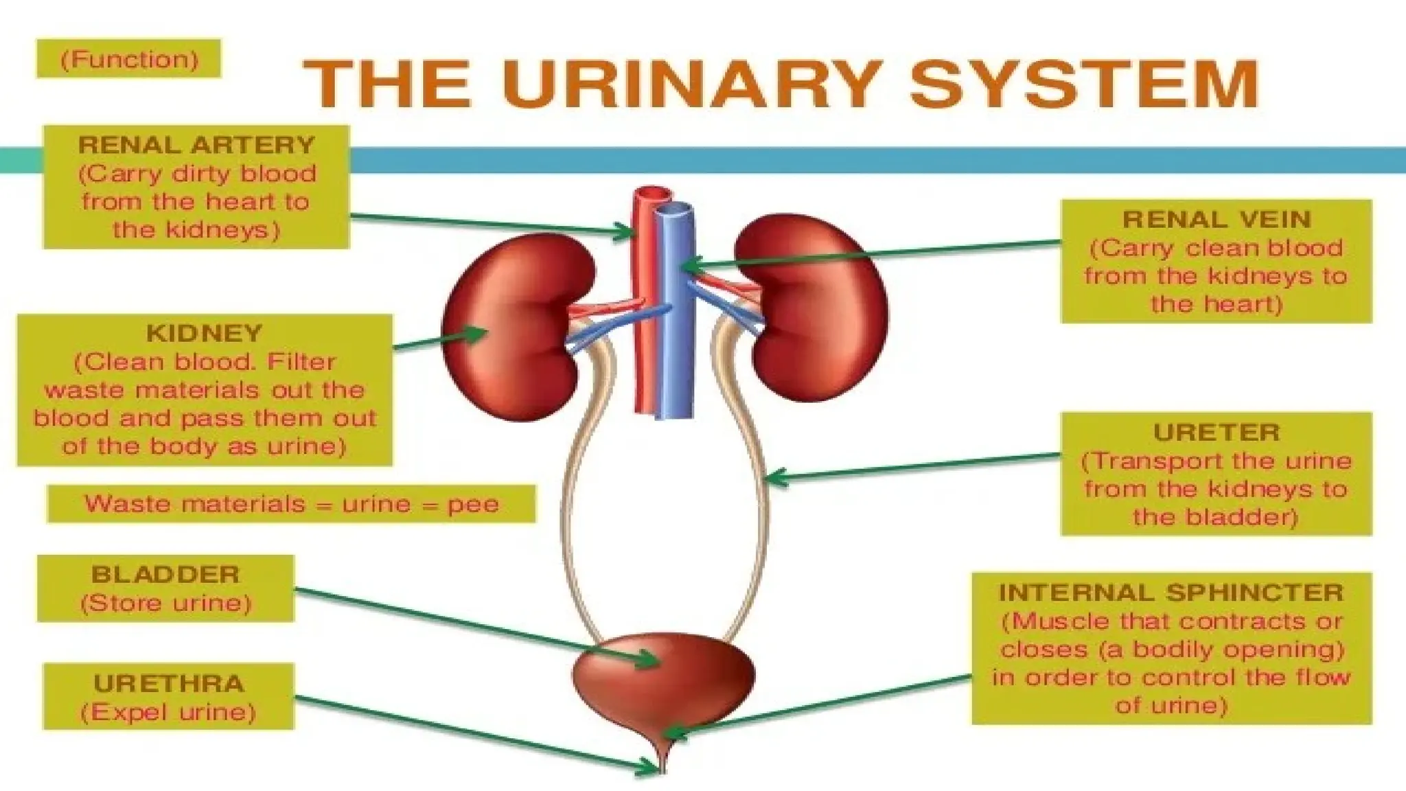
Organs of theUrinary
system
•Kidneys
•Ureters
•Urinary bladder
•Urethra
Figure 15.1a
5.
Functions of theUrinary System
1. Filter. Every day, the kidneys filter gallons of
fluid from the bloodstream. Filter blood and
remove waste products, toxins, extra water and
salt, in the form of urine.
2. Waste processing: The kidneys then process
this filtrate, allowing wastes and excess ions to
leave the body in urine while returning needed
substances to the blood in just the right
6.
3. Elimination: Althoughthe lungs and the
skin also play roles in excretion, the kidneys
bear the major responsibility for eliminating
nitrogenous wastes, toxins, and drugs from
the body.
7.
4. Regulation: Thekidneys also regulate the
blood’s volume and chemical makeup so that
the proper balance between water and salts
and between acids and bases is maintained
8.
5. Other regulatoryfunctions: By producing
the enzyme renin, they help regulate blood
pressure, and their hormone erythropoietin
stimulates red blood cell production in the bone
marrow.
6.Conversion. Kidney cells also convert vitamin
D to its active form.
7. Store and transport urine out of the body
9.
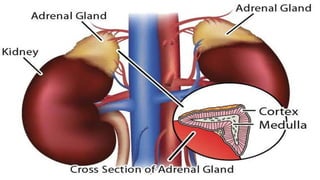
Location of theKidneys
•Against the dorsal body wall
•At the level of T12 to L3
•The right kidney is slightly lower than the left
•Attached to ureters, renal blood vessels, and
nerves at renal hilus
•top of each kidney is an adrenal gland
11.
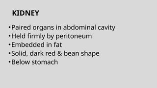
KIDNEY
•Paired organs inabdominal cavity
•Held firmly by peritoneum
•Embedded in fat
•Solid, dark red & bean shape
•Below stomach
12.
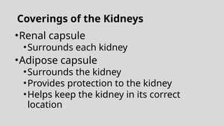
Coverings of theKidneys
•Renal capsule
•Surrounds each kidney
•Adipose capsule
•Surrounds the kidney
•Provides protection to the kidney
•Helps keep the kidney in its correct
location
14.
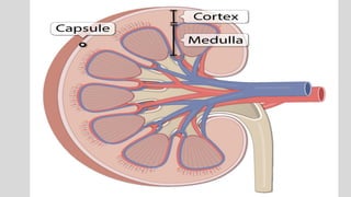
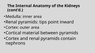
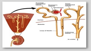
The Internal Anatomyof the Kidneys
(cont’d.)
•Medulla: inner area
•Renal pyramids: tips point inward
•Cortex: outer area
•Cortical material between pyramids
•Cortex and renal pyramids contain
nephrons
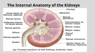
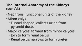
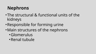
The Internal Anatomyof the Kidneys
(cont’d.)
•Nephrons: functional units of the kidney
•Minor calyx
•Funnel shaped, collects urine from
pyramid ducts
•Major calyces: formed from minor calyces
•Join to form renal pelvis
•Renal pelvis narrows to form ureter
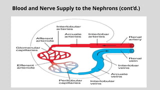
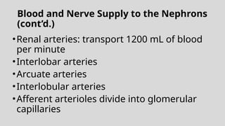
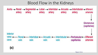
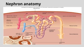
Blood and NerveSupply to the Nephrons
(cont’d.)
•Renal arteries: transport 1200 mL of blood
per minute
•Interlobar arteries
•Arcuate arteries
•Interlobular arteries
•Afferent arterioles divide into glomerular
capillaries
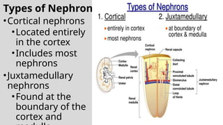
Types of Nephrons
•Corticalnephrons
•Located entirely
in the cortex
•Includes most
nephrons
•Juxtamedullary
nephrons
•Found at the
boundary of the
cortex and Figure 15.3a
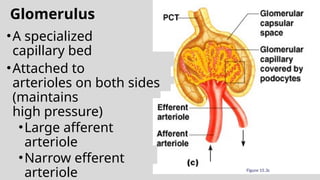
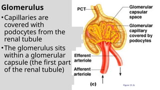
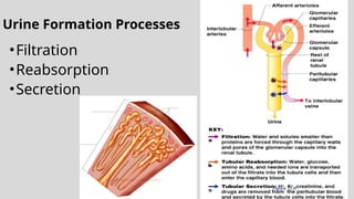
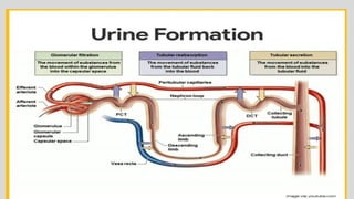
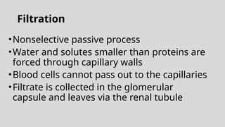
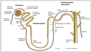
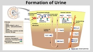
Filtration
•Nonselective passive process
•Waterand solutes smaller than proteins are
forced through capillary walls
•Blood cells cannot pass out to the capillaries
•Filtrate is collected in the glomerular
capsule and leaves via the renal tubule
30.
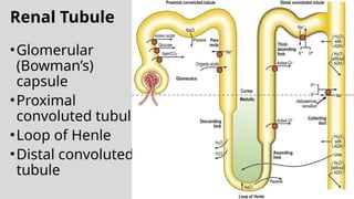
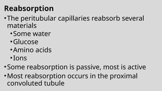
Reabsorption
•The peritubular capillariesreabsorb several
materials
•Some water
•Glucose
•Amino acids
•Ions
•Some reabsorption is passive, most is active
•Most reabsorption occurs in the proximal
convoluted tubule
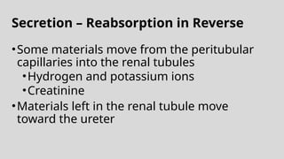
Secretion – Reabsorptionin Reverse
•Some materials move from the peritubular
capillaries into the renal tubules
•Hydrogen and potassium ions
•Creatinine
•Materials left in the renal tubule move
toward the ureter
Characteristics of UrineUsed for Medical
Diagnosis
•Colored somewhat yellow due to the pigment
urochrome (from the destruction of
hemoglobin) and solutes
•Sterile
•Slightly aromatic
•Normal pH of around 6
•Specific gravity of 1.001 to 1.035
36.
Ureters
•Slender tubes attachingthe kidney to the
bladder
•Continuous with the renal pelvis
•Enter the posterior aspect of the
bladder
•Runs behind the peritoneum
•Peristalsis aids gravity in urine transport
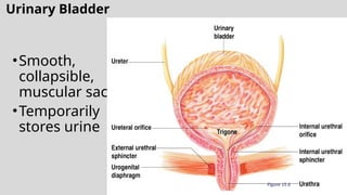
Urinary Bladder Wall
•Threelayers of smooth muscle (detrusor
muscle)
•Mucosa made of transitional epithelium
•Walls are thick and folded in an empty
bladder
•Bladder can expand significantly without
increasing internal pressure
40.
Urethra
•Thin-walled tube thatcarries urine from the
bladder to the outside of the body by
peristalsis
•Release of urine is controlled by two
sphincters
•Internal urethral sphincter (involuntary)
•External urethral sphincter (voluntary)
41.
Urethra Gender Differences
•Length
•Females– 3–4 cm (1 inch)
•Males – 20 cm (8 inches)
•Location
•Females – along wall of the vagina
•Males – through the prostate and penis
The Urinary Bladderand the Micturition
Reflex (cont’d.)
•Micturition: expulsion of urine from bladder
•External urinary sphincter: voluntary control
•Bladder capacity
•700-800 mL
•200-400 mL: conscious of need to urinate
•Stretch receptors initiate micturition reflex
44.
The Urinary Bladderand the Micturition
Reflex (cont’d.)
•During urination
•Detrusor muscle contracts
•Muscles of pelvic floor and abdominal
wall contract
•External urinary sphincter relaxes and
urine moves from bladder to the
outside
45.
The Link BetweenWater and Salt
•Changes in electrolyte balance
causes water to move from one
compartment to another
•Alters blood volume and blood
pressure
•Can impair the activity of cells
46.
Maintaining Water Balance
•Diluteurine is produced if water intake is
excessive
•Less urine (concentrated) is produced if
large amounts of water are lost
•Proper concentrations of various
electrolytes must be present
47.
Regulation of Waterand Electrolyte
Reabsorption
•Regulation is primarily by hormones
•Antidiuretic hormone (ADH) prevents
excessive water loss in urine
•Aldosterone regulates sodium ion content of
extracellular fluid
•Triggered by the rennin-angiotensin
mechanism
•Cells in the kidneys and hypothalamus are
active monitors
48.
Developmental Aspects ofthe Urinary
System
•Functional kidneys are developed by the third
month
•Urinary system of a newborn
•Bladder is small; urine cannot be
concentrated
•Control of the voluntary urethral sphincter
starts until age 18 months
•Urinary infections are the only common
problems before old age
49.
Aging and theUrinary System
•There is a progressive decline in urinary
function
•The bladder shrinks with aging
•Urinary retention is common in males


![Urinary System [45] (1).ppt anatomy and physiology](https://cdn.slidesharecdn.com/ss_thumbnails/urinarysystem451-250511124148-367634e6-thumbnail.jpg?width=640&height=640&fit=bounds)


