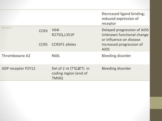
TataKelola dan KamSiber Kecerdasan Buatan v022.pdf
Genetics variations in gpcr
1. What is G-protein??
• GPCR also known as guanine nucleotide-binding proteins.
• Family of protein that act as a molecular switches inside the
cell.
• Activity regulated by factors that controls their ability to bind
to and hydrolyze GTP into GDP.
• When they are bound to GTP, they are 'on', and, when they
are bound to GDP, they are 'off'. G proteins belong to the
larger group of enzymes called GTPase.
• There are two classes of G proteins:
a. monomeric small GTPase
b. heterotrimeric G protein complexes (alpha (α), beta (β)
and gamma (γ) subunit)
2. G-Protein Coupled Receptor
7 trans membrane helices connected by alternating cytosolic and
extra cellular loop
C terminal: inside the cell
N terminal : extra cellular region
Extra cellular portion has unique messenger binding site
Cytosolic loop allow receptor to interact with G protein.
The eventual effect of agonist -induced activation is a change in the
relative orientations of the TM helices (likened to a twisting motion)
leading to a wider intracellular surface and "revelation" of residues
of the intracellular helices and TM domains crucial to signal
transduction function (i.e., G-protein coupling).
Inverse agonists and antagonists may also bind to a number of
different sites, but the eventual effect must be prevention of this TM
helix reorientation
3.
4. Genetic variations may be due to
1.Sequence variations of the human genome
• Introduces variability in genetic make-up
• Suspected to play a main role in diseases &
variable response in drug therapy
• Polymorphism- refers to sequence variation
leading to occurrence of two or more clearly
different forms.
• Single nucleotide polymorphism accounts for
approx. 80% of all sequence variations.
5. 2. Structure & function of GPCRs
• Comprises a large class of membrane proteins –
encoded by approx. 600 human genes.
• Molecular architecture might permit the
prediction of functionally relevant domains
where sequence variations are more likely to
alter receptor function.
• Normally, TM domains are highly conserved, the
loops are variable in sequence & length, &the C-
and N-terminals tails represents the most
diverse elements.
6. 3. GPCR coupling to G proteins and other signaling
pathways
• GPCR thought to be couple to heterotrimeric G
proteins composed of α, β and γ subunits. It display
considerable heterogenecity, with a predicted
number of 27 different α, 5 β and 13 γ subunits.
• Main sites of contact between receptor and G
proteins include the third intracellular loop, but i1, i2
and the C- terminus have also been reported to
contribute G protein coupling
• Proteins like protein kinases, arrestin &
phosphatases modulates receptor functions at
distinct domains that are possible targets for
polymorphic effects.
7. 4. GPCR binding pockets
• Ca++, acetyl choline, glutamate, bradykinin,
prostaglandins, & the large polypeptide FSH bind
to the same site.
• Distinct binding sites appear to exist, either
embedded within the pocket formed by the 7-
TMD bundle within the membrane, at pockets
formed by the extracellular loops, or in the N-
terminus.
• The thrombin receptor family represents a
special case whereas the protease activity of the
ligand thrombin cleaves a portion of the N-
terminus. The newly generated N-terminus then
serves as a tethered ligand.
8. • GPCRs appear to be activated by ligand binding to many
different sites of the protein . At the opioid receptors, peptide
endorphins bind primarily to the extracellular loops, whereas
opioid alkaloids dock deep into the 7-TMD core.
Human μ opioid receptor
9. • Sequence variation in the receptor protein can affect ligand
binding or the structural integrity of the receptor , indirectly
changing ligand binding .
5. Spontaneous GPCR signalling
• Exchange of single amino acid residues can lead
to constitutive receptor activation.
• Considerable number of human polymorphisms
enhance signalling (gain of function) or even
activate the receptor constitutively, causing
serious genetic disorders.
10. 6. Multiple receptor conformations with distinct
functions
• GPCRs are flexible structures and may
accommodate ligands in various ways. It exists in
multiple conformations. Discrete signalling
pathways are triggered by discrete
conformational states of GPCR
12. Impaired or enhanced agonist
signalling efficacy
Several inactivating sequence variants of peptide
receptors have been associated with congenital
disorders. For example,
1. A point mutation causing truncation of
thyrotropin stimulating hormone receptor leads to
leydig’s cell hyperplasia.(activating mutation)
Truncated TM5, D578G, T398M
13. 2. Inactivating mutations of the ACTH receptor are
associated with familial glucocorticoid deficiency .
The mutation occurs in the large N-terminus , the
binding site for glycoprotein hormone receptor,
leading to toxic multinodular goiter.
S120R, R201Stop, S74I, V254C
14. V2 vasopressin recptors
• A number of mutations in the gene encoding the
V2 vasopressin receptor leads to functionally
inactive receptor protein and are causative for
nephrogenic diabetes insipidus.(missense
mutations)
• This a clear indication that receptor activity
depends on intact signalling pathways.
(multiple SNPs; decreased ligand binding;
R113W; R137H)
15.
16. Thromboxane A2 Receptor
• This receptor performs an essential role in
haemostasis by inducing platelet aggregation. An
R60L amino acid substitution in the first
cytoplasmic loop of TBXA2 receptor causes a
dominantly inherited bleeding disorder
characterised by defective platelet response to
TBXA2. This leads to decreased agonist-induced
second messenger formation.
17.
18. P2Y 12ADP Receptor
• This receptor sub-type is shown to be the target
for anti-thrombotic drugs such as ticlodipine &
clopidogrel. 2-nucleotide deletion in a region
mapping to the end of TMD6, associated with a
rare bleeding disorder.
19. Chemokine reeptors
• Fusin and CKR5 have been identified as a co-
receptors for the cellular entry of HIV. Similarly ,
certain chemokines were found to block HIV
entry into cells.
• Natural resistance can be either by high
endogenous levels of chemokines or by
mutations of the receptors.
• A 32 bp deletion in CKR5 leading to a frame shift
and a non functional protein appeared to protect
homozygous carriers against HIV infection &
blocking its entry.
20. • Val 64 substitution with Ile was shown to result
in heterodimerisation of CCR2 with CCR5 or
CXCR4, thereby promoting resistance to AIDS.
21. Biogenic amine receptors
• The R16G substitution in the β2 adrenoreceptors
has been associated with nocturnal asthma
whereas W64R in the β3 receptor expressed in
adipocytes are involved in energy metabolism –
is linked with obesity.
23. receptors Variant/Allele Disease/ Phenotype
cellular mechanism/ Event
Β1 adrenergic receptor R16G Nocturnal asthma;
Enhanced agonist promoted
down regulation of receptor
Β3 adrenergic receptors W64R Obesity
Luteinising hormone Truncated TM5
D578G
Leydig’s cell hyperplasia;
Precocious puberty in male
children
FSH A189V Ovarian dysgenesis;
Altered protein folding;
inactivation of receptor
Thyrotropin (TSH) S120R, R201Stop, S74I,
V254C
Glucocorticoid deficiency;
altered/ loss of receptor
function/ reduced
expression
ACTH D727E Altered receptor
function/conformation;
Toxic multinodular goiter
Vasopressin V2 receptor Multiple SNPs Nephrogenic diabetes
24. Decreased ligand binding;
reduced expression of
receptor
Chemokine receptors CCR2
CCR3
CCR5
V64I
R275Q,L351P
CCR5P1 alleles
Delayed progression of AIDS
Unknown functional change
or influence on disease
Increased progression of
AIDS
Thromboxane A2 R60L Bleeding disorder
ADP receptor P2Y12 Del of 2 nt (TTCATT) in
coding region (end of
TMD6)
Bleeding disorder