Pre clinical screening for
Anti-asthmatics
DHINESHKUMAR V
IM.PHARM (PHARMACOLOGY)
Contents
Asthma – introduction
Test methods
1. In vitro methods
2. Test in isolated organs
3. In vivo methods
Asthma – Introduction
It is a complex inflammatory airway disease characterised by airflow
obstruction due to bronchi constriction, bronchi inflammation or due to
excessive secretion of mucus.
Triggered by allergen, cold air, moist air, exercise and emotional stress.
Symptoms include wheezing, shortness of breath, chest tightness and
coughing.
Animal models
To mimic bronchial asthma in animals, a wide variety of animal models have
been developed.
Each model has its own advantages and disadvantages, unfortunately no model
is identical to the conditions found in human disease.
Test methods
1. In vitro methods
i. CULTEX technique
ii. Binding assay
2. Test in isolated organs
i. Spasmolytic activity in guinea pig lungs
3. In vivo methods
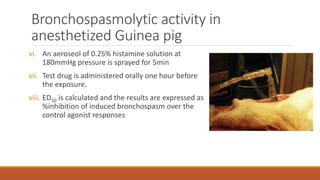
i. Bronchospasmolytic activity in anesthetized Guinea pig
ii. Arachidonic acid or PAF induced Respiratory and vascular dysfunction in guinea pig
iii. Anaphylactic Microshock in guinea pigs
iv. Serotonin Aerosol-induced Asphyxia in Guinea pig
v. Bronchial hyperactivity in guinea pigs
In vitro models
Cell culture methods :
CULTEX Technique:
CULTEX technique is a experimental system for cultivation and exposure of cells
intermediately at the air/liquid interface.
It allows direct exposure of bronchial epithelial cells to gases, ultrafine particles
or mixture of both
CULTEX Technique
CULTEX technique
Cultured bronchial epithelial cells in plates are washed with PBS (phosphate
buffer saline) and then transferred to cell exposure unit.
Then the cells are exposed to the clean air or different concentrations of side
stream smoke.
Test group bronchial epithelial cells are incubated with test drug for 24hours
and then exposed to diff. Conc. of sidestream smoke for 1 hour.
Parameters that can be estimated are number of cells, metabolic activity and
glutathione concentration.
Cell viability measurements are carried out using WST assay an electronic cell
counting
Binding Assays
Histamine receptor assay:
This method evaluates the affinity of test compound to histamine H1 receptor
It is done by measuring their inhibitory activities on the binding of 3H
pyrilamine to guinea pig brain plasma membrane.
Histamine receptor assay
Steps:
i. Male guinea pigs (300g-600g) are sacrificed by CO2 Necrosis
ii. Brain is homogenized in ice-cold tris buffer and homogenate is centrifuged
for 10 mins at 4°C at 50000g
iii. Supernatant is discarded and pellet is resuspended in buffer, centrifuged
again
iv. Pellet obtained is re-suspended in Tris-buffer and aliquots of 1ml are frozen
at -70 °C.
v. 50 µl of 3H pyrilamine and 50 µl of test compound is added in 100 µl of
membrane suspension is incubated for 30min
Histamine receptor assay
vi. Make 11 conc. Of 3H pyrilamine and perform saturation studies
vii. Add 3ml of scintillation cocktail to measure the radioactivity in scintillation
counter
viii. Radioactivity denotes the binding of pyrilamine binding
ix. % inhibition of 3H pyrilamine binding is measured using scintillation counter
reading
Tests in Isolated organs
Spasmolytic activity in Guinea pig Lungs:
Histamine and leukotrienes induce bronchoconstriction
Histamine induce bronchoconstriction by biding to H1 receptor
Calcium ionophores induce release of leukotrienes through 5-lipoxygenase
pathway which cause bronchoconstriction
In this method drugs are tested for their capability of inhibiting bronchospasm
induced by histamine or calcium ionophore.
Spasmolytic activity in guinea pig lungs
Steps:
i. Albino guinea pigs (300-450g) are sacrificed with an overdose of ether
ii. Chest is opened and the lungs are removed and cut into strips of 5cm each and
placed in physiological saline solution
iii. Lung strips are mounted in organ bath containing nutritive solution
iv. Prior to testing carbachol is added to the bath to test the lung strip’s ability to
contract
v. After 30 min spasmogens (histamine, ca-ionophore, leukotriene) are added to
induce constriction
vi. After 5min test drug is added and percentage inhibition of spasmogen induced
constriction is measured
In vivo models
Bronchospasmolytic activity in anesthetized Guinea pig:
Changes in air volume of a living animal in a closed system consisting of a
respiratory pump, the trachea and the lungs is measured here.
Bronchoconstriction – decreased inspired air, increased excess air.
Constriction of bronchi smooth muscle is achieved by administering
spasmogen.
This method evaluates the bronchoconstriction effect by measuring the volume
of air which is not taken up by the lungs after inducing bronchoconstriction
Bronchospasmolytic activity in
anesthetized Guinea pig
Steps:
i. Guinea pigs (250 to 500g) are anesthetized with 1.25g/kg urethane
intraperitonially
ii. Trachea is cannulated one arm is connected to a respiratory pump and the
other to statham P23 Db transducer (to measure the air volume)
iii. Animal is artificially ventilated at a frequency of 60strokes / min.
iv. Excess air which is not taken up by the lungs is measured and recorded in
polygraph
v. Jugular vein is cannulated for test drug administration and carotid artery for
blood pressure measurements
Bronchospasmolytic activity in
anesthetized Guinea pig
vi. An aeroseol of 0.25% histamine solution at
180mmHg pressure is sprayed for 5min
vii. Test drug is administered orally one hour before
the exposure.
viii. ED50 is calculated and the results are expressed as
%inhibition of induced bronchospasm over the
control agonist responses
In vivo methods
Arachidonic acid or PAF induced Respiratory and vascular dysfunction in guinea
pig :
Thromboxane, prostacyclin – products of arachidonic acid metabolism
Thromboxane- bronchoconstriction and thrombocytopenia
Prostacyclin – reduction in systolic and diastolic pressure
Arachidonic acid or PAF induced Respiratory
and vascular dysfunction in guinea pig
Steps:
i. Male guinea pigs (300-600g) are anesthetizd with 60mg/kg pentobarbitone
(intraperitonialy)
ii. Jugular vein is cannulated for administering spasmogen /test compound.
iii. Both carotid arteries are cannulated, one is connected to transducer (to
measure the Bp) and another is used for blood withdrawal
iv. Trachea is connected to respirator (70-75 strokes/min )
v. Excess air not taken up by the lungs is measured
vi. Changes in airflow and BP is recorded continuously
Arachidonic acid or PAF induced Respiratory
and vascular dysfunction in guinea pig
vii. Animals receive multiple intravenous injections of the same dose of
arachidonic acid (60 µg /kg) until two bronchospasms of equal intensity is
obtained
viii. Then the test drug compounds are administered intravenously and
spasmogen is given again
ix. Percent inhibition or increase of bronchospasm ,reduction of blood pressure,
thrombocytopenia following test drug administration is measured and
compared with control values before drug treatment
Anaphylactic Microshock in guinea pigs
Histamine is released from various sites in anaphylactic response
Introduction of foreign particle in the body can cause microshock.
A microshock is apparently one that is interrupted before death and is
repeatable
Anaphylactic Microshock in guinea pigs
Steps:
i. Guinea pigs (200-300g) are sensitized with subcutaneous injection of egg
albumin
ii. After 3 weeks the animals are exposed to an aerosol of 5% albumin.
iii. They are removed from the chamber as soon as they become dyspneic
iv. The time from the start of exposure to severe dyspnea is referred as
preconvulsion time.
v. If no shock is seen for more than 6min, the animal is considered as protected
and prconvulsion time is taken as infinite.
Anaphylactic Microshock in guinea pigs
v. After this control exposure, the guinea pig is treated with test drug and
preconvulsion time is measured
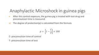
vi. The degree of protection(p) is calculated from the formula
𝑝 = 1 −
𝐶
𝑇
× 100
C- preconvulsion time of control
T- preconvulsion time of test
Serotonin Aerosol-induced Asphyxia in
Guinea pig
Guinea pigs when exposed to serotonin aerosol, develop constriction of
bronchi and leads to asphyxia in large doses
Here the test drug’s ability to reverse the serotonin induced
bronchoconstriction is measured
Serotonin Aerosol-induced Asphyxia in
Guinea pig
Steps:
i. Guinea pigs (200-300g) are placed in an anaesthetic box and 2% serotonin
aerosol is introduced .
ii. Animals shows progressive signs of difficulty in breathing and convulsion due
to the serotonin exposure.
iii. As soon as these signs are observed, the animals are removed from the box
and fresh air is given.
iv. Then the test drug is given either in orally or subcutaneously.
v. Again the test is performed after two days to calculate the preconvulsion
time
Serotonin Aerosol-induced Asphyxia in
Guinea pig
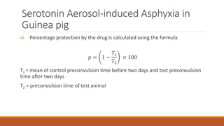
vi. Percentage protection by the drug is calculated using the formula
𝑝 = 1 −
𝑇1
𝑇2
× 100
T1 = mean of control preconvulsion time before two days and test preconvulsion
time after two days
T2 = preconvulsion time of test animal
Bronchial hyperactivity in guinea pigs
inhalation of histamine or other spasmogen cause symptoms like asphyctic
convulsion resembling bronchial asthma in guinea pigs.
Here spasmogens are applied as aerosols which is produced by ultrasound
nebuliser.
Here also the preconvulsion time is noted.
Bronchial hyperactivity in guinea pigs
Steps:
i. Male albino guinea pigs (300-400g) are used.
ii. The inhalation cages consists of 3 boxes each ventilated with an airflow of
1.5L/min
Box-A = test drug is applied using ultrasound nebuliser
Box- B= serves as sluice through which animal is passed to the BOX –C
Box-C = 0.1% histamine aerosol is given through ultrasonic nebuliser
Bronchial hyperactivity in guinea pigs
iii. Preconvulsion time is measured for both control and test groups
iv. Percentage increase of preconvulsion time is calculated against control
group.
v. ED50 (dose required to increase preconvulsion time by 50%) is also calculated
Reference
i. Gupta SK. (2016). Drug Screening Methods. 3rd ed. Jaypee
Brothers Medical Publisher (P) Ltd, pp.683-696.
pre clinical Screening  for anti asthmatic drugs