Download as PDF, PPTX


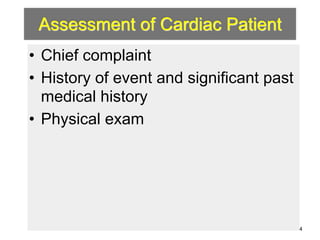

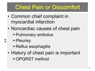
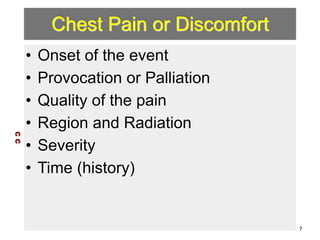

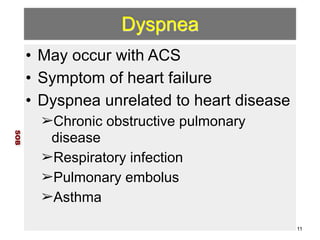


The document titled 'Cardiac Evaluation' by Joe Lex, MD, focuses on the assessment of cardiac patients, detailing the importance of understanding chief complaints such as chest pain, dyspnea, syncope, and palpitations. It emphasizes comprehensive histories, physical examinations, and specific diagnostic criteria for various cardiac conditions. Additionally, it addresses copyright information and guidelines for the use of the material, ensuring it is meant for educational purposes only.


