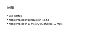
Left ventricular
noncompaction
Through ECHO window
1990-First diagnostic criteria
• LVNC
 X (distance between epicardial surface and trough of the intertrabecular
recesses)
 Y (distance between epicardial surface and peak of the trabeculations)
If X/Y< 0.5
if it progressively decreased from the papillary muscles toward the apex
Chin TK, Perloff JK, Williams RG, Jue K, Mohrmann R. Isolated noncompaction of left ventricular myocardiu.
A study of eight cases. Circulation 1990;82:507–13.
1999-2ND Criteria
• LVNC
Noncompacted to compacted myocardium ratio needs to exceed a cut-off of
2.3
Ichida F, Hamamichi Y, Miyawaki T, et al. Clinical features of isolated noncompaction
of the ventricular myocardium. Long-term clinical course, hemodynamic
properties, and genetic background. J Am Coll Cardiol 1999;34:233–40
Other three
• Oechslin EN, Attenhofer Jost CH, Rojas JR, Kaufmann PA, Jenni R. Long
term
• follow-up of 34 adults with isolated left ventricular noncompaction: A
distinct cardiomyopathy with poor prognosis. J Am Coll Cardiol
2000;36:493–500.
• Belanger AR, Miller MA, Donthireddi UR, Najovits AJ, Goldman ME.
New classification scheme of left ventricular noncompaction and
correlation with ventricular performance. Am J Cardiol 2008;102:92–
6.
• Stanton C, Bruce C, Connolly H, et al. Isolated left ventricular
noncompaction syndrome.Am J Cardiol 2009;104:1135–8.
2014-Latest:All 4 criteria to be fulfilled
LVNC
1. Prominent trabeculous formations along the left ventricular endocardial border visible
in end-diastole, distinct from papillary muscles, false tendons or aberrant bands
2. Trabeculations move synchronously with the compacted myocardium
3. Trabeculations form the noncompacted part of a two-layered myocardial structure, best
visible at end-systole
4. Perfusion of the intertrabecular spaces from the ventricular cavity is present at end-
diastole on colour Doppler echocardiography or contrast echocardiography
Refinement of echocardiographic criteria for left ventricular noncompaction Claudia Stöllberger,Birgit Gerecke,Josef
Finsterer,Rolf Engberding.International Journal of Cardiology 165 (2013) 463–467
Echocardiography
 LV short axis
 Mid systole
 Non compaction to compaction ratio >2
 More than 3 recesses
 Recesses freely communicates to LV in diastole directly
MRI
• End diastole
• Non compaction:compaction is >2.3
• Non compaction LV mass>20% of global LV mass
It is so beautiful !

Left ventricular noncompaction

  • 1.
  • 2.
  • 3.
    1990-First diagnostic criteria •LVNC  X (distance between epicardial surface and trough of the intertrabecular recesses)  Y (distance between epicardial surface and peak of the trabeculations) If X/Y< 0.5 if it progressively decreased from the papillary muscles toward the apex Chin TK, Perloff JK, Williams RG, Jue K, Mohrmann R. Isolated noncompaction of left ventricular myocardiu. A study of eight cases. Circulation 1990;82:507–13.
  • 4.
    1999-2ND Criteria • LVNC Noncompactedto compacted myocardium ratio needs to exceed a cut-off of 2.3 Ichida F, Hamamichi Y, Miyawaki T, et al. Clinical features of isolated noncompaction of the ventricular myocardium. Long-term clinical course, hemodynamic properties, and genetic background. J Am Coll Cardiol 1999;34:233–40
  • 5.
    Other three • OechslinEN, Attenhofer Jost CH, Rojas JR, Kaufmann PA, Jenni R. Long term • follow-up of 34 adults with isolated left ventricular noncompaction: A distinct cardiomyopathy with poor prognosis. J Am Coll Cardiol 2000;36:493–500. • Belanger AR, Miller MA, Donthireddi UR, Najovits AJ, Goldman ME. New classification scheme of left ventricular noncompaction and correlation with ventricular performance. Am J Cardiol 2008;102:92– 6. • Stanton C, Bruce C, Connolly H, et al. Isolated left ventricular noncompaction syndrome.Am J Cardiol 2009;104:1135–8.
  • 6.
    2014-Latest:All 4 criteriato be fulfilled LVNC 1. Prominent trabeculous formations along the left ventricular endocardial border visible in end-diastole, distinct from papillary muscles, false tendons or aberrant bands 2. Trabeculations move synchronously with the compacted myocardium 3. Trabeculations form the noncompacted part of a two-layered myocardial structure, best visible at end-systole 4. Perfusion of the intertrabecular spaces from the ventricular cavity is present at end- diastole on colour Doppler echocardiography or contrast echocardiography Refinement of echocardiographic criteria for left ventricular noncompaction Claudia Stöllberger,Birgit Gerecke,Josef Finsterer,Rolf Engberding.International Journal of Cardiology 165 (2013) 463–467
  • 7.
    Echocardiography  LV shortaxis  Mid systole  Non compaction to compaction ratio >2  More than 3 recesses  Recesses freely communicates to LV in diastole directly
  • 8.
    MRI • End diastole •Non compaction:compaction is >2.3 • Non compaction LV mass>20% of global LV mass
  • 9.
    It is sobeautiful !