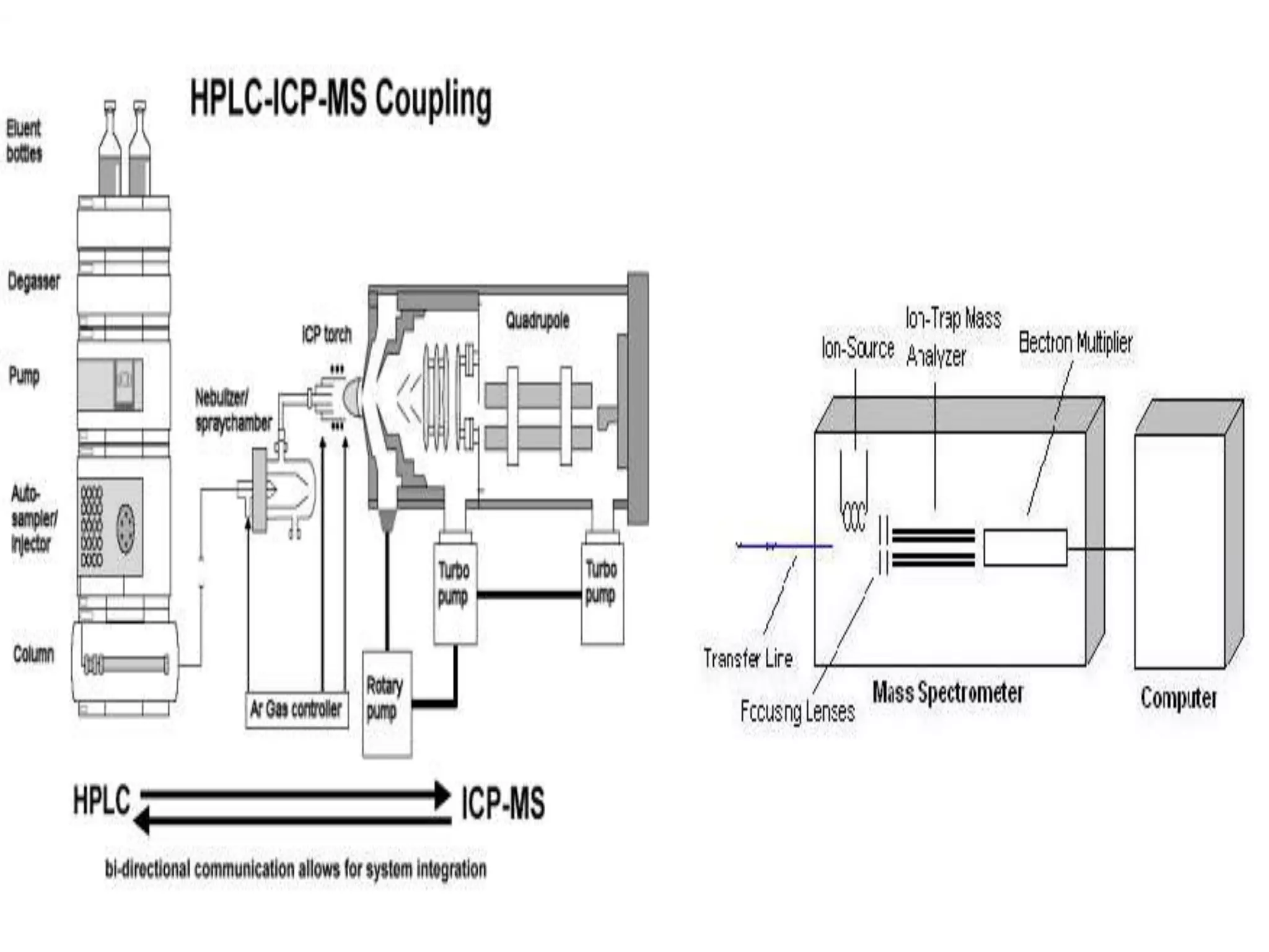
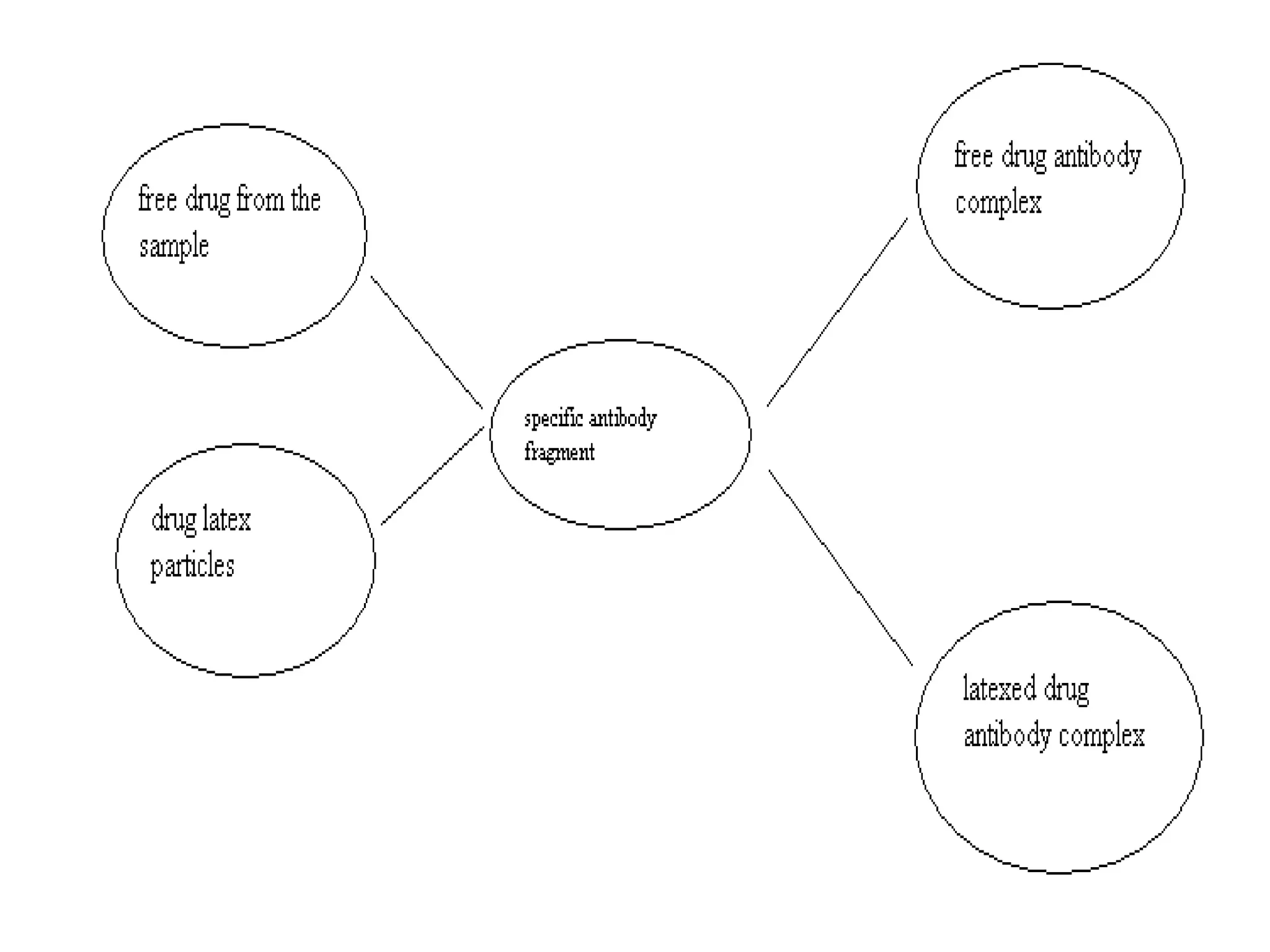
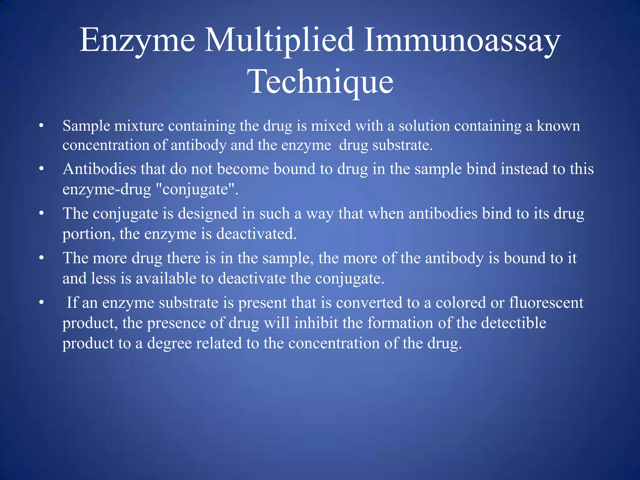
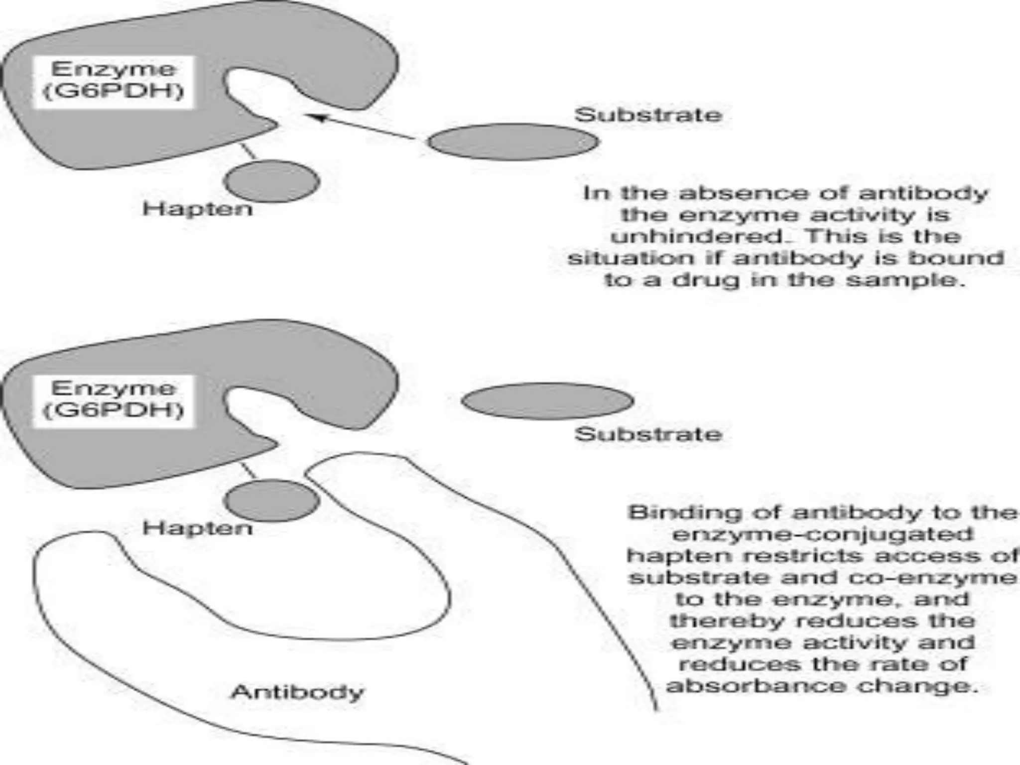
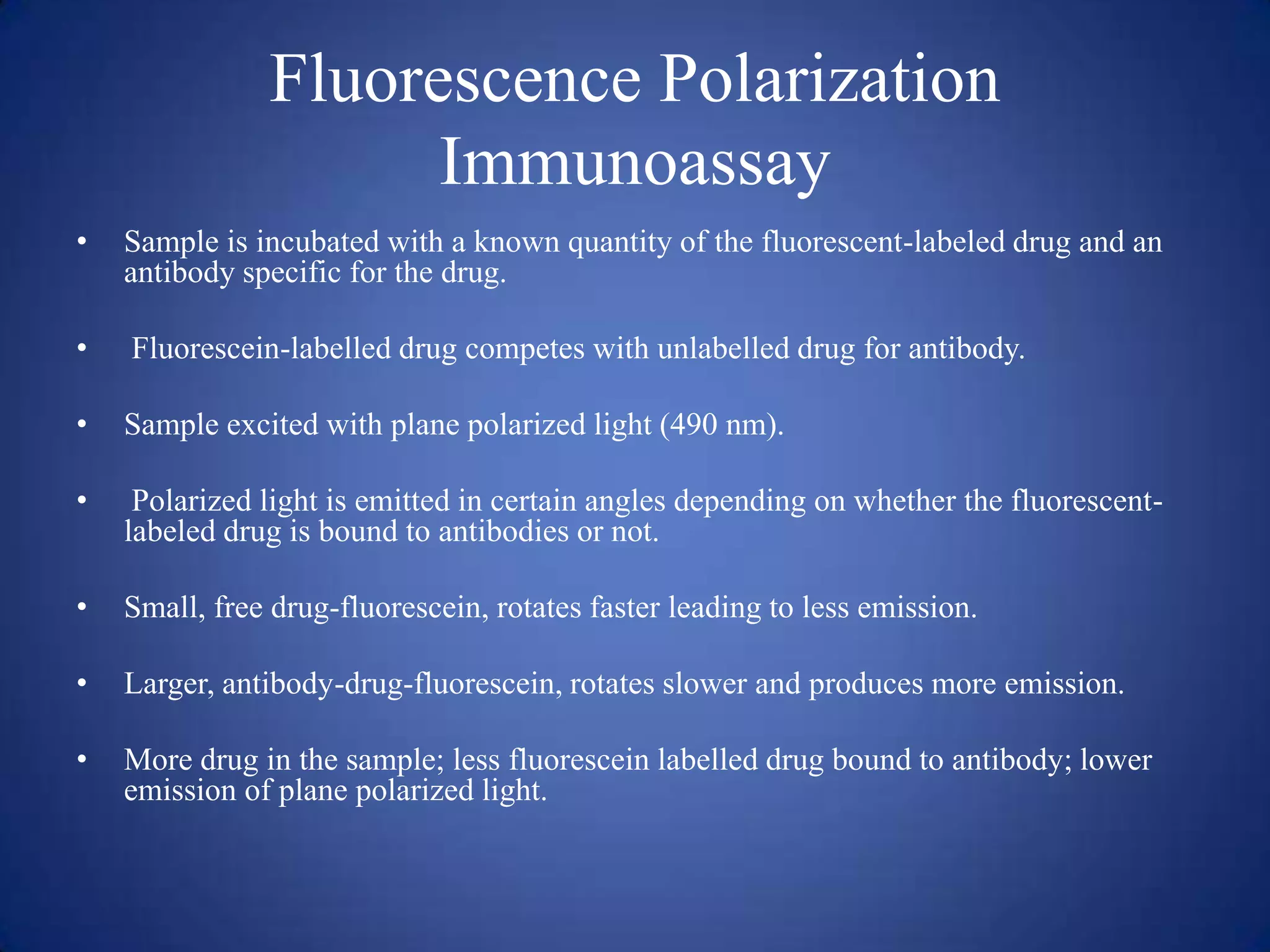
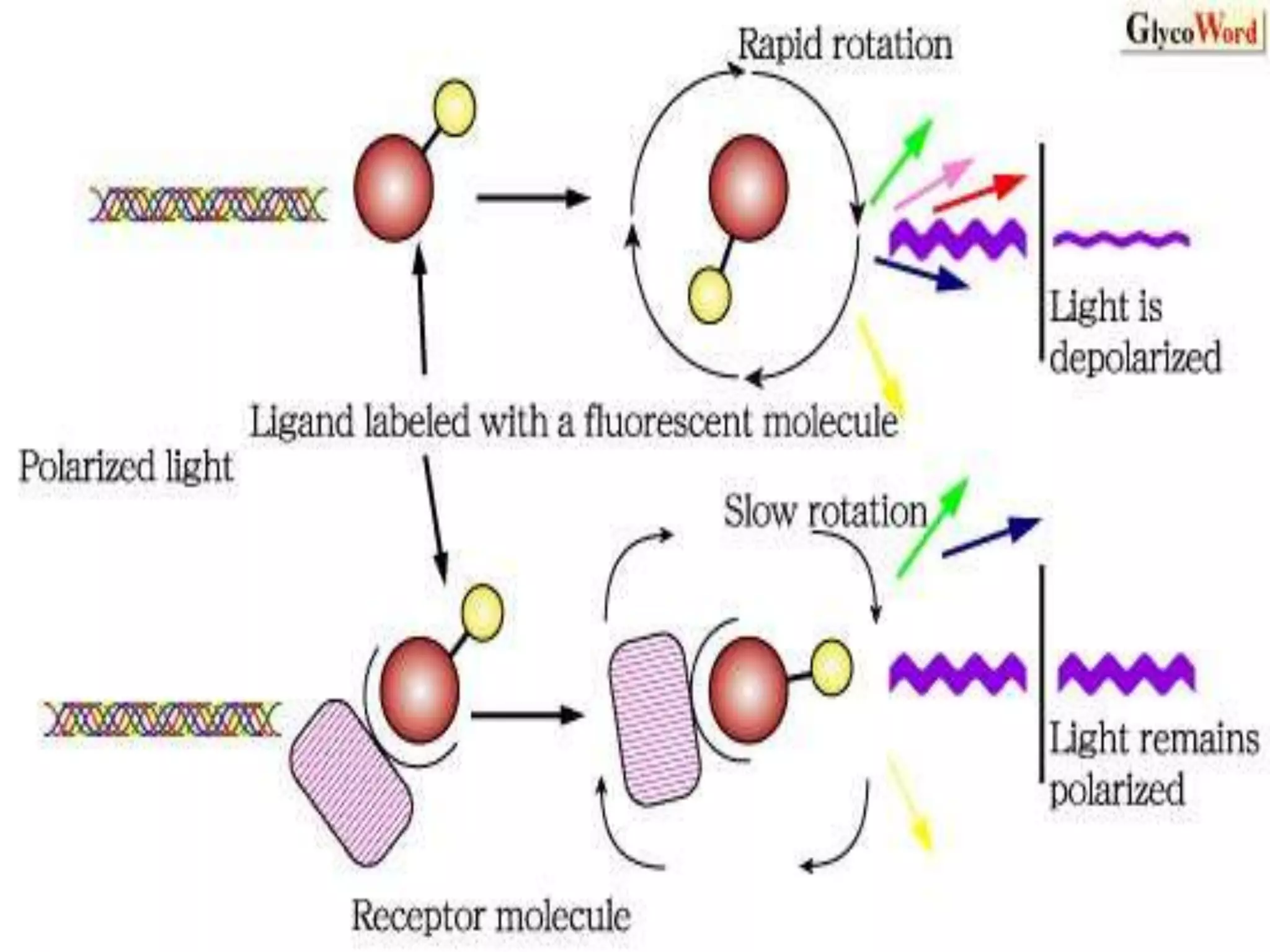
This document discusses several techniques for drug analysis including chromatography methods like high performance liquid chromatography (HPLC), gas chromatography (GC), and liquid chromatography-mass spectrometry (LC-MS). It also summarizes various immunoassay techniques such as radioimmunoassay, particle enhanced turbidimetric inhibition immunoassay, enzyme immunoassay, enzyme multiplied immunoassay technique, fluorescence polarization immunoassay, and chemiluminescence assays. Additional methods covered are affinity chrome-mediated immunoassay, and cloned enzyme donor immunoassay.
2. High Pressure Liquid Chromatography
PRINCIPLE:
•
HPLC column is filled with solid particles like silica and the sample mixture is
separated into compounds as it interacts with the column particles.
•
separation is influenced by liquid solvents condition, chemical interaction between
sample mixture and liquid solvent, and chemical interaction between sample
mixture and solid particles packed inside the separation column.
•
2 variants of HPLC:
– Normal phase HPLC
– Reverse phase HPLC
3. Normal Phase HPLC
• Stationary phase: polar silica particles
• Mobile phase: non polar hexane
• Polar compounds in the sample mixture will stick to the polar silica and the non
polar compounds will pass out more quickly through the column.
Reverse Phase HPLC
• Stationary phase: non polar silica
• Mobile phase: methanol
• Non polar compounds in the sample mixture will stick to the non polar silica
particles and the polar compounds will pass out more quickly through the column.
4.
5. Retention time
• Time taken for a particular compound to travel through the column to the detector.
• Its measured from the time at which the sample is injected to the point at which
display shows a maximum peak height for that compound.
Detection
• Common method used is UV absorption.
• Amount of light absorbed gives the amount of drug present in the sample mixture.
Interpretation of results
• UV spectroscopy: quantification
• Retention time: identification
• Chromatogram: identification, quantification
6. Gas-liquid chromatography
PRINCIPLE:
•
In GLC, the mobile phase is inert gas like helium and stationary phase is a
microscopic layer of liquid on an inert support.
•
The GLC column is placed within an oven. Very high temperatures is used to cause
sample vaporization. Vaporization separates the various molecules in the samples
into their different fractions.
•
The gaseous compounds being analyzed interact with the stationary phase
(adsorption) which causes each compound to elute at different time (retention time)
and the substances are identified qualitatively.
7. •
•
•
•
Commonly used detectors are flame ionization detector and thermal conductivity
detector.
thermal conductivity detector: identification
Retention time: identification
Chromatogram: identification, quantification
8.
9. Liquid Chromatography Mass
Spectrometry
PRINCIPLE:
•
column is filled with solid particles like silica and the sample mixture is separated
into compounds as it interacts with the column particles.
•
separation is influenced by liquid solvents condition, chemical interaction between
sample mixture and liquid solvent, and chemical interaction between sample
mixture and solid particles packed inside the separation column.
•
Elute is analyzed by mass spectrometry.
10. •
Elute is loaded into MS instrument and undergoes vaporization .
•
The components of the sample are ionized by a variety of methods like
impacting with electron beam resulting in the formation of ions.
•
Ions are then separated according to their mass- to- charge ratio in an analyzer
by electromagnetic fields.
11.
12. Radioimmunometric assays
PRINCIPLE:
•
The specimen is mixed with a specific antibody to the drug being analyzed as
well as a known quantity of the same drug labeled with radioactive iodine.
•
The drug in the sample and the radioactive labeled drug compete for sites on
the antibody.
•
The sample is washed to remove unbound radioactive drug. A gamma counter
is then used to measure the amount of radioactivity in the sample as counts per
minute (CPM).
•
This number is inversely proportional to the amount of drug in the sample.
13.
14. Particle Enhanced Turbidimetric
Inhibition Immunoassay
PRINCIPLE:
• PETINIA is a homogenous competitive immunoassay.
• The latex particle-bound drug binds to the drug-specific antibody, forming
insoluble light-scattering aggregates.
•
This causes an increase in the turbidity of the reaction mixture.
• The rate of particle aggregation (turbidity) is inversely proportional to the
concentration of drug in the sample.
15.
16. Enzyme Immunoassay
•
EIA uses a non-radioactive enzyme label.
•
This eliminates the need for special handling and reduces disposal costs.
•
Sample with unknown amount of antigen is immobilized on a solid support
usually a polystyrene microtiter plate.
•
After immobilization of the antigen, a specific antibody is added forming a
complex with the antigen.
•
the antigen in a sample competes for limited antibody binding sites with
antigen conjugated to a reporter enzyme.
•
In the final step a substance containing the enzymes substrate is added.
17. •
When chromogenic substrate is added to the assay to develop color, samples with
high antigen concentration generate a lower signal than those containing low
antigen concentration, yielding the inverse correlation between antigen
concentration in the sample and color development in the assay.
•
Subsequent reaction produces a detectable signal, usually a color change in the
substrate.
18.
19. Enzyme Multiplied Immunoassay
Technique
•
Sample mixture containing the drug is mixed with a solution containing a known
concentration of antibody and the enzyme drug substrate.
•
Antibodies that do not become bound to drug in the sample bind instead to this
enzyme-drug "conjugate".
The conjugate is designed in such a way that when antibodies bind to its drug
portion, the enzyme is deactivated.
The more drug there is in the sample, the more of the antibody is bound to it
and less is available to deactivate the conjugate.
If an enzyme substrate is present that is converted to a colored or fluorescent
product, the presence of drug will inhibit the formation of the detectible
product to a degree related to the concentration of the drug.
•
•
•
20.
21. Fluorescence Polarization
Immunoassay
•
Sample is incubated with a known quantity of the fluorescent-labeled drug and an
antibody specific for the drug.
•
Fluorescein-labelled drug competes with unlabelled drug for antibody.
•
Sample excited with plane polarized light (490 nm).
•
Polarized light is emitted in certain angles depending on whether the fluorescentlabeled drug is bound to antibodies or not.
•
Small, free drug-fluorescein, rotates faster leading to less emission.
•
Larger, antibody-drug-fluorescein, rotates slower and produces more emission.
•
More drug in the sample; less fluorescein labelled drug bound to antibody; lower
emission of plane polarized light.
22.
23. Chemiluminescence
•
Chemiluminescence is the emission of light, as the result of a chemical reaction.
• The most common chemiluminescent assay methods are either
1. enzyme-amplified (or)
2. direct chemiluminescent measurements.
24.
25. In direct chemiluminescence, such as the ADVIA Centaur system:
• The sample is incubated with the AE-labeled drug, and the drug specific
antibody.
• Once the AE is oxidized by hydrogen peroxide and the sample becomes
alkaline, a light emission occurs.
• The lower the drug concentration in the sample, the greater the light
emission.
26. • An example of an enzyme-amplified chemiluminescence method is the
IMMULITE system.
27. Affinity Chrome-Mediated
Immunoassay
• Magnetic particles coated with the drug are added to bind free antibodyenzyme conjugate.
• The reaction mixture is then separated magnetically.
• Following separation, the supernatant containing the drug-antibody-enzyme
complex is transferred by the instrument to another cuvette and mixed with
a substrate.
• B-galactosidase catalyzes the hydrolysis of CPRG (chloropenol red Bgalactopyranoside) to produce CPR (chloropenol red) that absorbs light at
577 nm.
• The change at 577 nm due to the formation of CRP is directly proportional
to the amount of drug present in the sample, and is measured by a
bichromatic (577, 700 nm) rate technique.
28. Cloned Enzyme Donor Immunoassay
• The polypeptide chain of the beta-galactosidase enzyme from Escherichia
coli is presented as two inactive fragments, the large fragment, termed
enzyme acceptor (EA), and a small fragment, enzyme donor (ED), which
contains the 5% of enzyme missing from the EA portion.
• By conjugating drug to the ED fragment, the addition of antibodies that
bind the drug can prevent the formation of an intact and therefore active
enzyme.
• Any drug present in the sample competes for antibody–binding sites, such
that an increase in drug concentrations yields less binding of antibody to
the ED fragment and therefore results in more enzyme activity which can
be monitored spectrophotometrically.