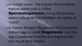
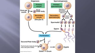
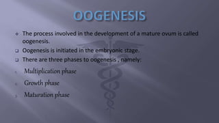
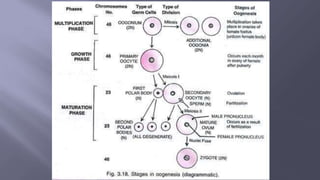
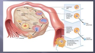
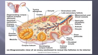
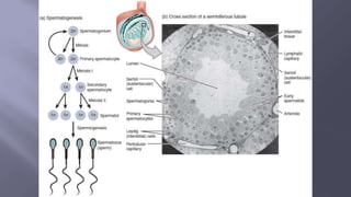
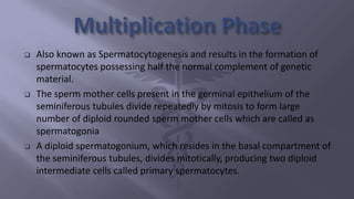
 Gametogenesis is a biological process by
which diploid or haploid precursor cells
undergo cell division and differentiation to
form mature haploid gametes.
 Depending on the biological life cycle of the
organism, gametogenesis occurs by meiotic
division of diploid gametocytes into various
gametes.
 At the end of meiosis, four haploid cells have been produced,
but the cells are not yet gametes.
 The cells need to develop before they become mature
gametes capable of fertilization.
 The development of haploid cells into gametes is called
gametogenesis.
DEFINATION :
 The process involved in the maturation of the two highly
specialized cells , spermatozoon in male and ovum in female
before they unite to form zygote, is called gametogenesis.
 In human males , the process that produces
mature sperm cells is called
Spermatogenesis.During this process,
sperm cells grow a tail and gain the ability to
“swim”.
 In human females, the process that produces
mature eggs is called Oogenesis. Just one
egg is produced from the four haploid cells
that result from meiosis.
 The process involved in the development of a mature ovum is called
oogenesis.
 Oogenesis is initiated in the embryonic stage.
 There are three phases to oogenesis , namely:
1. Multiplication phase
2. Growth phase
3. Maturation phase
A MATURE OVARIAN FOLLICLE
 The primitive germ cells take their origin from the yolk sac at about the
end of 3rd week and their migration to the developing gonadal ridge is
completed around about the end of 4th week.
 During foetal development, certain cells in the germinal epithelium of
the ovary are larger than others and also have larger nuclei.
 These cells undergo mitotic divisions, producing undifferentiated germ
cells called oogonia, or egg mother cells, in the ovary.
 The number of oogonia reaches its maximum at 20th week,numbering
about 7 million.
 The oogonia have 2n, or diploid, number of chromosomes,
46 in humans.
 The oogonia multiply by mitotic divisions, and project into
the stroma as a cord, the egg tube of Pfluger, which later
becomes a round mass, the egg nest.
 One cell in the egg nest grows and becomes the primary
oocyte. It is the future ovum.
 Other oogonia in the nest lose the potentiality to become
primary oocytes and form a regular layer, the follicular
epithelium, round the primary oocyte to protect and
nourish it.
Haematoxylin & Eosin staining of sections of
human gonads ,quiescent oogonia are
indicated by arrowheads.
 At birth, there is no more mitotic division and all the oogonia are
replaced by primary oocytes which have finished the prophase of
the first meiotic division and remain in resting phase (dictyotene
stage) between prophase and metaphase.
 Total number of primary oocytes at birth is estimated to be about
2 million.
 The primary oocytes do not finish the first meiotic division until
puberty is reached.
 At puberty,some 400,000 primary oocytes are left behind, the rest
being atretic. Out of these, some 400 are likely to ovulate during
the entire reproductive period.
 The growth phase of the oogenesis is comparatively longer than the
growth phase of the spermatogenesis , extending over many years
until puberty is reached.
 In this, the oogonium nurtures into bigger primary oocytes.
 After this, each primary oocyte gets surrounded by a granulosa cells
layer to create primary follicle.
 Later, a large number of follicles get debased during the duration from
birth to puberty. Therefore, at puberty around 60,000 to 80,000
primary follicles can be found in each ovary.
 In the primary oocyte, large amount of fats and proteins
becomes accumulated in the form of yolk.
 The cytoplasm of the oocyte becomes rich in RNA, DNA,
ATP and enzymes. Moreover, the mitochondria, Golgi
apparatus, ribosomes, etc.,become concentrated in the
cytoplasm of the oocyte.
 During the growth phase, tremendous changes also occur
in the nucleus of the primary oocyte.
 The nucleus becomes large due to the increased amount
of the nucleoplasm and is called germinal vesicle.
 The essence of maturation is reduction of the number of
chromosomes to half.
 Before the onset of first meiotic division, the primary oocytes double
its DNA by replication, so they contain double the amount of normal
protein content.
 The first stage of maturation occurs with full maturation of the ovarian
follicle just prior to ovulation but the final maturation occurs only after
fertilization.
 The primary oocyte undergoes first meiotic division giving rise to
secondary oocyte and one polar body.
 The two are of unequal size,
the secondary oocyte contains
haploid number of chromosomes(23, X),
but nearly all the cytoplasm and the
small polar body also contains half
of the chromosomes(23, X) but
with scanty cytoplasm.
 The secondary oocyte completes the second meiotic division (homotypical) only after
fertilization by the sperm in the Fallopian tube and results in the formation of two
unequal daughter cells, each possessing 23 chromosomes (23, X), the larger one is called
the mature ovum and the smaller one is the second polar body containing the same
number of chromosomes.
 In the absence of fertilization, the secondary oocyte
does not complete the second meiotic division and
degenerates as such.
 A fully mature ovum is the largest cell in the body
and is about 130 microns in diameter. It consists
of cytoplasm and a nucleus with its nucleolus
which is eccentric in position and contains 23
chromosomes (23, X).
 During fertilization, the nucleus is converted into
a female pronucleus.
A human egg cell with surrounding corona radiata
 Spermatogenesis is the process of formation of haploid, microscopic
and motile gametes, called spermatozoa, from the diploid
spermatogonia of the testis of male organism.
 Spermatozoa are produced in the seminiferous tubules of the testes.
 There are three phases to Spermatogenesis, namely:
1. Multiplication phase
2. Growth phase
3. Maturation phase
 Also known as Spermatocytogenesis and results in the formation of
spermatocytes possessing half the normal complement of genetic
material.
 The sperm mother cells present in the germinal epithelium of the
seminiferous tubules divide repeatedly by mitosis to form large
number of diploid rounded sperm mother cells which are called as
spermatogonia
 A diploid spermatogonium, which resides in the basal compartment of
the seminiferous tubules, divides mitotically, producing two diploid
intermediate cells called primary spermatocytes.
 Some of these sex cells move towards the lumen of
seminiferous tubules and enter the growth phase.
These cells are called primary spermatocytes.
 The primary spermatocytes are diploid and contain
(44 + XY) chromosomes.
 Some of the sex cells produced by the division of
spermatogonia remain in their original condition
and continue to divide giving rise to primary
spermatocytes. Such cells are known as stem cells.
 During phase of growth, the primary spermatocyte enlarges in size and
prepares to undergo maturation division.
 Some spermatogonia either due to growth or due to food storage
become 2 or 3 times of their original size.
 The remaining spermatogonia remain in the seminiferous tubules in
the form of reserved stock.
 Now these primary spermatocytes enter into the next phase namely,
maturation phase.
 The spermatogonia,which in turn,differentiate into primary
spermatocytes remain in the stage of prophase of the first meiotic
division for a long time (about 16 days).
 With the completion of the first meiotic division,two secondary
spermatocytes are formed having equal share of cytoplasm and
haploid number of chromosomes either 23, X or 23, Y.
 Then immediately follows the second meiotic division (homotypical)
with the formation of four spermatids, each containing haploid
number of chromosomes, two with 23, X and two with 23, Y.
 Immediately after their formation, extensive morphological differentiation
of the spermatids occurs without further cell division to convert them into
spermatozoa.
 The process is called spermiogenesis.In man, the time required for a
spermatogonium to develop into a mature spermatozoon is about 61 days.
Structure of a mature spermatozoon:
 It has got two parts, a head and a tail.
 The head consistsprincipally of the condensed nucleus and acrosomal
cap.Acrosome is rich in enzymes.
 The tail is divided into four zones — the neck, the middle piece, the principal
piece and the end piece.
GAMETOGENESIShhdhdhdhdhdhhdfhddhdhdhhdhd

GAMETOGENESIShhdhdhdhdhdhhdfhddhdhdhhdhd

  • 2.
     Gametogenesis isa biological process by which diploid or haploid precursor cells undergo cell division and differentiation to form mature haploid gametes.  Depending on the biological life cycle of the organism, gametogenesis occurs by meiotic division of diploid gametocytes into various gametes.
  • 3.
     At theend of meiosis, four haploid cells have been produced, but the cells are not yet gametes.  The cells need to develop before they become mature gametes capable of fertilization.  The development of haploid cells into gametes is called gametogenesis. DEFINATION :  The process involved in the maturation of the two highly specialized cells , spermatozoon in male and ovum in female before they unite to form zygote, is called gametogenesis.
  • 4.
     In humanmales , the process that produces mature sperm cells is called Spermatogenesis.During this process, sperm cells grow a tail and gain the ability to “swim”.  In human females, the process that produces mature eggs is called Oogenesis. Just one egg is produced from the four haploid cells that result from meiosis.
  • 6.
     The processinvolved in the development of a mature ovum is called oogenesis.  Oogenesis is initiated in the embryonic stage.  There are three phases to oogenesis , namely: 1. Multiplication phase 2. Growth phase 3. Maturation phase
  • 8.
  • 9.
     The primitivegerm cells take their origin from the yolk sac at about the end of 3rd week and their migration to the developing gonadal ridge is completed around about the end of 4th week.  During foetal development, certain cells in the germinal epithelium of the ovary are larger than others and also have larger nuclei.  These cells undergo mitotic divisions, producing undifferentiated germ cells called oogonia, or egg mother cells, in the ovary.  The number of oogonia reaches its maximum at 20th week,numbering about 7 million.
  • 10.
     The oogoniahave 2n, or diploid, number of chromosomes, 46 in humans.  The oogonia multiply by mitotic divisions, and project into the stroma as a cord, the egg tube of Pfluger, which later becomes a round mass, the egg nest.  One cell in the egg nest grows and becomes the primary oocyte. It is the future ovum.  Other oogonia in the nest lose the potentiality to become primary oocytes and form a regular layer, the follicular epithelium, round the primary oocyte to protect and nourish it.
  • 11.
    Haematoxylin & Eosinstaining of sections of human gonads ,quiescent oogonia are indicated by arrowheads.
  • 12.
     At birth,there is no more mitotic division and all the oogonia are replaced by primary oocytes which have finished the prophase of the first meiotic division and remain in resting phase (dictyotene stage) between prophase and metaphase.  Total number of primary oocytes at birth is estimated to be about 2 million.  The primary oocytes do not finish the first meiotic division until puberty is reached.  At puberty,some 400,000 primary oocytes are left behind, the rest being atretic. Out of these, some 400 are likely to ovulate during the entire reproductive period.
  • 13.
     The growthphase of the oogenesis is comparatively longer than the growth phase of the spermatogenesis , extending over many years until puberty is reached.  In this, the oogonium nurtures into bigger primary oocytes.  After this, each primary oocyte gets surrounded by a granulosa cells layer to create primary follicle.  Later, a large number of follicles get debased during the duration from birth to puberty. Therefore, at puberty around 60,000 to 80,000 primary follicles can be found in each ovary.
  • 15.
     In theprimary oocyte, large amount of fats and proteins becomes accumulated in the form of yolk.  The cytoplasm of the oocyte becomes rich in RNA, DNA, ATP and enzymes. Moreover, the mitochondria, Golgi apparatus, ribosomes, etc.,become concentrated in the cytoplasm of the oocyte.  During the growth phase, tremendous changes also occur in the nucleus of the primary oocyte.  The nucleus becomes large due to the increased amount of the nucleoplasm and is called germinal vesicle.
  • 16.
     The essenceof maturation is reduction of the number of chromosomes to half.  Before the onset of first meiotic division, the primary oocytes double its DNA by replication, so they contain double the amount of normal protein content.  The first stage of maturation occurs with full maturation of the ovarian follicle just prior to ovulation but the final maturation occurs only after fertilization.
  • 17.
     The primaryoocyte undergoes first meiotic division giving rise to secondary oocyte and one polar body.  The two are of unequal size, the secondary oocyte contains haploid number of chromosomes(23, X), but nearly all the cytoplasm and the small polar body also contains half of the chromosomes(23, X) but with scanty cytoplasm.
  • 18.
     The secondaryoocyte completes the second meiotic division (homotypical) only after fertilization by the sperm in the Fallopian tube and results in the formation of two unequal daughter cells, each possessing 23 chromosomes (23, X), the larger one is called the mature ovum and the smaller one is the second polar body containing the same number of chromosomes.  In the absence of fertilization, the secondary oocyte does not complete the second meiotic division and degenerates as such.
  • 21.
     A fullymature ovum is the largest cell in the body and is about 130 microns in diameter. It consists of cytoplasm and a nucleus with its nucleolus which is eccentric in position and contains 23 chromosomes (23, X).  During fertilization, the nucleus is converted into a female pronucleus.
  • 22.
    A human eggcell with surrounding corona radiata
  • 23.
     Spermatogenesis isthe process of formation of haploid, microscopic and motile gametes, called spermatozoa, from the diploid spermatogonia of the testis of male organism.  Spermatozoa are produced in the seminiferous tubules of the testes.  There are three phases to Spermatogenesis, namely: 1. Multiplication phase 2. Growth phase 3. Maturation phase
  • 25.
     Also knownas Spermatocytogenesis and results in the formation of spermatocytes possessing half the normal complement of genetic material.  The sperm mother cells present in the germinal epithelium of the seminiferous tubules divide repeatedly by mitosis to form large number of diploid rounded sperm mother cells which are called as spermatogonia  A diploid spermatogonium, which resides in the basal compartment of the seminiferous tubules, divides mitotically, producing two diploid intermediate cells called primary spermatocytes.
  • 26.
     Some ofthese sex cells move towards the lumen of seminiferous tubules and enter the growth phase. These cells are called primary spermatocytes.  The primary spermatocytes are diploid and contain (44 + XY) chromosomes.  Some of the sex cells produced by the division of spermatogonia remain in their original condition and continue to divide giving rise to primary spermatocytes. Such cells are known as stem cells.
  • 27.
     During phaseof growth, the primary spermatocyte enlarges in size and prepares to undergo maturation division.  Some spermatogonia either due to growth or due to food storage become 2 or 3 times of their original size.  The remaining spermatogonia remain in the seminiferous tubules in the form of reserved stock.  Now these primary spermatocytes enter into the next phase namely, maturation phase.
  • 28.
     The spermatogonia,whichin turn,differentiate into primary spermatocytes remain in the stage of prophase of the first meiotic division for a long time (about 16 days).  With the completion of the first meiotic division,two secondary spermatocytes are formed having equal share of cytoplasm and haploid number of chromosomes either 23, X or 23, Y.  Then immediately follows the second meiotic division (homotypical) with the formation of four spermatids, each containing haploid number of chromosomes, two with 23, X and two with 23, Y.
  • 29.
     Immediately aftertheir formation, extensive morphological differentiation of the spermatids occurs without further cell division to convert them into spermatozoa.  The process is called spermiogenesis.In man, the time required for a spermatogonium to develop into a mature spermatozoon is about 61 days. Structure of a mature spermatozoon:  It has got two parts, a head and a tail.  The head consistsprincipally of the condensed nucleus and acrosomal cap.Acrosome is rich in enzymes.  The tail is divided into four zones — the neck, the middle piece, the principal piece and the end piece.