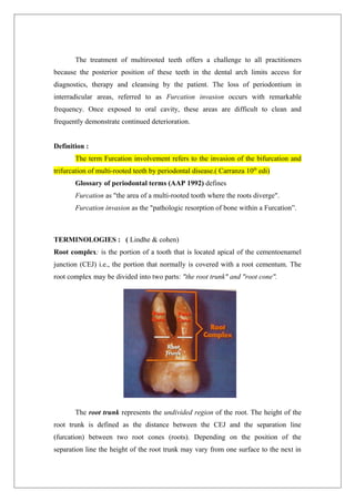
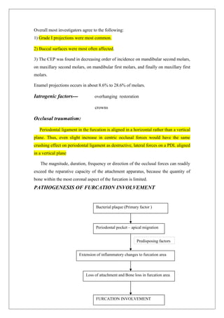
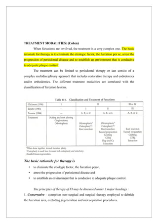
1. COLLEGE OF DENTAL SCIENCES
DAVANGERE
PRESENTED BY,
DEPT. OF PERIODONTICS
CONTENTS
SEMINAR ON
FURCATION INVOLVEMENT
2. Introduction
Definition
Anatomy of root
Morphology of root complex in multirooted teeth
Prevalence of Furcation Involvement
Etiology of Furcation Involvement
Pathogenesis of Furcation Involvement
Classification of Furcation Involvement
Diagnosis of Furcation Involvement
- Clinical examination
- Radiographic examination
- Intraoperative measurements
Differential Diagnosis
Treatment Modalities
Factors affecting clinical outcome
Prognosis of treated molar furcations
Conclusion
References
INTRODUCTION
3. The treatment of multirooted teeth offers a challenge to all practitioners
because the posterior position of these teeth in the dental arch limits access for
diagnostics, therapy and cleansing by the patient. The loss of periodontium in
interradicular areas, referred to as Furcation invasion occurs with remarkable
frequency. Once exposed to oral cavity, these areas are difficult to clean and
frequently demonstrate continued deterioration.
Definition :
The term Furcation involvement refers to the invasion of the bifurcation and
trifurcation of multi-rooted teeth by periodontal disease.( Carranza 10th
edi)
Glossary of periodontal terms (AAP 1992) defines
Furcation as "the area of a multi-rooted tooth where the roots diverge".
Furcation invasion as the "pathologic resorption of bone within a Furcation”.
TERMINOLOGIES : ( Lindhe & cohen)
Root complex: is the portion of a tooth that is located apical of the cementoenamel
junction (CEJ) i.e., the portion that normally is covered with a root cementum. The
root complex may be divided into two parts: "the root trunk" and "root cone".
The root trunk represents the undivided region of the root. The height of the
root trunk is defined as the distance between the CEJ and the separation line
(furcation) between two root cones (roots). Depending on the position of the
separation line the height of the root trunk may vary from one surface to the next in
4. one given molar or premolar.
The root cone is included in the divided region of the root complex. The root
cone (root) may vary in size and position and may at certain levels be connected to or
separated from other root cones. Two or more root cones make up the furcated region
of the root complex.
The furcation is the area located between individual root cones and it refers to
the anatomic area of a mulrirooted tooth where the roots divide or diverge from the
common root trunk. The Furcation can be divided into three parts.
The roof which is the base of the root trunk and may contains bifurcation
ridge.
The flute - usually concave or grooving of the root trunk, extending from the
cervical line and blending into the actual furca.
The inter-radicular area or the area of the root separation or furcation chamber,
bordered coronally by the base of the root trunk (or roof), laterally by the roots and
apically by the remaining periodontium. It contains the interradicular osseous
septum and pcriodontal ligament.
Furcation entrance: the transitional area between the undivided and the
divided part of the root.
Furcation fornix : the roof of the furcation.
Degree of separation : the angle of separation between two roots (cones).
Divergence : is the distance between two roots; this distance normally
increases in apical direction.
5. Coefficient of separation : the length of the root cones in relation to the length of the
root complex.
MORPHOLOGY OF THE ROOT COMPLEX OF MULTIROOTED TEETH
Maxillary molars :
The three furcation entrances of the maxillary first & second molars vary in
width and are positioned at varying distances apical to the CEJ. As a rule, the first
molar has a shorter root trunk than the second molar. In the first molar the mesial
furcation entrance is located about 3 mm from the CEJ, while the buccal is 3.5 mm
and the distal entrance about 5 mm apical of CEJ (Abrams & Trachtenberg 1974,
Rosenberg 1988). This implies that the furcation fornix is inclined; in the mesiodistal
plane the fornix is comparatively close to CEJ at the mesial but closer to apex at the
distal surface. The buccal furcation entrance is narrower than its distal & mesial
counterparts.
The degree of separation between the roots and their divergence decrease from
the first to the second, and from the second to the third maxillary molar.
Maxillary premolars:
In about 40% of cases the maxillary first premolars have two root cones; one
buccal and one palatal and hence a mesiodistal furcation. A concavity (about 0.5 mm
deep) is often present in the furcation aspect of the buccal root. The furcation is in
many cases located in the middle or in the apical third of the root complex. The mean
distance between CEJ and the furcation entrance is about 8 mm. The width of the
furcation entrance is about 0.7 mm.
6. Mandibular molars:
The mandibular first molar is larger than the second molar, which in turn is
larger than the third molar. In the first and second molars the root complex almost
always includes two root cones, one mesial and one distal. The mesial root is larger
than the distal. The mesial root has a position which is mainly vertical while the distal
root projects distally.
The root trunk of the first molar is often shorter than the trunk of the second
molar. The furcation entrance of the mandibular first molar similar to those of the
maxillary first molar are located at
different distances from the CEJ. Thus the
lingual entrance is frequently found more
apical of CEJ (> 4 mm) than the buccal
entrance (> 3 mm). Thus, the furcation
fornix is inclined in the buccolingual
direction.
The buccal furcation entrance is often < 0.75 mm wide while the lingual
entrance is > 0.75 mm in most cases (Bower 1979). The degree of separation &
divergence between the roots decreases from the first to the third molar.
Other teeth:
Furcations may be present also in teeth which normally have only one root. In
fact, two rooted incisors, canines and mandibular premolars may exist. Occasionally
three rooted maxillary premolars and three rooted mandibular molars can be found.
CLASSIFICATION :
Periodontal involvement of the furcation regions has been classified based on
degree & severity of involvement.
Glickman (1953) was one of the first to classify furcation invasions based on the
degree of lateral penetration of the periodontal destruction under the roof of the
furcation. He divided them into the following four grades:
Grade-I: When there is soft-tissue lesion or pocket extending into the flute of the
furcation, but the inter-radicular bone is intact. The pocket is suprabony. This
involvement of the periodontium in the furcation area is without manifested
radiographic evidence of bone loss.
7. Grade-II: Loss of inter-radicular bone
& pocket formation of varying depths
into the furcation but not completely
through the opposite side of the tooth.
This is radiographic evidence of
involvement.
Grade-III: Complete loss of inter-
radicular bone with radiographic
evidence presenting a small triangular radiolucency at the furcation area. There is a
pocket formation that is completely probable to the opposite side of the tooth.
However, the furcation is not visible clinically.
Grade-lV: Same features as those of Grade III except that loss of periodontal
attachment & gingival recession has made the furcation clearly visible to a clinical
examination.
Hamp, Nyman and Lindhe (1975) described a classification which defined the
horizontal extent of the furcation involvement.
Class 1: represents horizontal attachment loss of less than 3 mm within the furcation
involvement.
Class-11: represents horizontal loss greater than 3 mm but not encompassing the total
width of the furcation.
Class-III: denotes horizontal
through and through
destruction.
Lindhe (1983), Hamp and
8. Nyman (1989), Nyman & Lindhe (1997)
Horizontal classification:
Degree I / Grade I (Initial): Loss of inter radicular bone less than or equal to one third.
Degree II / Grade-II (Partial): Loss of inter radicular bone greater than one third but
not through and through.
Degree III / Grade-III(Total):Through& through loss of interradicular bone.
Eskow and Kapin (1984) classified vertical loss in thirds of inter radicular loss
Tarnow & Fletcher (1984) classified vertical attachment loss in millimeters as
follows:
Subclass A: Vertical destruction to one
third of the total inter radicular height
(1 to 3 mm).
Subclass B: Vertical destruction
reaching two thirds of the inter
radicular height (4 to 6 mm).
Subclass C: Inter radicular osseous
destruction into or beyond the apical third (> 7 mm).
Easley & Drennan’s classification (1969): This classification system provides
information relative to the vertical
component of furcation involvement. Class I
furcation involvement - there is attachment
loss that involves the root flutings, but there
is no horizontal component to the
involvement. In this classification system.
Class II and III furcations are separated into
subtypes 1 and 2 on the basis of the
configuration of the alveolar bone at the
entrance to the furcation. Horizontal
resorption into the furca is subtype 1,
whereas subtype 2 indicates a significant
9. vertical component to the defect. This separation contributes to treatment planning as
a vertical component may contribute to the ability to "fill" the deformity.
DIAGNOSIS OF FURCATION INVOLVEMENT : (Muller 1996)
A) CLINICAL EXAMINATION
Signs and symptoms of periodontal inflammation may be present to varying extent,
such as :
Redness
Swelling - Periodontal abscess
Increased temperature
Pain
Loss of function – tooth may be perceived elongated and mobile
Increased bleeding tendency
Suppuration
Clinical probing – using a curved graduated periodontal probe (Naber’s
probe), an explorer or a small curette. Furcation can best be evaluated with a curved
Nabers #2 probe. Also there are calibrated furcation probe ZA - 2 with calibrations in
2 mm increments, ZA - 3 with calibrations in 3 -mm increaments. Straight probes
detect only vertical attachment loss and are not accessible for determining horizontal
attachment loss.
Vertical attachment loss on the adjacent roots should be probed at the
furcation line angle of each root, angling the probe somewhat into the furca. A
probing depth deeper than the length of the root trunk should be immediately
evaluated. Horizontal attachment loss is measured from an imaginary tangent passing
through or applied at both roots or root cones at the level of the furcation entrance.
The buccal furcation of the maxillary molars and the buccal and lingual furcas
of the mandibular molars are normally accessible for examination by clinical probing.
The clinical examination of furcas on the approximal tooth surfaces may be more
difficult when neighboring teeth are present, especially if the contact area between the
teeth is large. This is particularly the case in maxillary molars. The mesial furcation
opens about 2/3rd of the way toward the palate and is easily probed from the palatal
aspect of the tooth. While the furcation in the distal surface, located midway bucco-
lingually is probed form either the buccal or the palatal aspect.
10. When periodontitis affected furcations should be diagnosed, a somewhat
different approach is required. Since the furcation entrance lies subgingivally in most
cases, the operator has to search for a concavity at the respective site and then to
penetrate into the area between the roots. Consequently, a pressure controlled,
flexible, plastic probe (Hunter et al. 1992) was shown to hamper a correct furcation
assessment especially in case of a through-and-through involvement (Kim et al. 1996,
Eickholz & Kim 1998). On the other hand, bleeding on probing, which has been
shown to negatively affect the reproducibility of furcation measurements
(Steinbrenner et al. 1997), is expected to be provoked more frequently with the
‘‘searching probe’’ trying to gain access to this area. Since the pocket has a vertical
and horizontal component, conventional straight probes may also underestimate the
severity of the furcation involvement.
The clinical examination of maxillary premolars is often difficult due to
limited access for probing. It may not always be possible to identify the presence and
the degree of furcation involvement in such teeth until flap is raised in an explorative
(surgical) procedure in the area.
B) RADIOGRAPHIC EXAMINATION ( Lindhe , Carranza, & Muller )
In practice, periapical or vertical bitewing exposures in combination with a long
cone as well as panoramic tomograms are commonly used for the diagnosis of the
periodontal condition. An initial furcal lesion, in particular in maxillary molars (and
correspondingly periapical lesions), may actually be better uncovered on a panoramic
tomogram (Rohlin et al. 1989), which frequently images the central plane of the
alveolar bone including the furcation area, whereas structures not in the interesting
plane are blurred.
Glickman found that clinically significant tissue changes in the furcation often
are not detected by radiographs.
X-rays are virtually the only diagnostic tool to reliably determine the height of
the alveolar bone crest in relation to the tooth length. In addition, limited but
important information may be gained with regard to the anatomy and topography of
the root complex, i.e., number and form of roots, separation degree, divergence of
roots etc., as well the neighbouring anatomical structures and teeth. In the radiographs
the location of the interdental bone as well as the bone level within the root complex
should be examined. Radiographs may be taken with probes, explores or curettes in
11. place and a more accurate assessment of the extent of the lesion can be determined.
On x-ray films of maxillary molars, a small, triangular, radiolucent shadow is
sometimes seen over the mesial or distal roots in the proximal furcation areas, which
has been called furcation arrow. The association of this image with deep grade II or
grade III FI was significant compared with uninvolved furcations, but the arrow was
not seen in more than half of the sites with a deep grade II FI and in slightly less than
half of grade III sites. However, a radiographic image of a furcation arrow should
induce the periodontist to carefully further assess the severity of furcation invasions
clinically and, if need be, intra-operatively.
To assist in the radiographic detection of FI, the following three diagnostic
criteria suggested: (Carranza 10th
ed)
1) The slightest radiographic change in the furcation area should be investigated
clinically, especially if there is bone loss on adjacent roots.
2) Diminished radiodensity in the furcation area in which outlines of bony trabeculae
are visible suggests furcation involvement.
3) Whenever there is marked bone loss in relation to a single molar root, it may be
assumed that the furcation is also involved.
Radiographs alone do not detect FI with any predictable accuracy and that
probing the furcation area is necessary to confirm the presence & severity of FI.
With the aid of high-resolution computed tomography a more detailed, 3-
dimensional interpretation of bony lesions and tooth structures especially in the
furcation area seems possible (Fuhrmann et al. 1997). Direct axial scanning parallel
to the occlusal plane has been recommended for detection of infrabony pockets,
furcation involvements and buccal or lingual bone dehiscences (Fuhrmann et al.
1997). A serious drawback of computed tomography and other radiographic
techniques is the exposure with ionizing radiation.
Evidence for healing in furcations may be provided by computer- assisted
densitometric image analysis (CADIA) as well as qualitative and quantitative digital
subtraction radiography
C) INTRAOPERATIVE MEASUREMENTS :(Muller)
Comprehensive information with regard to the morphology of a periodontal
defect may be further achieved by intrasurgical inspection. Not infrequently, a
definite conclusion for one or the other treatment modality can therefore only be
12. drawn intra-operatively. After surgically opening of the defect, removal of the
granulation tissue and careful debridement of the root surfaces within and outside the
interradicular projection, the actual extent of periodontal destruction may be visible.
DIFFERENTIAL DIAGNOSIS: (Lindhe)
The treatment of a furcation involved tooth should not be initiated until a proper
differential diagnosis of the lesion has been made.
1. Problems originating from the root canal
Pulpal pathosis may at times cause a lesion in the periodontal tissues of the furcation.
The radiographic appearance of such a defect may have some features in common
with a plaque associated furcation lesion. In order to difference between the two
lesions the vitality of the affected tooth must always be tested. If the tooth is vital, a
plaque-associated lesion should be suspected. If the tooth is non vital, the furcation
involvement may have an endodontic origin & in such case proper endodontic
treatment must always precede periodontal therapy. In fact, endodontic therapy may
resolve the inflammatory lesion, soft & hard tissue healing occur & the furcation
defect disappear. If signs of healing of a furcation defect fail to appear within 2
months following endodontic treatment, the furcation involvement is probably
associated with marginal periodontitis.
2. The result of occlusal overload - Trauma from occlusion :
Forces elicited by occlusal interferences eg; bruxers and clenchers may cause
inflammation and tissue destruction or adaptations within inter radicular area of a
multi-rooted tooth. In such a tooth radiolucency may be seen in the radiograph of the
root complex. The tooth may exhibit increased mobility. Probing however fails to
detect in involvement of the furcation. In this particular situation, occlusal adjustment
must always precede periodontal therapy. If the defects seen within the root complex
are of "occlusal" origin, the tooth will become stabilized and the defects disappear
within weeks following correction of the occlusal overload.
PREVALENCE OF FURCATION INVOLVEMENT: (MARCELLO CATTABRIGA)
Sparse information is available regarding the prevalence of naturally occurring
involvement of the molar furcation area in the general population in epidemiological
surveys. Most of the available data are derived from studies based on observations
performed in dry skulls.
13. In the study by Volkansky and Cleatan Jones on 43 dry mandibles of South
African Bantu People, 30.9% of molar teeth present had furcation involvement. Tal
examined (1982) 100 dry mandibles from South African skulls and found that 85.4%
of mandibular molars presented osseous resorption in the furcation area. In a
subsequent study on mandibular molars, Tal and Lemmer (1982) confirmed the
finding that moderate and severe involvements predominant in older adults, first
molars being more affected than second molars.
Bjorn & Hjort (1982) assessed longitudinally the radiographic prevalence,
degree and development of bone destruction in mandibular molar furcations in a
sample of 221 factory workers observed over a period of 13 years. The prevalence of
FI steadily increased from an initial value of 18% to 32% at the end of the observation
period. Third and second molars had higher frequencies of advanced destruction than
first molars. Therefore, the prevalence of involvement in the furcation area in
maxillary & mandibular molars range from 25% to 52% and from 16% to 35%
respectively. Svadstrom and Wennstrm (1996) studied in detail the prevalence of FI in
a group of 222 patients referred for periodontal treatment. They reported that, from
the age of 30 years onward, about 50% of molars in the maxilla show at least 1
furcation site with deep involvement, while in the mandible a similar prevalence was
first observed after the age of 40. Furcation lesions were more prevalent in the maxilla
than in the mandible and Periodontal destruction was most frequently observed at the
distal aspect of the first and second maxillary molars (53% and 35% respectively). In
the mandible the buccal and lingual entrances of furcations were affected with similar
frequencies.
Furcation involvement is more frequently detected in smokers (72%) than in
nonsmokers (36%). The calculated odds ratio for a smoker to have furcation
involvement in one molar is 4.6. It has also been demonstrated that molars with
crowns or proximal restorations have significantly higher percentage of FI (52-63%)
compared with molars without restorations (39%).
ETIOLOGY OF FURCATION INVOLVEMENT: (Abdallah et al)
Primary factor – Bacterial Plaque
Predisposing factors:
a. Anatomic factors –
b. Iatrogenic factors – overhanging reatorations
14. C. Local developmental anamolies
d. Occlusal trauma.
Anatomic factors :
Local anatomic factors are important to be identified because they not
only affect the development and treatment of furcation involvement (FI), but also
the prognosis of the therapy.
1. Root trunk length
2. Interradicular dimension
3. Root length
4. Root form
5. Anatomy of furcation :
a. Root concavities
b. Bifurcation ridges
c. Accessory pulp canals
d. Cervical Enamel pearls and projection
e. Location and diameter of furcation entrance
1. Root trunk length: This is the key factor in both the development and treatment of
furcation involvement (FI). When the root trunk is short, the less attachment needs to
be lost before the furcation is involved early in the disease process. Once the furcation
is exposed, teeths with short root trunks may be more accessible to therapy. When the
root trunk is long, the furcation will be invaded later but will be more difficult to
reach and instrument.
First molars generally have shorter root trunks than second molars. This may
account for their having a higher prevalence of furcation involvements than second
molars.
2. Interradicular dimension : The degree of separation of roots is also important.
Closely approximated or fused roots can preclude adequate instrumentation during
scaling, root planing and surgery. Teeth with widely separated roots present more
treatment options and are readily treated. First molar furcations are frequently wider
than those of second molars because of greater spread of their roots and hence allow
for better access during treatment.
Root length:
Directly related to the quantity of attachment supporting The tooth
15. Root form:
The mesial root of mandibular first and second molars and
mesiobuccal root of maxillary first molar are typically curved on distal side.
In addition, the distal aspect of this root is usually heavilly fluted.
Increase the potential for root perforation during endodontic therapy
complicates post placement during restoration
Increase the incidence of vertical root fracture.
3. Anatomy of furcation :
a. Root concavities : Furcation root surface morphology has been extensively studied
by many authors. Bower (1979) found that all the root surfaces facing the furcation
exhibit some degree of concavity or depression in an occluso-apical direction.
Mandibular molars had deeper concavity in relation to the mesial root (mean
concavity 0.7 mm) than in the distal root (mean concavity 0.5 mm). The maxillary
first molar had deepest concavity in the furcal aspect of the mesiobuccal root (mean
concavity 0.3 mm); 94% of mesiobuccal roots; 31% of distobuccal roots & 17%) of
palatal roots.
In the same study, he noted that the furcation concavities are covered by more
cementum than the adjacent convexities. This may have clinical significance due to
cementum's ability to absorb and/or adsorb toxic bacterial products such as
endotoxins when altered by the inflammatory process. Incomplete removal of this
diseased thicker cementum would thus discourage intimate union of healthy
fibroblasts and epithelial cells after treatment, resulting in persistence of pathological
pockets.
This may make instrumentation for calculus removal and root planing almost
impossible. After surgical exposure chances for complete removal improve; in narrow
furcations, ultrasonic instrumentation is more effective than hand instrumentation.
16. b. Bifurcation ridges: Everett (1958) described a ridge on mandibular molars that
originated from the mesial surface of the distal root, ran across the bifurcation and
ended high up on the mesial root, where it blended into the commonly occurring
concavity of the distal surface of the mesial root. He called it an "intermediate
bifurcation ridge" and found an overall incidence of 73% Burch & Hulen (1974) in
another study, found an incidence of 76%. Histologic examination revealed that the
ridge is formed mostly of cementum. The presence of an intermediate bifurcation
ridge may demand special consideration in determining the method of treatment of a
periodontal lesion within the bifurcation areas of the mandibular molars, as this
anatomical structure creates niches where plaque can accumulate undisturbed when
the furcation is exposed to the oral environment.
Buccal and lingual ridges were found in 63% of the mandibular molars. These
ridges resulted in the roof of the furcation being located more coronally than the
entrances.
C .Accessory pulp canals : The presence of accessory pulpal canals in the furcation
area may extend pulpal inflammation to the furcation. The high percentage of molar
teeth with patent accessory canal opening into the furcation suggests that pulpal
disease could be an initiating cofactor in the development of furcation involvement.
Several cases have been reported of clinically & radiographically detectable furcation
involvements that were attributable to pulpal pathosis. Furcation involvement thus
becomes a combined endodontic periodontic defect. Accessory canals connecting the
pulp chamber floor to the furcation have been found in 36% maxillary first molars,
12% of maxillary second molars, 32% of mandibular first molars and 24% of
mandibular second molars.
D .Location and diameter of furcation entrance: The distal maxillary is the most
frequently involved as corroborated by Klavan (1975).
The furcation entrance diameter of first permanent molar teeth was extensively
studied by Bower (1979). In a sample of 114 maxillary & 103 mandibular teeth, he
found that 81% of furcation entrance diameters was 1.0 mm or less, and 58% of the
diameter was 1.75 mm. Overall furcation entrance diameter was smaller than the
blade face width of commonly used periodontal curettes in 58% of the furcation
examined.
17. Bower (1979) noted that the buccal furcation entrance diameter of the
maxillary first molar teeth examined was smaller than either the mesiopalatal or
distopalatal. Similarly the buccal entrance diameter in the mandibular first molar
teeth examined was smaller than the lingual.
Local developmental anamolies:
American Academy of Periodontology's (AAP) 1986 Glossary of periodontal terms,
defines an enamel pearl as "a small focal mass of enamel formed apical to the CEJ",
while enamel projection is "an extension of the cervical enamel margin either toward
or into the root furcation area ".
.
Enamel pearls differ from enamel projections in that they are isolated islands
of ectopic enamel rather than extensions of coronal enamel and also differ from
enamelomas in that enamelomas are not attached to the root but are found in the
periodontal ligament.
Enamel pearls can be found on any lateral surface of the root. Cavanah (1965)
studied molars with enamel pearls and concluded that the only situation in which
enamel pearls may become contributors or predispose to periodontal breakdown
occurs when the inflammatory process contacts their coronal aspect. Therefore, the
closer the enamel pearl is to the CEJ, the greater is its chance of becoming a
contributory factor.
Cervical Enamel pearls and projection :
Master & Hoskins (1964) classified the CEP into the following grades
according to severity of the projections:
grade I: The enamel projection extends from CEJ of the tooth towards the
furcation entrance
grade II, The enamel projection approaches the entrance to the furcation. It
does not enter the furcation, and therefore no horizontal component is present.
grade III: The enamel projection extends horizontally into the furcation.
Master & Hoskins (1964) studied the incidence & prevalence of CEP on 474
extracted molars. They found that CEP occurred primarily on the buccal surfaces of
mandibular molars, 28.6% and 17% on maxillary molars, with highest incidence of
occurrence on the buccal surface of second mandibular molars. They also observed
the presence of CEP in more than 90% of isolated furca lesions of mandibular teeth.
18. Overall most investigators agree to the following:
1) Grade I projections were most common.
2) Buccal surfaces were most often affected.
3) The CEP was found in decreasing order of incidence on mandibular second molars,
on maxillary second molars, on mandibular first molars, and finally on maxillary first
molars.
Enamel projections occurs in about 8.6% to 28.6% of molars.
Iatrogenic factors--- overhanging restoration
crowns
Occlusal traumatism:
Periodontal ligament in the furcation is aligned in a horizontal rather than a vertical
plane. Thus, even slight increase in centric occlusal forces would have the same
crushing effect on periodontal ligament as destructive, lateral forces on a PDL aligned
in a vertical plane
The magnitude, duration, frequency or direction of the occlusal forces can readily
exceed the reparative capacity of the attachment apparatus, because the quantity of
bone within the most coronal aspect of the furcation is limited.
PATHOGENESIS OF FURCATION INVOLVEMENT
Predisposing factors
Periodontal pocket – apical migration
Extension of inflammatory changes to furcation area
Loss of attachment and Bone loss in furcation area
Bacterial plaque (Primary factor )
FURCATION INVOLVEMENT
19. TREATMENT MODALITIES: (Cohen)
When furcations are involved, the treatment is a very complex one. The basic
rationale for therapy is to eliminate the etiologic factor, the furcation per se, arrest the
progression of periodontal disease and to establish an environment that is conducive
to adequate plaque control.
The treatment can be limited to periodontal therapy or can consist of a
complex multidisciplinary approach that includes restorative therapy and endodontics
and/or orthodontics. The different treatment modalities are correlated with the
classification of furcation lesions.
The basic rationale for therapy is
• to eliminate the etiologic factor, the furcation perse,
• arrest the progression of periodontal disease and
• to establish an environment that is conducive to adequate plaque control.
The principles of therapy of FI may be discussed under 3 major headings :
1. Conservative – comprises non-surgical and surgical therapy employed to debride
the furcation area, excluding regeneration and root separation procedures.
20. 2. Resective – includes root resection, hemisection and tooth extraction
3. Regenerative – includes GTR, bone grafting
However the borderline between conservative and resective terms sometimes
does not lend itself to a sharp definition. Resective procedures must sometimes be
performed in order to attain a result which can eventually be considered more
conservative. For instance, tunnel preparation is an example of conservative therapy
carried out to avoid more radical and resective forms of treatment for class II and III
FI.
Three broad strategies of furcation therapy are:
I. Maintenance of the existing Furcation:
Scaling and root planing
Obstruction of the Furcation
II. Increasing access to the Furcation:
Gingivectoiny/Apical positioned flap
Odontoplasty .
Osteoplasty/ostectomy
III. Elimination of the Furcation
Root amputation/ Tooth resection
Bicuspidization
SCALING AND ROOT PLANING :
Plaque Gingivitis Periodontitis
Therefore, the removal of plaque and calculus by scaling and root planing and
the prevention of their recurrence by good patient oral hygiene is mandatory. It can be
closed or open.
I. Non- surgical (Closed) scaling and Root Planing: is sufficient for Grade I and
shallow Grade II furcations. But healing must result in a furcation morphology that is
optimal for good patient plaque control, otherwise other therapeutic methods must be
used. Closed scaling and root planing may be the treatment of choice if surgery is
contraindicated for medical or psychological reasons.
II. Surgical (Open) Scaling and Root planing: If sufficient subgingival access is not
possible with a closed approach, for furcated molars with deep lesions, then open
scaling using flap procedure such as modified Widman flap yields more effective
calculus removal with Furcation involvement. Thus replaced flap results in some
21. pocket reduction by formation of a long junctional epithelial adhesion.
Factors affecting the efficacy of SRP depend on:
Open or closed SRP
Experience of the operator
Instruments used
Bowers (1979) found that in 58% of upper and lower first molars, the furcation
entrance diameter is narrower (<0.75 mm) than the width of conventional periodontal
curette. Hence the use of curettes alone would result in inadequate debridement of
many furcation areas. The use of ultrasonic scalers is more effective than hand scaling
in close debridement of advanced furcations.
The various instruments that can be used are :
Furcation curettes
Mini Five curettes
Ultrasonic Furcation inserts
Rotary diamond burs
The large dimensions of conventional ultrasonic-tips inhibit entry into the
furcation in some cases. So many new designs of furcation tips are designed and
developed and were shown in vitro to be superior to conventional sonic/ultrasonic
inserts with greater accessibility and ease of instrumentation in furcation areas.
The intraoperative, careful re-assessment of the lesion within the furcation is by
no means dispensable. Based on intrasurgical findings, the periodontist may be forced
to thoroughly alter his or her treatment plan.
Adjunctive chemotherapeutic agents have been used locally. Agents such as
1% metronidazole gel
50mg/ml Tetracycline irrigation
Tetracycline impregnated fibers
Povidone iodine
The sporadic, uncontrolled local delivery of antibiotic substance did not
have any significant advantage.
22. Del Peloso Ribeiro E (2006) found that use of topically applied povidone-
iodine as an adjunct to subgingival instrumentation does not provide additional
benefits.
OBLITERATION OF THE FURCATION - OCCLUSIVE BARRIER
This involves the filling of advanced furcation defects with biocompatible
material to eliminate anatomic niches in which bacteria accumulate. These occlusive
barrier not only seal the furcation entrance from epithelial, bacterial and food debris
invasion, but enable easier home care due to less surface area of the Furcation.
The various materials tried are Zinc Phosphate cement, amalgam, Glass
ionomer cement (GIC) and resin ionomer cement. However, only GIC and resin
ionomer have been found to be effective.
Von Swol et al (1989) compared Zinc Phosphate cement, amalgam and GIC in
the treatment of surgically created furcation in non-human primates. Radiographs
taken at various time intervals demonstrated only slight radiolucent changes that were
less likely to progress with time when using GIC. They reported greatest
biocompatibility with GIC via histologic evaluation.
The potential advantage of using GIC as an occlusive barrier in the treatment
of Furcation defects has the following advantages :
GIC restoration provides a barrier seal to furcation entrance from epithelial,
bacterial and food debris invasion.
Enable easier home care due to reduced furcation area of the furcation left to
clean.
Ease of placement and does not require suture for stability
Does not interefere with epithelial attachment.
Does not require complete coverage by the gingival flap
Bacteriostatic due to fluoride release
GIC has been used in orthopaedics for joint replacement therapy and has been
found to have potential osteogenic properties.
Many authors (Reddy KP et al 2005,have found that molars with grade III FI,
especially maxillary molars, when restored with GIC, showed a reduction of probing
depth, tooth mobility over a period of time.
23. GINGIVECTOMY/APICAL POSITIONED FLAP:
Reducing or eliminating the soft tissue pockets over the furcation region
increases access for plaque control and allows resolution of periodontal inflammation.
Gingivectomy or apically repositioned flaps may be used depending on the pocket
depth, the fibrosity of the tissue and the amount of the attached gingiva.
FURCATION OPERATION / FURCATIONPLASTY : MASSIMO DESANCTIS
It includes the following procedures ;
- Reflection of a mucoperiosteal flap to obtain proper access to the inter-radicular
area.
- Removal of soft and hard bacterial deposits and inflammatory soft tissue from the
furcation area.
- Odontoplasty i.e. removal of tooth substance in the furcation area in order to widen a
narrow entrance of the furca and to reduce the horizontal depth of the involvement. It
eliminates the dentinal hood of tooth structure over the furcation and provide a
smooth surface from the seam of the root surface and bone to occlusal surface of the
tooth.
- Osteoplasty - recontouring of bony defects in the furcation area, if indicated.
- Repositioning and suturing of the flap
The purpose of the procedure is to establish a condition in the dentogingival
region which facilitates self performed plaque control. It results in the establishment
of a soft tissue papilla which covers the entrance to the inter-radicular periodontal
tissues.
This technique, however is limited by tooth sensitivity, therefore when
odontoplasty leaves exposed dentin, care must be taken to select only those patients
who demonstrate adequate plaque control capabilities. However, in some cases topical
application of fluoride for the treatment of hypersensitive dentin and caries control
may be indicated.
TUNNEL PROCEDURE :
It is the intentional creation of a Class III furcation with its entrance accessible
24. for oral hygiene procedure. It is a very conservative approach in the treatment of
Class II and Class III furcation involvement. The objective of this treatment is to
obtain the possibility of cleaning the furcal area by the patient using an interdental
tooth brush.
The main advantage of this technique is the avoidance of prosthetic
reconstruction and for mandibular molars, endodontic therapy. It can be utilized only
when the furcation entrance dimension is wide enough and coronally located to allow
for an easy utilization of cleaning devices. A degree of divergence longer than 30o
is
required.
It can be implemented sometimes in maxillary molars (Hellden et al 1989). In
this situation, however one of the three roots may have to be resected to improve
accessibility to the furcation area.
During surgery, bone is reshaped to obtain a scalloped morphology and the
soft tissues are apically positioned, care must be taken that the space obtained under
the roof of the furcation will allow proper plaque removal. Surgical packs may be
applied to prevent excessive granulation tissue forming in the tunnel space during
healing, which may interfere with accessibility to oral hygiene devices.
Tunneled teeth appear to be at higher risk for the development of caries
(Hellden et al 1989). Furcations treated with resective osseous surgery for tunnel
preparation are expected to result in a slight loss in attachment as a consequence of
the therapy.
BICUSPIDIZATION :
• Separation of a two-rooted tooth (mandibular molar) & restoration of the
crown portion of each section has been described to enhance plaque control &
to convert the part of the tooth most susceptible to caries attack (dentin &
cementum in the furcation) into metal.
Indication : Grade III furcation involvement & divergent well supported
roots.
Disadvantages : Time, expense & attention to detail required for successful
completion of the case.
• Three disciplines of dentistry are involved. They have been described at
endodontic, restorative and surgical.
• Minor tooth movement is effective to separate the two roots orthodontically
25. and open the embrasure space.
RESECTIVE PERIODONTAL SURGERIES : (DESANCTIS M & MURPHY K G)
Resective techniques are designed to eliminate the morphological
characteristics and create an area conducive to good oral hygiene. Several definitions
for root resection and root amputation have been proposed. The 1986 Glossary of
periodontic terms lists the following clinical situations and definitions.
Root resection (root amputation, radectomy, radiectomy) : The process by which one
or more roots are removed at the level of the furcation while leaving the crown and
remaining roots in function.
Hemisection : The surgical separation of a multi rooted tooth through the furcation
area in such a way that a root or roots may be removed with the associated portion of
the crown.
The guidelines for periodontal therapy produced by the AAP in 1992 list root
resection and tooth hemisection as resective treatment of multi-rooted teeth. Root
separation is indicated as the sectioning of the root complex & maintenance of all.
Key periodontal anatomical factors include the lack of fusion between the roots,
the length of the root trunk, the position of the root separation, the divergence
between the roots as well as their shape and length, the amount of residual attachment,
the anticipated stability of the individual roots, and the access for oral hygiene
procedures.
Objective of Root Resection is the obliteration of the furcation as a problem in
periodontal maintainence. The ideal tooth is the one with well developed long roots
that have adequate divergence and a narrow root trunk. The furcation area should
have a good deal of remaining bone, and the remaining roots should have adequate
support and a favourable crown-root ratio.
Disadvantages of root resection:
1. Time consuming
2. Costly
3. often requires interdisciplinary approach
4. technique-sensitive procedure
26. Which root and Why?
1. Remove the root(s) that will eliminate the furcation and allow
the production of a maintainable architecture on the remaining
roots.
2. Remove the tooth with the greatest amount of bone and
attachment loss.
3. Remove the root that best contributes to the elimination of
periodontal problems on adjacent teeth.
4. Remove the root with the greatest number of anatomic
problems, such as severe curvature, developmental grooves,
root flutings, or accessory and multiple root canals.
27. 5. Remove the root that least complicates future periodontal
maintenance.
Procedure :
All tooth sectioning procedures require the use of buccal and lingual(palatal)
flaps for access and visibility both for sectioning and osseous surgery. Osseous
surgery should always be performed at the time of sectioning.
If resection of the root requires cutting through metallic restorations, it is
generally wise to reduce or section through the metal before incision and reflection of
the flap. This prevents metallic fragments from becoming embedded in the soft
tissues.
Insertion of a curved furcation probe, such as Naber’s probe or explorer into
the furcation can serve to guide the plane of the surgical section. To prevent damage
to the adjacent roots, toothpicks or orthodontic wire are sometimes inserted into the
open furcations to act as stops or guides.
The removal of the portion of the crown of the tooth immediately coronal to
the root (odontoplasty) and the placement of a long beveled section into the dome of
furca also facilitates subsequent removal of the root and increase access to and
visibility of the internal furcation. The root is gently removed. Attention must be paid
to removing any residual spurs of pulpal floor left attached to the root or undercuts
and checking for and removing any residual internal furcations. The remaining tooth
structure in the furcation is smoothed to prevent any overhangs, and the coronal
portion of tooth is blended. Leaving residual tooth structure in the dome of the
furcation provides a niche for plaque v
Subsequent to the removal of the root, ostectomy and osteoplasty are required to
eliminate remaining bony deformities and for establishing final physiologic form with
adjacent teeth. Not every sectioned tooth requires a crown and, if stable, it does not
have to be splinted.
Reported failures are categorized as caries, root fracture, endodontics, and
periodontal disease, but the etiology of most failures is nonperiodontal. This
underscores the need to evaluate the patency of the root canal system for successful
28. root canal therapy; the occlusal forces relating to the opposing arch; the length of the
edentulous span; and the length, width, and shape of the root.
The 1989 World Workshop in Periodontics stated that root resection therapy is a
procedure which should still remain as part of the periodontal armamentarium to treat
very specific problems which cannot be solved by any other therapeutic approach and
when the tooth in question has a very high strategic value.
Procedure for Hemisection :
It is often convinent to draw a straight line in pencil on the tooth from both the
buccal and lingual furcations to the occlusal surface where they are joined. The line is
drawn with the same axial inclination as the tooth. This gives the clinician a
perspective on sectioning the tooth, particularly if the molar is tilted.
A single sectioned root of a mandibular molar will offer a better prognosis
when a short edentulous span is being restored. The longer span is more problematic
and should be reevaluated for osseointegrated implants unless the remaining root is
large.
REGENERATIVE PROCEDURES
It is always preferable to regenerate the lost periodontium; however, the
predictability for regeneration in the case of a through-and-through furcation is low.
It is time consuming and expensive. The defects with the most promising prospects
for regeneration have a vertical wall of bone buccal to the furcation and divergent
roots to allow proper root debridement.
Regenerative Procedure for the Treatment of Furcation defects includes a
variety of surgical approaches :
Root conditioning combined with coronally advanced flap procedure.
Placement of bone grafts or bone substitute implants.
Guided Tissue Regeneration.
Root conditioning is intended to decontaminate, detoxify and demineralize the
root surface, removing the smear layer and exposing collagen matrix.
Agents commonly used are:
- Citric acid
- Tetracycline HC1
29. - Fibronectin
Others - EDTA, Detergents, Phosphoric acid, Bile salts.
Acid etching of the debrided planed root surface removes the smear layer on the
denuded root surface and exposes Type I collagen chemotactic to fibroblasts. Polson
and Proye 1983 suggested that a fibrin linkage to the exposed collagen fibrils is a
precursor to the connective tissue attachment. This fibrin network may serve to
prevent apical migration of epithelium allowing migration of periodontal precursor
cells to the root.
Acid treated defects have been predictably closed with new connective tissue
attachment, while nonacid treated controls regularly showed failure by re-
epithelization. The research has also demonstrated the need for adequate wound
closure during treatment of more extensive defects. Either extreme coronal
positioning of the surgical flaps covering most of the tooth crown, or a more modest
coronal positioning of the flaps combined with use of crown-attached sutures, was
necessary for furcation closure
CORONALLY POSITIONED FLAPS AND CITRIC ACID
Stahl and Froum (1991) have confirmed histologically the ability to achieve new
attachment using this technique.
Indications
1. Class II or Class III mandibular furcations
2. Class II maxillary buccal furcations
Advantages
1. Simple
2. Predictable
3. Cost effective
Disadvantage
Complicated suturing for coronal flap position.
BONE GRAFTING :
The use of various types of bone grafts to enhance periodontal regenerative
30. procedures has been investigated in several clinical studies. Among these are
autografts, allografts, xenografts and alloplasts. The primary rationale for the use of
bone grafts is to enhance the regenerative capability of the bone and achieve a new
attachment apparatus. It is the assumption that the material may either.
i) Contain bone forming cells (osteogenesis)
ii) Serve as a scaffold for bone formation (osteoconduction)
iii) Matrix of the grafting material contains bone inductive substances
(osteoinduction).
Indication : Grade II furcation of mandibular molars
Various bone grafts and alloplastic materials are being evaluated in furcation
defects in clinical trials and in experimental animals. Schallhorn O. (1967) observed
probing depth reduction and bone fill of degree II furcation objects following
transplantation of illiac grafts.
Kenney et al. (1988) used porous hydroxylapatite as a grafting material in treating
Class II furcations (a 2.0-mm gain of vertical fill in bone graft sites versus a 0.3-mm
loss of height in OFD sites).
The effect of bone replacement graft (BRG) with demineralized freeze-dried bone
allograft (DFDBA) and citric acid in treating Class II furcations reported by Gantes et
al. (1988) showed a similar vertical bone fill of 2.4 mm.
Tsao et al (2006) evaluated the effects of newly introduced mineralized human
cancellous bone allograft (MBA), with and without a bioabsorbable collagen
membrane, for the treatment of mandibular class II furcation defects. Results obtained
from this study indicate that solvent-preserved, mineralized human cancellous
allograft, with or without collagen membrane, can significantly improve bone fill in
mandibular Class II furcation defects. In addition, initial vertical defect depth was
found to be the only factor that was associated with a higher probability of clinical
improvement.
A major question is whether partial improvement in mandibular Class II
furcations, improves the prognosis of the tooth. Higher chances of bone fill were
found in moderate defects, rather than deep Class II defects. Similar trends were also
reported by others. However, the treatment outcome facilitating the ability of patients
to perform better plaque control or the ability of clinicians to maintain the tooth
remains undecided.
31. The furcation area is characterized by defects, the walls of which are made
primarily of the tooth structure. Therefore, although the area is capable of holding a
graft, it has little or no vascularity to support one. For this reason, the success of
grafts is limited in furcations. Grafts may be indicated where the destruction of the
furcation is only partial or where deep vertical lesions have still left some bone on the
inner aspects of the roots.
GUIDED TISSUE REGENERATION: (COHEN)
American Academy of Periodontology's (AAP) Glossary of Periodontal Terms
(1992) defined Guided Tissue Regeneration (GTR) as procedures wherein
regeneration of lost periodontal structures (i:e, cementum, PDL and alv. bone)
is sought via selective cell and tissue repopulation of the periodontal wound.
Karring et al. 1993 in a series of experiments showed that only cells residing
in the periodontal ligament are capable of forming a layer of new cementum on the
diseased root surface. This observation resulted in the development of the biological
principle of ‘‘guided tissue regeneration’’ (GTR).
Indication : Grade II furcation of mandibular molars
Contrandication : Grade III furcation of mandibular and maxillary molars
It was suggested that the size of the furcation defect and the degree of bone
loss adjacent to the defect are also determining factors for the outcome of GTR
treatment of degree III furcation defects. Other investigators, on the other hand,
reported on complete healing in larger degree III furcation involvements when the
membrane was kept completely covered by the flap during healing. These results
indicate that defect morphology might be less important than postsurgical flap
dehiscence and consequent exposure of the membrane.
From the systematic review by Jepsen et al. (2002), the mean difference of
horizontal defect fill between the GTR and OFD groups was 0.87 mm when combined
with mandibular and maxillary molars; however, it was 1.51 mm when only
mandibular molars were included. In this systematic review, only one case out of
eight reentry studies showed complete furcation fill among cited randomized
controlled trials of GTR in furcations. New connective tissue attachment may form
without a concomitant buildup of the alveolar bone. Those studies indicated that bone
changes at furcations cannot be properly assessed without reentry, and conclusions
32. drawn from clinical measurements of gingival recession, probing depth, and
attachment level may be problematical.
More recently, techniques aimed at using bone grafts and barrier materials
have been evaluated in regenerating furcation defects. Encouraging results have been
shown in some studies. Additional placement of collagen membrane over bone graft
could promote PDL fibroblast and osteoblast cell adhesion and enhance periodontal
regeneration via collagen membrane hemostatic and chemotactic function.
Machtei and Schallhorn (1995) in a metaanalysis stated that in the
regeneration of mandibular class II defects, GTR, used alone or in combination with
bone replacement grafts, had the highest overall ranking. A combination of GTR and
bone replacement grafts yielded better results than did GTR alone.
Recent systematic reviews by Jepsen et al 2002 and Reynolds et al 2003 stated
that it is not possible to compare the outcomes from different barrier and grafting
materials for the treatment of furcations due to the limited controlled clinical trials
available.
Recently, the use of enamel matrix derivatives (EMD) was introduced as a
new treatment alternative for periodontal regeneration (Hammarstrom, 1997). It was
suggested that the application of such proteins on a previously diseased root surface
promotes periodontal regeneration because they mimic events that take place during
the development of the periodontal tissues.
Donos et al (2003) evaluate the healing of mandibular degree III furcation
involvements histologically following treatment with EMD, GTR or a combination of
EMD and GTR. The histological analysis revealed that with GTR or combined EMD
and GTR treatment, new attachment formation (new cementum with inserting
collagen fibers) had occurred on almost the entire circumference of the furcation and
new bone was almost filling the defect in the situations where the membrane was not
exposed. The sites treated only with EMD exhibited new attachment and new bone
formation to a varying extent, while the control sites presented only limited new
attachment and bone formation. The results provided histological evidence suggesting
that both GTR and EMD may result in true periodontal regeneration.
FACTORS AFFECTING THE CLINICAL OUTCOME: (Lindhe)
These factors are related to:
I. Patient factors
33. 1. Proper plaque control
2. Smoking
3. Genetics
4. Presence of systemic diseases or conditions
5. Innate wound healing potential
6. Succeptibility to caries
7. Stress
8. Recall maintainence visits
II. Defect factors
1. Anatomy of the furcation with its complex morphology
2. Size of the defect - vertical height, width, depth and volume
3. Thickness of gingival tissue
4. Tooth mobility
Maxillary molars show less improvement than mandibular molars
III. Technical factors
1. Operator skill and experience
2. Flap management technique – placing the wound margins away from
the healing defect is essential for positive outcome
3. Accesibility to the defect
4. Post-operative infection control
5. Adjunctive antibiotic therapy – essential during regenerative
procedures
TOOTH EXTRACTION :
The extraction of a periodontally involved multirooted tooth will of course
predictably eliminate the disease in this particular area. Indications are :
1. The extraction of tooth must always be considered for molars with advanced
attachment loss in the furcation or the presence of unfavourable tooth
anatomy, or when the destruction of the periodontium has progressed to such
a level that no root can be maintained.
2. may also be performed when the maintenance of the affected tooth will not
improve the overall treatment
34. 3. When the treatment will not result in a tooth / gingival anatomy which allows
proper self-performed plaque control measures.
4. When, due to endodontic or caries-related lesions, the preservation of tooth
will represent a risk factor for the long term prognosis of the overall treatment.
5. Although molars with advanced class II or class III FI can be maintained for
significant periods by frequent instrumentations, they should be removed
before allowing the development of bony deformities that would require major
surgical repair or would preclude routine prosthesis.
6. When the patient is financially compromised to undergo regenerative or
resective therapy (which involves the disciplines of endodontics and
restorative dentistry).
Saxe and Carmen (1969) had stated that the indications for removal of a tooth
with a Grade III furcal defects are:
1) The existence of an unopposed molar which is the terminal tooth in the arch.
2) A first molar with adjacent second premolar and second molar each with
adequate bone support.
3) A solitary distal abutment tooth which exhibits mobility
Deep furcal invasions of maxillary bicuspids usually indicate a hopeless
prognosis (Goldman and Cohen 1980).
PROGNOSIS OF TREATMENT OF MOLAR FURCATIONS
Molars with varying degrees of FI can be effectively treated and preserved in
function for periods equal to that of other teeth with intact furcations. The long-term
prognosis of teeth with FI treated with conventional therapy demonstrates a higher
frequency of tooth loss than non-furcated teeth. The significant difference between
mandibular and maxillary FI is that the mandibular furcations open in the buccal or
lingual directions. Therefore, no matter how advanced the loss of bone, it does not
damage the interdental bone for the adjacent teeth. The maxillary molars have mesial
and distal furcations, and inflammatory lesions can result in the loss of interdental
bone on adjacent teeth.
McGuire and Nunn (1996) reported that the risk of periodontitis progression in
the Furcation lesions increases with the severity of the Furcation involvement and
35. thus Class I Furcation lesions have better prognosis than Class II and class III lesions
The factor which determines the success or failure in the long perspective
following surgery is the degree of plaque control that can be obtained and maintained.
Successful treatment requires that all the tooth surfaces are properly cleaned during
the active phase, of treatment and that patients are thereafter enrolled in a
professionally supervised maintenance program including measures which prevent
recurrence of the disease.
CONCLUSION:
The keys to successful treatment of molar FI are the same as for any other
periodontal problem – that is, early diagnosis, thorough treatment planning, good oral
hygiene by the patient, careful technical execution of the therapeutic modality, and
well-designed and implemented program of periodontal maintainence.