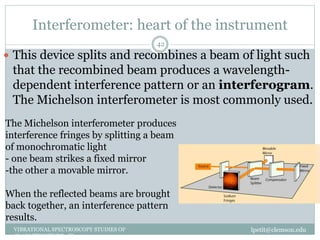
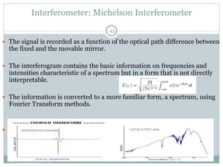
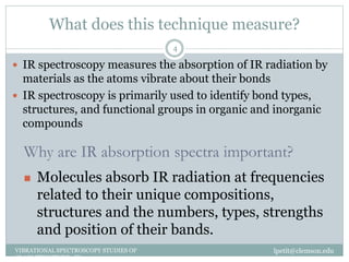
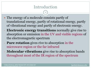
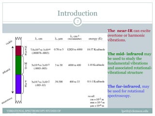
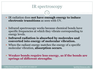
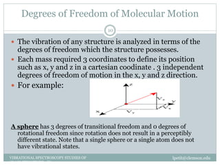
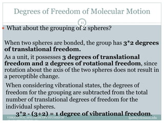
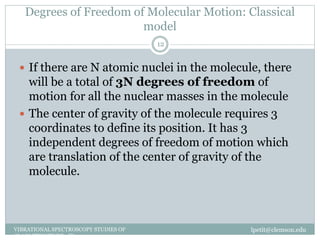
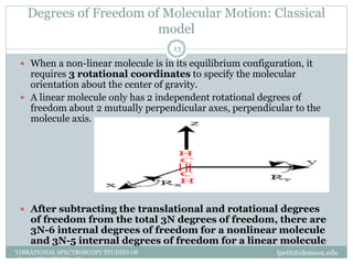
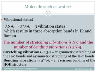
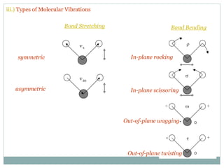
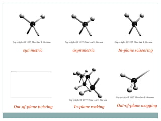
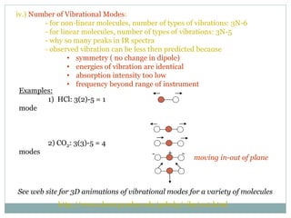
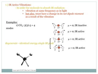
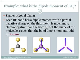
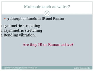
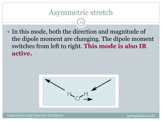
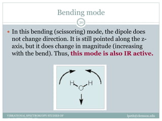
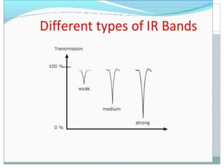
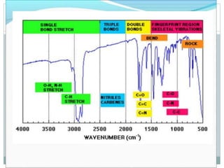
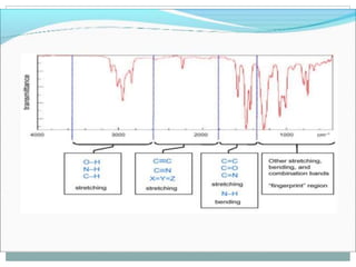
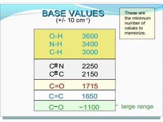
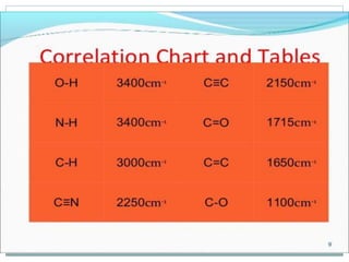
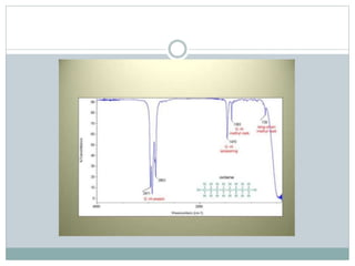
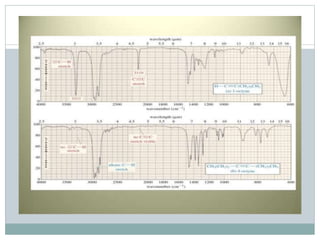
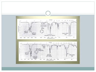
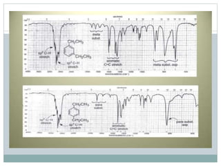
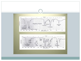
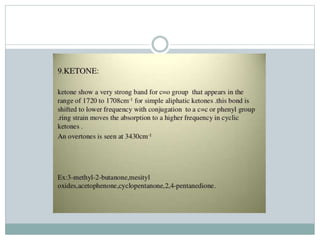
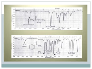
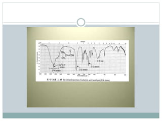
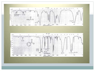
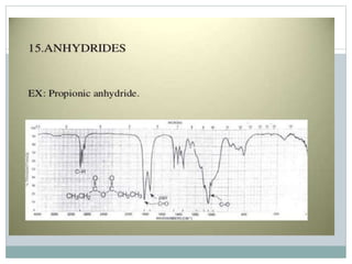
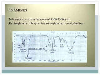
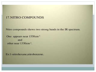
HOW THE FTIR WORKS
WHAT IS IR RADIATION
What does this technique measure?
lpetit@clemson.eduVIBRATIONAL SPECTROSCOPY STUDIES OF
GLASS STRUCTURE : IR spectroscopy
4
 IR spectroscopy measures the absorption of IR radiation by
materials as the atoms vibrate about their bonds
 IR spectroscopy is primarily used to identify bond types,
structures, and functional groups in organic and inorganic
compounds
Why are IR absorption spectra important?
 Molecules absorb IR radiation at frequencies
related to their unique compositions,
structures and the numbers, types, strengths
and position of their bands.
lpetit@clemson.eduVIBRATIONAL SPECTROSCOPY STUDIES OF
GLASS STRUCTURE : IR spectroscopy
5
Infrared refers to that
part of the
electromagnetic
spectrum between the
visible and microwave
regions.
Introduction
lpetit@clemson.eduVIBRATIONAL SPECTROSCOPY STUDIES OF
GLASS STRUCTURE : IR spectroscopy
6
 The energy of a molecule consists partly of
translational energy, partly of rotational energy, partly
of vibrational energy and partly of electronic energy.
- Electronic energy transitions normally give rise to
absorption or emission in the UV and visible regions of
the electromagnetic spectrum
- Pure rotation gives rive to absorption in the
microwave region or the far infrared
- Molecular vibrations give rise to absorption bands
throughout most of the IR region of the spectrum
Introduction
lpetit@clemson.eduVIBRATIONAL SPECTROSCOPY STUDIES OF
GLASS STRUCTURE : IR spectroscopy
7
The near-IR can excite
overtone or harmonic
vibrations.
The mid- infrared may
be used to study the
fundamental vibrations
and associated rotational-
vibrational structure
The far-infrared, may
be used for rotational
spectroscopy.
Definition
lpetit@clemson.eduVIBRATIONAL SPECTROSCOPY STUDIES OF
GLASS STRUCTURE : IR spectroscopy
8
 Wavelength and frequency are inversely related:
 Energy is related to wavelength and frequency :
 Note that energy is directly proportional to
frequency and inversely proportional to
wavelength.
IR spectroscopy
lpetit@clemson.eduVIBRATIONAL SPECTROSCOPY STUDIES OF
GLASS STRUCTURE : IR spectroscopy
9
 IR radiation does not have enough energy to induce
electronic transitions as seen with UV.
 Infrared spectroscopy works because chemical bonds have
specific frequencies at which they vibrate corresponding to
energy levels.
 Infrared radiation is absorbed by molecules and
converted into energy of molecular vibration.
 When the radiant energy matches the energy of a specific
molecular vibration, absorption occurs.
 Weaker bonds require less energy, as if the bonds are
springs of different strengths.
Degrees of Freedom of Molecular Motion
lpetit@clemson.eduVIBRATIONAL SPECTROSCOPY STUDIES OF
GLASS STRUCTURE : IR spectroscopy
10
 The vibration of any structure is analyzed in terms of the
degrees of freedom which the structure possesses.
 Each mass required 3 coordinates to define its position
such as x, y and z in a cartesian coodinate . 3 independent
degrees of freedom of motion in the x, y and z direction.
 For example:
A sphere has 3 degrees of transitional freedom and 0 degrees of
rotational freedom since rotation does not result in a perceptibly
different state. Note that a single sphere or a single atom does not
have vibrational states.
Degrees of Freedom of Molecular Motion
lpetit@clemson.eduVIBRATIONAL SPECTROSCOPY STUDIES OF
GLASS STRUCTURE : IR spectroscopy
11
 What about the grouping of 2 spheres?
When two spheres are bonded, the group has 3*2 degrees
of translational freedom.
As a unit, it possesses 3 degrees of translational
freedom and 2 degrees of rotational freedom, since
rotation about the axis of the two spheres does not result in
a perceptible change.
When considering vibrational states, the degrees of
freedom for the grouping are subtracted from the total
number of translational degrees of freedom for the
individual spheres.
3*2 - (3+2) = 1 degree of vibrational freedom.
Degrees of Freedom of Molecular Motion: Classical
model
lpetit@clemson.eduVIBRATIONAL SPECTROSCOPY STUDIES OF
GLASS STRUCTURE : IR spectroscopy
12
 If there are N atomic nuclei in the molecule, there
will be a total of 3N degrees of freedom of
motion for all the nuclear masses in the molecule
 The center of gravity of the molecule requires 3
coordinates to define its position. It has 3
independent degrees of freedom of motion which
are translation of the center of gravity of the
molecule.
Degrees of Freedom of Molecular Motion: Classical
model
lpetit@clemson.eduVIBRATIONAL SPECTROSCOPY STUDIES OF
GLASS STRUCTURE : IR spectroscopy
13
 When a non-linear molecule is in its equilibrium configuration, it
requires 3 rotational coordinates to specify the molecular
orientation about the center of gravity.
 A linear molecule only has 2 independent rotational degrees of
freedom about 2 mutually perpendicular axes, perpendicular to the
molecule axis.
 After subtracting the translational and rotational degrees
of freedom from the total 3N degrees of freedom, there are
3N-6 internal degrees of freedom for a nonlinear molecule
and 3N-5 internal degrees of freedom for a linear molecule
Molecule such as water?
lpetit@clemson.eduVIBRATIONAL SPECTROSCOPY STUDIES OF
GLASS STRUCTURE : IR spectroscopy
14
 Vibrational states?
3N-6  3*3-6 = 3 vibration states
which results in three absorption bands in IR and
Raman.
The number of stretching vibrations is N-1 and the
number of bending vibrations is 2N-5.
Stretching vibrations  3-1 = 2: symmetric stretching of
the H-0 bonds and asymmetric stretching of the H-O bonds
Bending vibration  2*3-5 = 1: 1 scissors bending of the
HOH structure.
Vibrations
lpetit@clemson.eduVIBRATIONAL SPECTROSCOPY STUDIES OF
GLASS STRUCTURE : IR spectroscopy
15
 Stretching: Change in inter-atomic distance along bond axis
 Bending: Change in angle between two bonds. There are four
types of bend:
Rocking
Scissoring
Wagging
Twisting
iii.) Types of Molecular Vibrations
Bond Stretching Bond Bending
symmetric
asymmetric
In-plane rocking
In-plane scissoring
Out-of-plane wagging
Out-of-plane twisting
symmetric asymmetric
In-plane rocking
In-plane scissoring
Out-of-plane waggingOut-of-plane twisting
Another Illustration of Molecular Vibrations
iv.) Number of Vibrational Modes:
- for non-linear molecules, number of types of vibrations: 3N-6
- for linear molecules, number of types of vibrations: 3N-5
- why so many peaks in IR spectra
- observed vibration can be less then predicted because
• symmetry ( no change in dipole)
• energies of vibration are identical
• absorption intensity too low
• frequency beyond range of instrument
http://www.chem.purdue.edu/gchelp/vibs/co2.html
See web site for 3D animations of vibrational modes for a variety of molecules
Examples:
1) HCl: 3(2)-5 = 1
mode
2) CO2: 3(3)-5 = 4
modes - -
moving in-out of plane
+
v.) IR Active Vibrations:
- In order for molecule to absorb IR radiation:
• vibration at same frequency as in light
• but also, must have a change in its net dipole moment
as a result of the vibration
Examples:
1) CO2: 3(3)-5 = 4
modes
- -+
m = 0; IR inactive
m > 0; IR active
m > 0; IR active
m > 0; IR active
d-
d-
2d+
d-
d-2d+
d-
d-2d+
d-
d-2d+
degenerate –identical energy single IR peak
MODES OF VIBRATIONS
Vibrations
lpetit@clemson.eduVIBRATIONAL SPECTROSCOPY STUDIES OF
GLASS STRUCTURE : IR spectroscopy
22
 The symmetric stretch/bend is an easier
deformation than the asymmetric
stretch/bend
 The bending vibration is much easier than
stretching
Can you find the absorption band related to:
lpetit@clemson.eduVIBRATIONAL SPECTROSCOPY STUDIES OF
GLASS STRUCTURE : IR spectroscopy
23
 Gas Phase Infrared Spectrum of Formaldehyde,
H2C=O
 The symmetric stretch is an easier
deformation than the asymmetric
stretch
 occurs at lower wavenumbers
 The bending vibration is much easier
than stretching so it occurs at lower
wavenumber
Dipole moment
lpetit@clemson.eduVIBRATIONAL SPECTROSCOPY STUDIES OF
GLASS STRUCTURE : IR spectroscopy
24
 A dipole moment is a vector quantity - it has both
size and direction.
where μ is the bond dipole moment in coulomb-
meters, δ is the amount of charge at either end of the
dipole, given in coulombs, and d is the distance
between the charges in meters
Dipole moment
lpetit@clemson.eduVIBRATIONAL SPECTROSCOPY STUDIES OF
GLASS STRUCTURE : IR spectroscopy
25
 Any molecule where the overall centre of the positive charges and the overall
centre of the negative charges coincide will have zero dipole moment. This
happens in molecules with sufficiently symmetric shape.
 So it is the shape of a molecule that is important in determining
whether it has a dipole moment.
 For a complete molecule the overall dipole moment is calculated as the vector
sum of individual dipole moments
Example: what is the dipole moment of BF3?
lpetit@clemson.eduVIBRATIONAL SPECTROSCOPY STUDIES OF
GLASS STRUCTURE : IR spectroscopy
26
 Shape: trigonal planar
 Each BF bond has a dipole moment with a partial
negative charge on the fluorine (it is much more
electronegative than the boron), but the shape of the
molecule is such that the bond dipole moments add
up to zero.
Molecule such as water?
lpetit@clemson.eduVIBRATIONAL SPECTROSCOPY STUDIES OF
GLASS STRUCTURE : IR spectroscopy
27
 3 absorption bands in IR and Raman
1 symmetric stretching
1 asymmetric stretching
1 Bending vibration.
Are they IR or Raman active?
Symmetrical stretch
lpetit@clemson.eduVIBRATIONAL SPECTROSCOPY STUDIES OF
GLASS STRUCTURE : IR spectroscopy
28
 In this mode the dipole moment for the molecule does
not change in direction, but it does change in
magnitude. As the molecule stretches, the dipole
moment increases. So the dipole moment changes and
it does so along the z-axis. This vibration is IR
active
Asymmetric stretch
lpetit@clemson.eduVIBRATIONAL SPECTROSCOPY STUDIES OF
GLASS STRUCTURE : IR spectroscopy
29
 In this mode, both the direction and magnitude of
the dipole moment are changing. The dipole moment
switches from left to right. This mode is also IR
active.
Bending mode
lpetit@clemson.eduVIBRATIONAL SPECTROSCOPY STUDIES OF
GLASS STRUCTURE : IR spectroscopy
30
 In this bending (scissoring) mode, the dipole does
not change direction. It is still pointed along the z-
axis, but it does change in magnitude (increasing
with the bend). Thus, this mode is also IR active.
What about CO2?
lpetit@clemson.eduVIBRATIONAL SPECTROSCOPY STUDIES OF
GLASS STRUCTURE : IR spectroscopy
31
 Linear Molecule! O=C=O
Vibrational states: 3n-5 = 3*3-5= 4
Theory: Hooke’s law
lpetit@clemson.eduVIBRATIONAL SPECTROSCOPY STUDIES OF
GLASS STRUCTURE : IR spectroscopy
32
 the frequency of the vibration of the spring is related
to the mass and the force constant of the spring,k, by
the following formula:
where k is the force constant, m is the mass, ν is the
frequency of the vibration
Theory: Hooke’s law
lpetit@clemson.eduVIBRATIONAL SPECTROSCOPY STUDIES OF
GLASS STRUCTURE : IR spectroscopy
33
 The stretching frequency of a bond can be
approximated by Hooke’s Law.
 In this approximation, two atoms and the connecting
bond are treated as a simple harmonic oscillator
composed of 2 masses (atoms) joined by a spring:
Theory: Classical vibrational Frequency for a diatomic
molecule
lpetit@clemson.eduVIBRATIONAL SPECTROSCOPY STUDIES OF
GLASS STRUCTURE : IR spectroscopy
34
 Diatomic molecule is represented by 2 masses: m1 and
m2, connected by a massless spring. For simplicity,
the masses may be allowed to move only along the
molecular axis
 The displacement of each mass from equilibrium
along the axis is X1 and X2. In this case, (X2-X1) is the
amount the bond length differs from the equilibrium
length.
Theory: Classical vibrational Frequency for a diatomic
molecule
lpetit@clemson.eduVIBRATIONAL SPECTROSCOPY STUDIES OF
GLASS STRUCTURE : IR spectroscopy
35
 Each mass will experience a force equal to a
constant F.
  2
1
2
112
dt
Xd
mXXF    2
2
2
212
dt
Xd
mXXF 
)cos( tX  )sin( t
dt
dX
 )cos(2
2
2
t
dt
Xd

Acceleration along the X axis
Source
lpetit@clemson.eduVIBRATIONAL SPECTROSCOPY STUDIES OF
GLASS STRUCTURE : IR spectroscopy
36
 All the manufacturers use a heated ceramic
source.
 The composition of the ceramic and the method of
heating vary but the idea is always the same, the
production of a heated emitter operating at as high a
temperature as with a very long life.
 The manufacturers tend to go for either a conducting
ceramic or a wire heater coated with ceramic.
C) Instrumentation
1.) Basic Design
- normal IR instrument similar to UV-vis
- main differences are light source & detector
2.) Fourier Transfer IR (FTIR) – alternative to Normal IR
- Based on Michelson Interferometer
Principal:
1) light from source is split by central mirror into 2 beams of equal intensity
2) beams go to two other mirrors, reflected by central mirror, recombine
and pass through sample to detector
3) two side mirrors. One fixed and other movable
a) move second mirror, light in two-paths travel different distances
before recombined
b) constructive & destructive interference
c) as mirror is moved, get a change in signal
Destructive Interference can be created when two waves from the
same source travel different paths to get to a point.
This may cause a difference in the phase between the two waves.
• If the paths differ by an integer multiple of a wavelength, the waves will also be in
phase.
• If the waves differ by an odd multiple of half a wave then the waves will be 180
degrees out of phase and cancel out.
- observe a plot of Intensity vs. Distance (interferograms)
- convert to plot of Intensity vs. Frequency by doing a Fourier
Transform
- resolution Dn = 1/Dd (interval of distance traveled by mirror)
Advantages of FTIR compared to Normal IR:
1) much faster, seconds vs. minutes
2) use signal averaging to increase signal-to-noise (S/N)
3) higher inherent S/N – no slits, less optical equipment, higher light
intensity
4) high resolution (<0.1 cm-1)
Disadvantages of FTIR compared to Normal IR:
1) single-beam, requires collecting blank
2) can’t use thermal detectors – too slow
In normal IR, scan through frequency range.
In
FTIR collect all frequencies at once.
/increase S N number scans
Interferometer: heart of the instrument
lpetit@clemson.eduVIBRATIONAL SPECTROSCOPY STUDIES OF
GLASS STRUCTURE : IR spectroscopy
42
 This device splits and recombines a beam of light such
that the recombined beam produces a wavelength-
dependent interference pattern or an interferogram.
The Michelson interferometer is most commonly used.
The Michelson interferometer produces
interference fringes by splitting a beam
of monochromatic light
- one beam strikes a fixed mirror
-the other a movable mirror.
When the reflected beams are brought
back together, an interference pattern
results.
Interferometer: Michelson Interferometer
lpetit@clemson.eduVIBRATIONAL SPECTROSCOPY STUDIES OF
GLASS STRUCTURE : IR spectroscopy
43
 The signal is recorded as a function of the optical path difference between
the fixed and the movable mirror.
 The interferogram contains the basic information on frequencies and
intensities characteristic of a spectrum but in a form that is not directly
interpretable.
 The information is converted to a more familiar form, a spectrum, using
Fourier Transform methods.
 With an infrared data station, the computer acquires, processes, stores
and retrieves spectral data.
Infrared Detector
lpetit@clemson.eduVIBRATIONAL SPECTROSCOPY STUDIES OF
GLASS STRUCTURE : IR spectroscopy
44
 It is a device which measured the infrared energy of
the source which has passed through the
spectrometer. These devices change radiation
energy into electrical energy which can be
processed to generate a spectrum.
 2 basic types:
- Thermal detectors which measure the heating
effect of radiation and respond equally well to all
wavelengths
- Selective detectors whose response is markedly
dependent on the wavelength
Infrared detectors
lpetit@clemson.eduVIBRATIONAL SPECTROSCOPY STUDIES OF
GLASS STRUCTURE : IR spectroscopy
45
 Examples of thermal detectors:
Thermocouples: when incident radiation is
absorbed at the junction, the temperature rise causes
an increase in the electromotive potential developed
across the junction leads
Bolometers: detecting device which depends on a
change of resistance with temperature
Pyroelectric: it consists of a thin pyroelectric crystal.
If it is electrically polarized in an electric field, it
retains a residual electric polarization after the field
is removed.
Nicolet Continuµm Infrared Microscope
lpetit@clemson.eduVIBRATIONAL SPECTROSCOPY STUDIES OF
GLASS STRUCTURE : IR spectroscopy
46
 Acquisition of mid-IR spectra from
very small (>10 µm) samples with
diffraction limited spatial resolution.
 Material science applications
including:
• Forensic analysis
• Surface analysis
• Art conservation
• Mineralogy
• Biochemistry
 SAMPLE
PREPARATION
Measurement of IR spectra
lpetit@clemson.eduVIBRATIONAL SPECTROSCOPY STUDIES OF
GLASS STRUCTURE : IR spectroscopy
48
 IR spectra are acquired on a special instrument,
called an IR spectrometer.
 IR is used both to gather information about the
structure of a compound and as an analytical tool to
assess the purity of a compound or the structure of
the glasses
 IR spectra are quick and easy to run
 Cells for holding samples must be made of infrared
transmitting material.
Infrared transmitting materials
lpetit@clemson.eduVIBRATIONAL SPECTROSCOPY STUDIES OF
GLASS STRUCTURE : IR spectroscopy
49
Infrared transmitting materials
lpetit@clemson.eduVIBRATIONAL SPECTROSCOPY STUDIES OF
GLASS STRUCTURE : IR spectroscopy
50
 Some of the most useful window materials for the IR
are quite soluble in water: NaCl, KCl, KBr, CsBr and
CsI
 A typical cell for liquid which is made of NaCl has a
transmission that starts to drop at about 700cm-1, is
roughly 50% at 600cm-1 and is nearly opaque at
500cm-1.
Sample Requirements
lpetit@clemson.eduVIBRATIONAL SPECTROSCOPY STUDIES OF
GLASS STRUCTURE : IR spectroscopy
51
 The detection limit for routine analysis is ~0.1 wt%;
under ideal conditions greater sensitivity can be
achieved
 Typically only a few milligrams of material are
needed for analysis.
 Samples may be in liquid, solid, or gaseous form.
Preparation of the samples: gas
lpetit@clemson.eduVIBRATIONAL SPECTROSCOPY STUDIES OF
GLASS STRUCTURE : IR spectroscopy
52
 Gaseous samples require little preparation beyond
purification!
 To obtain an infrared spectrum of a gas requires the
use of a cylindrical gas cell with windows at each end
composed of an infrared inactive material such as
KBr, NaCl or CaF2. The cell usually has an inlet and
outlet port with a tap to enable the cell to be easily
filled with the gas to be analyzed.
Preparation of the samples: liquid
lpetit@clemson.eduVIBRATIONAL SPECTROSCOPY STUDIES OF
GLASS STRUCTURE : IR spectroscopy
53
 Sandwiched between two plates of a high purity salt
(commonly sodium chloride, or common salt,
although a number of other salts such as potassium
bromide or calcium fluoride are also used).
 The plates are transparent to the infrared light (not
introduce any lines onto the spectra).
Preparation of the samples: solid
lpetit@clemson.eduVIBRATIONAL SPECTROSCOPY STUDIES OF
GLASS STRUCTURE : IR spectroscopy
54
 Solid samples can be prepared in two major ways.
1) Crush the sample with a mulling agent (usually nujol) in a
marble or agate mortar, with a pestle. A thin film of the mull
is applied onto salt plates and measured.
2) Grind a quantity of the sample with a specially purified salt
(usually potassium bromide) finely (to remove scattering
effects from large crystals). This powder mixture is then
crushed in a mechanical die press to form a translucent pellet
through which the beam of the spectrometer can pass.
Samples dispersed in powder must be homogenously dispersed, with a
particle size small enough not to cause scatter (theoretically < 2
microns).

 INTERPRITATION
USES
FTIR AND ITS INTERPRETATION BY KARAN AJAY GUPTA

FTIR AND ITS INTERPRETATION BY KARAN AJAY GUPTA

  • 2.
  • 3.
    WHAT IS IRRADIATION
  • 4.
    What does thistechnique measure? lpetit@clemson.eduVIBRATIONAL SPECTROSCOPY STUDIES OF GLASS STRUCTURE : IR spectroscopy 4  IR spectroscopy measures the absorption of IR radiation by materials as the atoms vibrate about their bonds  IR spectroscopy is primarily used to identify bond types, structures, and functional groups in organic and inorganic compounds Why are IR absorption spectra important?  Molecules absorb IR radiation at frequencies related to their unique compositions, structures and the numbers, types, strengths and position of their bands.
  • 5.
    lpetit@clemson.eduVIBRATIONAL SPECTROSCOPY STUDIESOF GLASS STRUCTURE : IR spectroscopy 5 Infrared refers to that part of the electromagnetic spectrum between the visible and microwave regions.
  • 6.
    Introduction lpetit@clemson.eduVIBRATIONAL SPECTROSCOPY STUDIESOF GLASS STRUCTURE : IR spectroscopy 6  The energy of a molecule consists partly of translational energy, partly of rotational energy, partly of vibrational energy and partly of electronic energy. - Electronic energy transitions normally give rise to absorption or emission in the UV and visible regions of the electromagnetic spectrum - Pure rotation gives rive to absorption in the microwave region or the far infrared - Molecular vibrations give rise to absorption bands throughout most of the IR region of the spectrum
  • 7.
    Introduction lpetit@clemson.eduVIBRATIONAL SPECTROSCOPY STUDIESOF GLASS STRUCTURE : IR spectroscopy 7 The near-IR can excite overtone or harmonic vibrations. The mid- infrared may be used to study the fundamental vibrations and associated rotational- vibrational structure The far-infrared, may be used for rotational spectroscopy.
  • 8.
    Definition lpetit@clemson.eduVIBRATIONAL SPECTROSCOPY STUDIESOF GLASS STRUCTURE : IR spectroscopy 8  Wavelength and frequency are inversely related:  Energy is related to wavelength and frequency :  Note that energy is directly proportional to frequency and inversely proportional to wavelength.
  • 9.
    IR spectroscopy lpetit@clemson.eduVIBRATIONAL SPECTROSCOPYSTUDIES OF GLASS STRUCTURE : IR spectroscopy 9  IR radiation does not have enough energy to induce electronic transitions as seen with UV.  Infrared spectroscopy works because chemical bonds have specific frequencies at which they vibrate corresponding to energy levels.  Infrared radiation is absorbed by molecules and converted into energy of molecular vibration.  When the radiant energy matches the energy of a specific molecular vibration, absorption occurs.  Weaker bonds require less energy, as if the bonds are springs of different strengths.
  • 10.
    Degrees of Freedomof Molecular Motion lpetit@clemson.eduVIBRATIONAL SPECTROSCOPY STUDIES OF GLASS STRUCTURE : IR spectroscopy 10  The vibration of any structure is analyzed in terms of the degrees of freedom which the structure possesses.  Each mass required 3 coordinates to define its position such as x, y and z in a cartesian coodinate . 3 independent degrees of freedom of motion in the x, y and z direction.  For example: A sphere has 3 degrees of transitional freedom and 0 degrees of rotational freedom since rotation does not result in a perceptibly different state. Note that a single sphere or a single atom does not have vibrational states.
  • 11.
    Degrees of Freedomof Molecular Motion lpetit@clemson.eduVIBRATIONAL SPECTROSCOPY STUDIES OF GLASS STRUCTURE : IR spectroscopy 11  What about the grouping of 2 spheres? When two spheres are bonded, the group has 3*2 degrees of translational freedom. As a unit, it possesses 3 degrees of translational freedom and 2 degrees of rotational freedom, since rotation about the axis of the two spheres does not result in a perceptible change. When considering vibrational states, the degrees of freedom for the grouping are subtracted from the total number of translational degrees of freedom for the individual spheres. 3*2 - (3+2) = 1 degree of vibrational freedom.
  • 12.
    Degrees of Freedomof Molecular Motion: Classical model lpetit@clemson.eduVIBRATIONAL SPECTROSCOPY STUDIES OF GLASS STRUCTURE : IR spectroscopy 12  If there are N atomic nuclei in the molecule, there will be a total of 3N degrees of freedom of motion for all the nuclear masses in the molecule  The center of gravity of the molecule requires 3 coordinates to define its position. It has 3 independent degrees of freedom of motion which are translation of the center of gravity of the molecule.
  • 13.
    Degrees of Freedomof Molecular Motion: Classical model lpetit@clemson.eduVIBRATIONAL SPECTROSCOPY STUDIES OF GLASS STRUCTURE : IR spectroscopy 13  When a non-linear molecule is in its equilibrium configuration, it requires 3 rotational coordinates to specify the molecular orientation about the center of gravity.  A linear molecule only has 2 independent rotational degrees of freedom about 2 mutually perpendicular axes, perpendicular to the molecule axis.  After subtracting the translational and rotational degrees of freedom from the total 3N degrees of freedom, there are 3N-6 internal degrees of freedom for a nonlinear molecule and 3N-5 internal degrees of freedom for a linear molecule
  • 14.
    Molecule such aswater? lpetit@clemson.eduVIBRATIONAL SPECTROSCOPY STUDIES OF GLASS STRUCTURE : IR spectroscopy 14  Vibrational states? 3N-6  3*3-6 = 3 vibration states which results in three absorption bands in IR and Raman. The number of stretching vibrations is N-1 and the number of bending vibrations is 2N-5. Stretching vibrations  3-1 = 2: symmetric stretching of the H-0 bonds and asymmetric stretching of the H-O bonds Bending vibration  2*3-5 = 1: 1 scissors bending of the HOH structure.
  • 15.
    Vibrations lpetit@clemson.eduVIBRATIONAL SPECTROSCOPY STUDIESOF GLASS STRUCTURE : IR spectroscopy 15  Stretching: Change in inter-atomic distance along bond axis  Bending: Change in angle between two bonds. There are four types of bend: Rocking Scissoring Wagging Twisting
  • 16.
    iii.) Types ofMolecular Vibrations Bond Stretching Bond Bending symmetric asymmetric In-plane rocking In-plane scissoring Out-of-plane wagging Out-of-plane twisting
  • 17.
    symmetric asymmetric In-plane rocking In-planescissoring Out-of-plane waggingOut-of-plane twisting
  • 18.
    Another Illustration ofMolecular Vibrations
  • 19.
    iv.) Number ofVibrational Modes: - for non-linear molecules, number of types of vibrations: 3N-6 - for linear molecules, number of types of vibrations: 3N-5 - why so many peaks in IR spectra - observed vibration can be less then predicted because • symmetry ( no change in dipole) • energies of vibration are identical • absorption intensity too low • frequency beyond range of instrument http://www.chem.purdue.edu/gchelp/vibs/co2.html See web site for 3D animations of vibrational modes for a variety of molecules Examples: 1) HCl: 3(2)-5 = 1 mode 2) CO2: 3(3)-5 = 4 modes - - moving in-out of plane +
  • 20.
    v.) IR ActiveVibrations: - In order for molecule to absorb IR radiation: • vibration at same frequency as in light • but also, must have a change in its net dipole moment as a result of the vibration Examples: 1) CO2: 3(3)-5 = 4 modes - -+ m = 0; IR inactive m > 0; IR active m > 0; IR active m > 0; IR active d- d- 2d+ d- d-2d+ d- d-2d+ d- d-2d+ degenerate –identical energy single IR peak
  • 21.
  • 22.
    Vibrations lpetit@clemson.eduVIBRATIONAL SPECTROSCOPY STUDIESOF GLASS STRUCTURE : IR spectroscopy 22  The symmetric stretch/bend is an easier deformation than the asymmetric stretch/bend  The bending vibration is much easier than stretching
  • 23.
    Can you findthe absorption band related to: lpetit@clemson.eduVIBRATIONAL SPECTROSCOPY STUDIES OF GLASS STRUCTURE : IR spectroscopy 23  Gas Phase Infrared Spectrum of Formaldehyde, H2C=O  The symmetric stretch is an easier deformation than the asymmetric stretch  occurs at lower wavenumbers  The bending vibration is much easier than stretching so it occurs at lower wavenumber
  • 24.
    Dipole moment lpetit@clemson.eduVIBRATIONAL SPECTROSCOPYSTUDIES OF GLASS STRUCTURE : IR spectroscopy 24  A dipole moment is a vector quantity - it has both size and direction. where μ is the bond dipole moment in coulomb- meters, δ is the amount of charge at either end of the dipole, given in coulombs, and d is the distance between the charges in meters
  • 25.
    Dipole moment lpetit@clemson.eduVIBRATIONAL SPECTROSCOPYSTUDIES OF GLASS STRUCTURE : IR spectroscopy 25  Any molecule where the overall centre of the positive charges and the overall centre of the negative charges coincide will have zero dipole moment. This happens in molecules with sufficiently symmetric shape.  So it is the shape of a molecule that is important in determining whether it has a dipole moment.  For a complete molecule the overall dipole moment is calculated as the vector sum of individual dipole moments
  • 26.
    Example: what isthe dipole moment of BF3? lpetit@clemson.eduVIBRATIONAL SPECTROSCOPY STUDIES OF GLASS STRUCTURE : IR spectroscopy 26  Shape: trigonal planar  Each BF bond has a dipole moment with a partial negative charge on the fluorine (it is much more electronegative than the boron), but the shape of the molecule is such that the bond dipole moments add up to zero.
  • 27.
    Molecule such aswater? lpetit@clemson.eduVIBRATIONAL SPECTROSCOPY STUDIES OF GLASS STRUCTURE : IR spectroscopy 27  3 absorption bands in IR and Raman 1 symmetric stretching 1 asymmetric stretching 1 Bending vibration. Are they IR or Raman active?
  • 28.
    Symmetrical stretch lpetit@clemson.eduVIBRATIONAL SPECTROSCOPYSTUDIES OF GLASS STRUCTURE : IR spectroscopy 28  In this mode the dipole moment for the molecule does not change in direction, but it does change in magnitude. As the molecule stretches, the dipole moment increases. So the dipole moment changes and it does so along the z-axis. This vibration is IR active
  • 29.
    Asymmetric stretch lpetit@clemson.eduVIBRATIONAL SPECTROSCOPYSTUDIES OF GLASS STRUCTURE : IR spectroscopy 29  In this mode, both the direction and magnitude of the dipole moment are changing. The dipole moment switches from left to right. This mode is also IR active.
  • 30.
    Bending mode lpetit@clemson.eduVIBRATIONAL SPECTROSCOPYSTUDIES OF GLASS STRUCTURE : IR spectroscopy 30  In this bending (scissoring) mode, the dipole does not change direction. It is still pointed along the z- axis, but it does change in magnitude (increasing with the bend). Thus, this mode is also IR active.
  • 31.
    What about CO2? lpetit@clemson.eduVIBRATIONALSPECTROSCOPY STUDIES OF GLASS STRUCTURE : IR spectroscopy 31  Linear Molecule! O=C=O Vibrational states: 3n-5 = 3*3-5= 4
  • 32.
    Theory: Hooke’s law lpetit@clemson.eduVIBRATIONALSPECTROSCOPY STUDIES OF GLASS STRUCTURE : IR spectroscopy 32  the frequency of the vibration of the spring is related to the mass and the force constant of the spring,k, by the following formula: where k is the force constant, m is the mass, ν is the frequency of the vibration
  • 33.
    Theory: Hooke’s law lpetit@clemson.eduVIBRATIONALSPECTROSCOPY STUDIES OF GLASS STRUCTURE : IR spectroscopy 33  The stretching frequency of a bond can be approximated by Hooke’s Law.  In this approximation, two atoms and the connecting bond are treated as a simple harmonic oscillator composed of 2 masses (atoms) joined by a spring:
  • 34.
    Theory: Classical vibrationalFrequency for a diatomic molecule lpetit@clemson.eduVIBRATIONAL SPECTROSCOPY STUDIES OF GLASS STRUCTURE : IR spectroscopy 34  Diatomic molecule is represented by 2 masses: m1 and m2, connected by a massless spring. For simplicity, the masses may be allowed to move only along the molecular axis  The displacement of each mass from equilibrium along the axis is X1 and X2. In this case, (X2-X1) is the amount the bond length differs from the equilibrium length.
  • 35.
    Theory: Classical vibrationalFrequency for a diatomic molecule lpetit@clemson.eduVIBRATIONAL SPECTROSCOPY STUDIES OF GLASS STRUCTURE : IR spectroscopy 35  Each mass will experience a force equal to a constant F.   2 1 2 112 dt Xd mXXF    2 2 2 212 dt Xd mXXF  )cos( tX  )sin( t dt dX  )cos(2 2 2 t dt Xd  Acceleration along the X axis
  • 36.
    Source lpetit@clemson.eduVIBRATIONAL SPECTROSCOPY STUDIESOF GLASS STRUCTURE : IR spectroscopy 36  All the manufacturers use a heated ceramic source.  The composition of the ceramic and the method of heating vary but the idea is always the same, the production of a heated emitter operating at as high a temperature as with a very long life.  The manufacturers tend to go for either a conducting ceramic or a wire heater coated with ceramic.
  • 37.
    C) Instrumentation 1.) BasicDesign - normal IR instrument similar to UV-vis - main differences are light source & detector
  • 38.
    2.) Fourier TransferIR (FTIR) – alternative to Normal IR - Based on Michelson Interferometer Principal: 1) light from source is split by central mirror into 2 beams of equal intensity 2) beams go to two other mirrors, reflected by central mirror, recombine and pass through sample to detector 3) two side mirrors. One fixed and other movable a) move second mirror, light in two-paths travel different distances before recombined b) constructive & destructive interference c) as mirror is moved, get a change in signal
  • 39.
    Destructive Interference canbe created when two waves from the same source travel different paths to get to a point. This may cause a difference in the phase between the two waves. • If the paths differ by an integer multiple of a wavelength, the waves will also be in phase. • If the waves differ by an odd multiple of half a wave then the waves will be 180 degrees out of phase and cancel out.
  • 40.
    - observe aplot of Intensity vs. Distance (interferograms) - convert to plot of Intensity vs. Frequency by doing a Fourier Transform - resolution Dn = 1/Dd (interval of distance traveled by mirror)
  • 41.
    Advantages of FTIRcompared to Normal IR: 1) much faster, seconds vs. minutes 2) use signal averaging to increase signal-to-noise (S/N) 3) higher inherent S/N – no slits, less optical equipment, higher light intensity 4) high resolution (<0.1 cm-1) Disadvantages of FTIR compared to Normal IR: 1) single-beam, requires collecting blank 2) can’t use thermal detectors – too slow In normal IR, scan through frequency range. In FTIR collect all frequencies at once. /increase S N number scans
  • 42.
    Interferometer: heart ofthe instrument lpetit@clemson.eduVIBRATIONAL SPECTROSCOPY STUDIES OF GLASS STRUCTURE : IR spectroscopy 42  This device splits and recombines a beam of light such that the recombined beam produces a wavelength- dependent interference pattern or an interferogram. The Michelson interferometer is most commonly used. The Michelson interferometer produces interference fringes by splitting a beam of monochromatic light - one beam strikes a fixed mirror -the other a movable mirror. When the reflected beams are brought back together, an interference pattern results.
  • 43.
    Interferometer: Michelson Interferometer lpetit@clemson.eduVIBRATIONALSPECTROSCOPY STUDIES OF GLASS STRUCTURE : IR spectroscopy 43  The signal is recorded as a function of the optical path difference between the fixed and the movable mirror.  The interferogram contains the basic information on frequencies and intensities characteristic of a spectrum but in a form that is not directly interpretable.  The information is converted to a more familiar form, a spectrum, using Fourier Transform methods.  With an infrared data station, the computer acquires, processes, stores and retrieves spectral data.
  • 44.
    Infrared Detector lpetit@clemson.eduVIBRATIONAL SPECTROSCOPYSTUDIES OF GLASS STRUCTURE : IR spectroscopy 44  It is a device which measured the infrared energy of the source which has passed through the spectrometer. These devices change radiation energy into electrical energy which can be processed to generate a spectrum.  2 basic types: - Thermal detectors which measure the heating effect of radiation and respond equally well to all wavelengths - Selective detectors whose response is markedly dependent on the wavelength
  • 45.
    Infrared detectors lpetit@clemson.eduVIBRATIONAL SPECTROSCOPYSTUDIES OF GLASS STRUCTURE : IR spectroscopy 45  Examples of thermal detectors: Thermocouples: when incident radiation is absorbed at the junction, the temperature rise causes an increase in the electromotive potential developed across the junction leads Bolometers: detecting device which depends on a change of resistance with temperature Pyroelectric: it consists of a thin pyroelectric crystal. If it is electrically polarized in an electric field, it retains a residual electric polarization after the field is removed.
  • 46.
    Nicolet Continuµm InfraredMicroscope lpetit@clemson.eduVIBRATIONAL SPECTROSCOPY STUDIES OF GLASS STRUCTURE : IR spectroscopy 46  Acquisition of mid-IR spectra from very small (>10 µm) samples with diffraction limited spatial resolution.  Material science applications including: • Forensic analysis • Surface analysis • Art conservation • Mineralogy • Biochemistry
  • 47.
  • 48.
    Measurement of IRspectra lpetit@clemson.eduVIBRATIONAL SPECTROSCOPY STUDIES OF GLASS STRUCTURE : IR spectroscopy 48  IR spectra are acquired on a special instrument, called an IR spectrometer.  IR is used both to gather information about the structure of a compound and as an analytical tool to assess the purity of a compound or the structure of the glasses  IR spectra are quick and easy to run  Cells for holding samples must be made of infrared transmitting material.
  • 49.
    Infrared transmitting materials lpetit@clemson.eduVIBRATIONALSPECTROSCOPY STUDIES OF GLASS STRUCTURE : IR spectroscopy 49
  • 50.
    Infrared transmitting materials lpetit@clemson.eduVIBRATIONALSPECTROSCOPY STUDIES OF GLASS STRUCTURE : IR spectroscopy 50  Some of the most useful window materials for the IR are quite soluble in water: NaCl, KCl, KBr, CsBr and CsI  A typical cell for liquid which is made of NaCl has a transmission that starts to drop at about 700cm-1, is roughly 50% at 600cm-1 and is nearly opaque at 500cm-1.
  • 51.
    Sample Requirements lpetit@clemson.eduVIBRATIONAL SPECTROSCOPYSTUDIES OF GLASS STRUCTURE : IR spectroscopy 51  The detection limit for routine analysis is ~0.1 wt%; under ideal conditions greater sensitivity can be achieved  Typically only a few milligrams of material are needed for analysis.  Samples may be in liquid, solid, or gaseous form.
  • 52.
    Preparation of thesamples: gas lpetit@clemson.eduVIBRATIONAL SPECTROSCOPY STUDIES OF GLASS STRUCTURE : IR spectroscopy 52  Gaseous samples require little preparation beyond purification!  To obtain an infrared spectrum of a gas requires the use of a cylindrical gas cell with windows at each end composed of an infrared inactive material such as KBr, NaCl or CaF2. The cell usually has an inlet and outlet port with a tap to enable the cell to be easily filled with the gas to be analyzed.
  • 53.
    Preparation of thesamples: liquid lpetit@clemson.eduVIBRATIONAL SPECTROSCOPY STUDIES OF GLASS STRUCTURE : IR spectroscopy 53  Sandwiched between two plates of a high purity salt (commonly sodium chloride, or common salt, although a number of other salts such as potassium bromide or calcium fluoride are also used).  The plates are transparent to the infrared light (not introduce any lines onto the spectra).
  • 54.
    Preparation of thesamples: solid lpetit@clemson.eduVIBRATIONAL SPECTROSCOPY STUDIES OF GLASS STRUCTURE : IR spectroscopy 54  Solid samples can be prepared in two major ways. 1) Crush the sample with a mulling agent (usually nujol) in a marble or agate mortar, with a pestle. A thin film of the mull is applied onto salt plates and measured. 2) Grind a quantity of the sample with a specially purified salt (usually potassium bromide) finely (to remove scattering effects from large crystals). This powder mixture is then crushed in a mechanical die press to form a translucent pellet through which the beam of the spectrometer can pass. Samples dispersed in powder must be homogenously dispersed, with a particle size small enough not to cause scatter (theoretically < 2 microns).
  • 55.
  • 91.