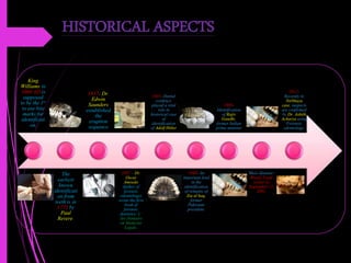
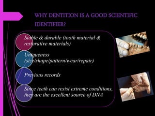
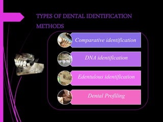
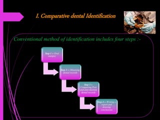
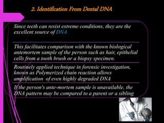
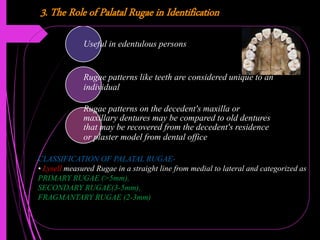
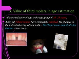
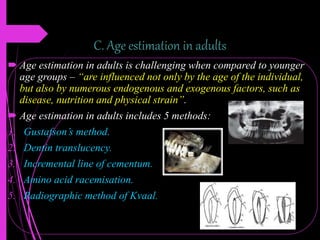
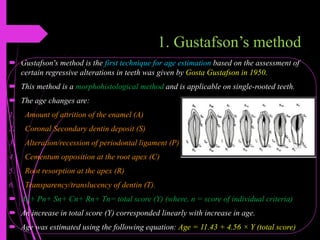
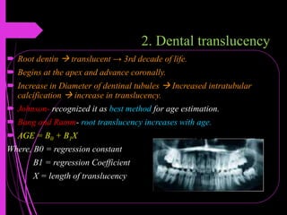
Sam Higginbottom University of Agriculture, Technology and Sciences
(University in Allahabad, Uttar Pradesh)
Seminar
On
“FORENSIC ODONTOLOGY”
Presented by:
Name- Pallavi Kumari
MSc. Forensic science
Contents
1. Definition
2. Historical aspects
3. When identification is required?
4. Scope of Forensic Odontology
5. Why dentition is a good scientific identifier?
6. Types of dental identification methods:-
a. Comparative dental Identification
b. Identification From Dental DNA
c. Edentulous identification
d. Dental Profiling :-
i) Identifying Ethnic Origin From Teeth
ii) Sex determination from teeth
iii) Age estimation from dentition :-
A) Age estimation in Prenatal, neonatal and early postnatal children
B) Age estimation in children and adolescents
C) Age estimation in adults
7. Bite marks: Introduction
8. Classification of bite-mark
9. Bite mark appearance
10. Bite mark investigation
11. Histopathological changes in bite marks.
DEFINITION
FDI defined Forensic odontology as “that branch of
dentistry which in the interest of justice, deals with the
proper handling and examination of dental evidences and
also with the proper evaluation and presentation of
dental findings in criminal or civil proceedings”.
.
HISTORICAL ASPECTS
King
Williams in
1066 AD is
supposed
to be the 1st
to use bite
marks for
identificati
on.
The
earliest
known
identificati
on from
teeth is in
1775 by
Paul
Revere.
1837- Dr.
Edwin
Saunders
established
the
eruption
sequence
1897 – Dr.
Oscar
Amoedo
(father of
forensic
odontology)
wrote the first
book of
forensic
dentistry- L’
Art Dentaire
en Medicine
Legale.
1945- Dental
evidence
played a vital
role in
historical case
of
identification
of Adolf Hitler
1988- An
important lead
in the
identification
of remains of
Zia ul haq,
former
Pakistani
president.
1989-
Identification
of Rajiv
Gandhi,
former Indian
prime minister
Mass disaster-
World Trade
center in
September 11,
2001.
2012-
Recently in
Nirbhaya
case, suspects
are confirmed
by Dr. Ashith
Acharya using
Forensic
odontology.
WHEN IDENTIFICATION IS REQUIRED?
Decomposing remains.
Skeletonized remains.
Charred remains.
Intact remains without any putative victim.
Scientific verification of identity is anticipated.
Multiple bodies recovered from a common location.
Mass disaster.
SCOPE OF FORENSIC ODONTOLOGY
• Identifying unknown human remains through dental records & cranio- facial bones.
• Age estimation of both the living and deceased.
• Recognition and analysis of bite marks found on victims
• Analysis of oro-facial trauma associated with person abuse
• Determining the gender of an unidentified individual.
• Eliciting the ethnicity
• Assisting in building up a picture of lifestyle and diet of skeletal remains.
• Analysis of dental malpractice claims
• Presenting evidence in court as an expert witness.
WHY DENTITION IS A GOOD SCIENTIFIC
IDENTIFIER?
Stable & durable (tooth material &
restorative materials)
Uniqueness
(size/shape/pattern/wear/repair)
Previous records
Since teeth can resist extreme conditions,
they are the excellent source of DNA
TYPES OF DENTAL IDENTIFICATION
METHODS
Comparative identification
DNA identification
Edentulous identification
Dental Profiling
I. Comparative dental Identification
Conventional method of identification includes four steps :-
Step 1 :- Oral
autopsy
Step 2 :- Obtaining
dental records
Step 3 :-
Comparing Post
and ante-mortem
dental records
Step 4 :- Writing a
report and
drawing
conclusions
2. Identification From Dental DNA
Since teeth can resist extreme conditions, they are the
excellent source of DNA
This facilitates comparison with the known biological
antemortem sample of the person such as hair, epithelial
cells from a tooth brush or a biopsy specimen.
Routinely applied technique in forensic investigation,
known as Polymerized chain reaction allows
amplification of even highly degraded DNA
If the person's ante-mortem sample is unavailable, the
DNA pattern may be compared to a parent or a sibling
3. The Role of Palatal Rugae in Identification
Useful in edentulous persons
Rugae patterns like teeth are considered unique to an
individual
Rugae patterns on the decedent's maxilla or
maxillary dentures may be compared to old dentures
that may be recovered from the decedent's residence
or plaster model from dental office
CLASSIFICATION OF PALATAL RUGAE-
• Lysell measured Rugae in a straight line from medial to lateral and categorized as
PRIMARY RUGAE (>5mm),
SECONDARY RUGAE(3-5mm),
FRAGMANTARY RUGAE (2-3mm)
4. Dental Profiling (Reconstructive post-mortem)
When ante-mortem records are not available
Dental Profiling includes extracting a triad of information –
the decedent’s ethnic origin, gender, and age
According to Sweet and Pretty – “The information from this
process will enable a more focused search for ante-mortem
records”.
• Identifying Ethnic Origin From Teeth
• Sex determination from teeth
• Age determination from dentition
There are a total of three steps in a dental Profiling:-
Identifying Ethnic Origin From Teeth
• Anthropologists have divided race into following broad groups:- Caucasoid, Negroid,
Mongoloid.
• Dental features used to describe population differences are broadly categorized as
metric (tooth size) and non metric (tooth shape).
Metric features are based on measurements, which include Size of teeth
1. Larger teeth are seen in Australian, South American Indian tribes.
2. Lateral incisor is relatively large compared to central incisor in mongoloids.
3. Intermediate sized teeth are seen in Taiwanese Aborigines and Australian whites
respectively.
Non metric features are based in terms of presence or absence of a particular feature,
e.g: whether carabelli's cusp is present or absent, shoveling, Enamel extension, assessory
ridge, etc.
Sex Determination From Teeth
• Gender can be determined based on data from---
Cranio-facial morphology and dimensions; Sex differences in tooth dimensions; Tooth morphology; Sex
determination by DNA analysis.
• Amelogenin (AMEL) is one of the major matrix proteins secreted by the ameloblasts of the enamel.
• Thus the females (XX) have two identical AMEL genes but the males (XY) have two non identical genes.
• Mandibular cuspid shows the maximum sexual dimorphism
• Mesio-distal diameter: < 6.7 mm (in females) & > 7 mm (in male).
• Root length of maxillary cuspid is >3 mm more in males than females.
• Distance b/w tips of coronoid processes(cm) x distance b/w angles of jaw…
• if > 90 = then male ; if < 78 = then female.
Age estimation from dentition
• Dental age estimation makes use of morphologic, radiographic, histologic, and biochemical
methods to examine the age dependent changes in teeth.
• It is grouped into three phases:
1. Ageing in prenatal, neonatal and early postnatal.
2. Age estimation in children and adolescents.
3. Age estimation in adults.
Phases in tooth development and eruption
1. Utero ( from 16 weeks) to eruption of 1st tooth at 6 months.
2. Primary dentition (milk teeth): from 6 months to 6 years.
3. Mixed dentition: 6 to 12 years.
4. Permanent dentition: From 12 years on
A. Age estimation in Prenatal, neonatal and early postnatal children
 Primary tooth germ forms → 7th week in utero (IU)·
 Enamel formation of deciduous teeth 1st year.
 Permanent first molar → germ formation →3.5 - 4 months IU.
 Age estimation in this group - very accurate.
 The neonatal line - indicator of birth.
B. Age estimation in children and adolescents
 Two events –
A) tooth emergence or eruption and
B) tooth calcification.
 Deciduous teeth: emergence genetic control6M- 2.5 years.
 Permanent teeth: under the influence of the intra oral environment, being affected
by infection, arch space and premature tooth loss.
 Methods:
1) tooth calcification
2) Demirjian's method
3) value of 3rd molars
1. Tooth calcification method
1. Calcification can be observed from radiographs for a period
of several years.
2. It is not altered by local factors.
3. The study of tooth calcification also let us assess age at
periods when no emergence takes place (2.5-6 years and > 12 yrs).
 Age estimation - accurate.
 Dental calcification - better indicator of age in first two decades of life
2. Demirjian’s method
 The development of 7 mandibular teeth on the left side was divided into 8 stages
each.
 These stages were named ‘A’to ‘H’.
3. Value of third molars in age estimation
 Valuable indicator of age in the age group of 16- 23 years.
 When all 4 third molars have completely calcified, the chances of
the individual being 18 years old is 96.3% for males and 95.1% for
females respectively.
C. Age estimation in adults
 Age estimation in adults is challenging when compared to younger
age groups – “are influenced not only by the age of the individual,
but also by numerous endogenous and exogenous factors, such as
disease, nutrition and physical strain”.
 Age estimation in adults includes 5 methods:
1. Gustafson’s method.
2. Dentin translucency.
3. Incremental line of cementum.
4. Amino acid racemisation.
5. Radiographic method of Kvaal.
1. Gustafson’s method
 Gustafson's method is the first technique for age estimation based on the assessment of
certain regressive alterations in teeth was given by Gosta Gustafson in 1950.
 This method is a morphohistological method and is applicable on single-rooted teeth.
 The age changes are:
1. Amount of attrition of the enamel (A)
2. Coronal Secondary dentin deposit (S)
3. Alteration/recession of periodontal ligament (P)
4. Cementum opposition at the root apex (C)
5. Root resorption at the apex (R)
6. Transparency/translucency of dentin (T).
 An+ Pn+ Sn+ Cn+ Rn+ Tn= total score (Y) (where, n = score of individual criteria)
 An increase in total score (Y) corresponded linearly with increase in age.
 Age was estimated using the following equation: Age = 11.43 + 4.56 × Y (total score)
2. Dental translucency
 Root dentin  translucent → 3rd decade of life.
 Begins at the apex and advance coronally.
 Increase in Diameter of dentinal tubules  Increased intratubular
calcification  increase in translucency.
 Johnson- recognized it as best method for age estimation.
 Bang and Ramm- root translucency increases with age.
 AGE = B0 + B1X
Where, B0 = regression constant
B1 = regression Coefficient
X = length of translucency
3. Incremental lines of cementum
 Kagerer and Grupe- Acellular cementum incremental lines are used in
estimation.
 Mineralized unstained cross sections of teeth,
preferably mandibular central incisors and
third molars are used.
 Author Claims accuracy to within 2-3 years
of actual age.
 Hypomineralized bands in the incremental line
– indicates pregnancy, skeletal trauma and renal
disorders which can be related to persons life-history
 facilitating identification.
4. Amino acid racemisation
 All humans use amino acids exclusively in protein synthesis.
 Aspartic acid is an amino acid that has a rapid rate of
racemisation i.e., it gets spontaneously converted from one type (L-
Aspartic acid) to another (D-Aspartic acid) with increasing age.
 Constant change in the ratio of L and D aspartic acid at different
ages and this ratio may be used for age estimation.
 Racemisation rate of aspartic acid is high in
root dentin.
 Age estimates within +_ 3 years of actual age.
5. Radiographic method of Kvaal
 Kvaal and his associates developed a method that used pulp size measurements of 6
teeth.
 Maxillary:- Central incisor, Lateral incisor and Second premolars.
 Mandibular:- Lateral incisor, Canine, and First premolar.
 The measurements included are
1. Pulp-root length (P).
2. Pulp-tooth length (R).
3. Tooth-root length (T).
4. Pulp-root width at CEJ (A).
5. Pulp-root width at the mid root level (C).
6. Pulp-root width at mid point between level C and A (B).
AGE = 129.8 – 316.4 (M) – 66.8 (W – L)
Where, M = Mean values of all ratios excluding T.
W = Mean values of width ratios B and C.
L = Mean values of length ratios P and R.
Identification in mass disaster
 Disasters: refers to natural calamities such as earthquakes,
floods and tsunami and accidental or man-made events such
as airplane crashes or terrorist attacks that result in multiple
human fatalities. Such incidents require identification of the
post-mortem remains due to severe mutilation.
 The process of dental identification involves examining and
comparing hundreds, sometimes thousands, of ante and post
mortem data.
 Human remains in such events may be highly fragmented
and, hence, only part of the body may be recovered. The
bodies may be incinerated or commingled i.e. parts of two
bodies are mixed with each other.
Cont………
 According to Clark, almost 50% of identifications in
disasters are from dental evidence. Therefore, most disaster
identifications have an odontology section.
 Vale and Noguchi suggest the division of the dental section
into three sub sections:- post- mortem unit, ante- mortem
and comparison and identification unit.
 Clark states that dental examination is usually done after
most other procedures such as photography, fingerprinting
and medical autopsy.
Bite marks: Introduction
 Bite marks have been defined by MacDonald as a “mark caused by the teeth
either alone or in combination with other mouth parts”.
 AFBO defines bite marks as a “a pattern left in an object or tissue by the dental
structures of an animal or human”.
 During sexual attacks including sexual homicide, rape and child sexual abuse,
bite marks are clustered around parts of body associated with sexuality.
 Sweet and Pretty: “The size, shape and pattern of the biting edges of the anterior
teeth in the upper and lower dental arches are considered to be specific to an
individual”.
Bite mark appearance
 Type of injury
 Identifying the injury as a bite mark
a. Gross features:- Circular or elliptical with central area of ecchymosis.
b. Class features :- Incisors  Rectangular ; Canines  Triangular or Rectangular ;
Premolars and molars  Spherical or pointed shaped.
c. Individual features :- Characteristics such as fractures/ rotations/ spacing, etc.
 Site of bite marks :-
Females  On breasts, abdomen, buttocks, pubis, legs (inner part of thighs) – sexual assault
Male children  Genitals – child abuse
Adult males  Chest, Fingers, arms and shoulders – fight.
Bite mark investigation
Bite mark cases have to be dealt step by step in the following way:-
1. Description of bite marks:- the following vital information should be recorded 
Demographics, Location, shape, size, color of bite marks, Type of injury.
2. Collection of evidence from the victim:- Steps include – Visual examination  Photography 
Salivary swabbing  Impression of bite marks.
3. Collection of evidence from the suspect:- Photography  Examination  Saliva swabbing
should be performed  Upper and lower dental models should be prepared.
4. Bite mark analysis and comparison:- Jaw movement , Flexibility Of bitten tissue, Individual
features of bite marks, Metric analysis, Computer software program.
Forensic odontology ppt

Forensic odontology ppt

  • 1.
    Sam Higginbottom Universityof Agriculture, Technology and Sciences (University in Allahabad, Uttar Pradesh) Seminar On “FORENSIC ODONTOLOGY” Presented by: Name- Pallavi Kumari MSc. Forensic science
  • 2.
    Contents 1. Definition 2. Historicalaspects 3. When identification is required? 4. Scope of Forensic Odontology 5. Why dentition is a good scientific identifier? 6. Types of dental identification methods:- a. Comparative dental Identification b. Identification From Dental DNA c. Edentulous identification d. Dental Profiling :- i) Identifying Ethnic Origin From Teeth ii) Sex determination from teeth iii) Age estimation from dentition :- A) Age estimation in Prenatal, neonatal and early postnatal children B) Age estimation in children and adolescents C) Age estimation in adults 7. Bite marks: Introduction 8. Classification of bite-mark 9. Bite mark appearance 10. Bite mark investigation 11. Histopathological changes in bite marks.
  • 3.
    DEFINITION FDI defined Forensicodontology as “that branch of dentistry which in the interest of justice, deals with the proper handling and examination of dental evidences and also with the proper evaluation and presentation of dental findings in criminal or civil proceedings”. .
  • 4.
    HISTORICAL ASPECTS King Williams in 1066AD is supposed to be the 1st to use bite marks for identificati on. The earliest known identificati on from teeth is in 1775 by Paul Revere. 1837- Dr. Edwin Saunders established the eruption sequence 1897 – Dr. Oscar Amoedo (father of forensic odontology) wrote the first book of forensic dentistry- L’ Art Dentaire en Medicine Legale. 1945- Dental evidence played a vital role in historical case of identification of Adolf Hitler 1988- An important lead in the identification of remains of Zia ul haq, former Pakistani president. 1989- Identification of Rajiv Gandhi, former Indian prime minister Mass disaster- World Trade center in September 11, 2001. 2012- Recently in Nirbhaya case, suspects are confirmed by Dr. Ashith Acharya using Forensic odontology.
  • 5.
    WHEN IDENTIFICATION ISREQUIRED? Decomposing remains. Skeletonized remains. Charred remains. Intact remains without any putative victim. Scientific verification of identity is anticipated. Multiple bodies recovered from a common location. Mass disaster.
  • 6.
    SCOPE OF FORENSICODONTOLOGY • Identifying unknown human remains through dental records & cranio- facial bones. • Age estimation of both the living and deceased. • Recognition and analysis of bite marks found on victims • Analysis of oro-facial trauma associated with person abuse • Determining the gender of an unidentified individual. • Eliciting the ethnicity • Assisting in building up a picture of lifestyle and diet of skeletal remains. • Analysis of dental malpractice claims • Presenting evidence in court as an expert witness.
  • 7.
    WHY DENTITION ISA GOOD SCIENTIFIC IDENTIFIER? Stable & durable (tooth material & restorative materials) Uniqueness (size/shape/pattern/wear/repair) Previous records Since teeth can resist extreme conditions, they are the excellent source of DNA
  • 8.
    TYPES OF DENTALIDENTIFICATION METHODS Comparative identification DNA identification Edentulous identification Dental Profiling
  • 9.
    I. Comparative dentalIdentification Conventional method of identification includes four steps :- Step 1 :- Oral autopsy Step 2 :- Obtaining dental records Step 3 :- Comparing Post and ante-mortem dental records Step 4 :- Writing a report and drawing conclusions
  • 10.
    2. Identification FromDental DNA Since teeth can resist extreme conditions, they are the excellent source of DNA This facilitates comparison with the known biological antemortem sample of the person such as hair, epithelial cells from a tooth brush or a biopsy specimen. Routinely applied technique in forensic investigation, known as Polymerized chain reaction allows amplification of even highly degraded DNA If the person's ante-mortem sample is unavailable, the DNA pattern may be compared to a parent or a sibling
  • 11.
    3. The Roleof Palatal Rugae in Identification Useful in edentulous persons Rugae patterns like teeth are considered unique to an individual Rugae patterns on the decedent's maxilla or maxillary dentures may be compared to old dentures that may be recovered from the decedent's residence or plaster model from dental office CLASSIFICATION OF PALATAL RUGAE- • Lysell measured Rugae in a straight line from medial to lateral and categorized as PRIMARY RUGAE (>5mm), SECONDARY RUGAE(3-5mm), FRAGMANTARY RUGAE (2-3mm)
  • 12.
    4. Dental Profiling(Reconstructive post-mortem) When ante-mortem records are not available Dental Profiling includes extracting a triad of information – the decedent’s ethnic origin, gender, and age According to Sweet and Pretty – “The information from this process will enable a more focused search for ante-mortem records”. • Identifying Ethnic Origin From Teeth • Sex determination from teeth • Age determination from dentition There are a total of three steps in a dental Profiling:-
  • 13.
    Identifying Ethnic OriginFrom Teeth • Anthropologists have divided race into following broad groups:- Caucasoid, Negroid, Mongoloid. • Dental features used to describe population differences are broadly categorized as metric (tooth size) and non metric (tooth shape). Metric features are based on measurements, which include Size of teeth 1. Larger teeth are seen in Australian, South American Indian tribes. 2. Lateral incisor is relatively large compared to central incisor in mongoloids. 3. Intermediate sized teeth are seen in Taiwanese Aborigines and Australian whites respectively. Non metric features are based in terms of presence or absence of a particular feature, e.g: whether carabelli's cusp is present or absent, shoveling, Enamel extension, assessory ridge, etc.
  • 14.
    Sex Determination FromTeeth • Gender can be determined based on data from--- Cranio-facial morphology and dimensions; Sex differences in tooth dimensions; Tooth morphology; Sex determination by DNA analysis. • Amelogenin (AMEL) is one of the major matrix proteins secreted by the ameloblasts of the enamel. • Thus the females (XX) have two identical AMEL genes but the males (XY) have two non identical genes. • Mandibular cuspid shows the maximum sexual dimorphism • Mesio-distal diameter: < 6.7 mm (in females) & > 7 mm (in male). • Root length of maxillary cuspid is >3 mm more in males than females. • Distance b/w tips of coronoid processes(cm) x distance b/w angles of jaw… • if > 90 = then male ; if < 78 = then female.
  • 15.
    Age estimation fromdentition • Dental age estimation makes use of morphologic, radiographic, histologic, and biochemical methods to examine the age dependent changes in teeth. • It is grouped into three phases: 1. Ageing in prenatal, neonatal and early postnatal. 2. Age estimation in children and adolescents. 3. Age estimation in adults. Phases in tooth development and eruption 1. Utero ( from 16 weeks) to eruption of 1st tooth at 6 months. 2. Primary dentition (milk teeth): from 6 months to 6 years. 3. Mixed dentition: 6 to 12 years. 4. Permanent dentition: From 12 years on
  • 16.
    A. Age estimationin Prenatal, neonatal and early postnatal children  Primary tooth germ forms → 7th week in utero (IU)·  Enamel formation of deciduous teeth 1st year.  Permanent first molar → germ formation →3.5 - 4 months IU.  Age estimation in this group - very accurate.  The neonatal line - indicator of birth.
  • 17.
    B. Age estimationin children and adolescents  Two events – A) tooth emergence or eruption and B) tooth calcification.  Deciduous teeth: emergence genetic control6M- 2.5 years.  Permanent teeth: under the influence of the intra oral environment, being affected by infection, arch space and premature tooth loss.  Methods: 1) tooth calcification 2) Demirjian's method 3) value of 3rd molars
  • 18.
    1. Tooth calcificationmethod 1. Calcification can be observed from radiographs for a period of several years. 2. It is not altered by local factors. 3. The study of tooth calcification also let us assess age at periods when no emergence takes place (2.5-6 years and > 12 yrs).  Age estimation - accurate.  Dental calcification - better indicator of age in first two decades of life
  • 19.
    2. Demirjian’s method The development of 7 mandibular teeth on the left side was divided into 8 stages each.  These stages were named ‘A’to ‘H’.
  • 20.
    3. Value ofthird molars in age estimation  Valuable indicator of age in the age group of 16- 23 years.  When all 4 third molars have completely calcified, the chances of the individual being 18 years old is 96.3% for males and 95.1% for females respectively.
  • 21.
    C. Age estimationin adults  Age estimation in adults is challenging when compared to younger age groups – “are influenced not only by the age of the individual, but also by numerous endogenous and exogenous factors, such as disease, nutrition and physical strain”.  Age estimation in adults includes 5 methods: 1. Gustafson’s method. 2. Dentin translucency. 3. Incremental line of cementum. 4. Amino acid racemisation. 5. Radiographic method of Kvaal.
  • 22.
    1. Gustafson’s method Gustafson's method is the first technique for age estimation based on the assessment of certain regressive alterations in teeth was given by Gosta Gustafson in 1950.  This method is a morphohistological method and is applicable on single-rooted teeth.  The age changes are: 1. Amount of attrition of the enamel (A) 2. Coronal Secondary dentin deposit (S) 3. Alteration/recession of periodontal ligament (P) 4. Cementum opposition at the root apex (C) 5. Root resorption at the apex (R) 6. Transparency/translucency of dentin (T).  An+ Pn+ Sn+ Cn+ Rn+ Tn= total score (Y) (where, n = score of individual criteria)  An increase in total score (Y) corresponded linearly with increase in age.  Age was estimated using the following equation: Age = 11.43 + 4.56 × Y (total score)
  • 23.
    2. Dental translucency Root dentin  translucent → 3rd decade of life.  Begins at the apex and advance coronally.  Increase in Diameter of dentinal tubules  Increased intratubular calcification  increase in translucency.  Johnson- recognized it as best method for age estimation.  Bang and Ramm- root translucency increases with age.  AGE = B0 + B1X Where, B0 = regression constant B1 = regression Coefficient X = length of translucency
  • 24.
    3. Incremental linesof cementum  Kagerer and Grupe- Acellular cementum incremental lines are used in estimation.  Mineralized unstained cross sections of teeth, preferably mandibular central incisors and third molars are used.  Author Claims accuracy to within 2-3 years of actual age.  Hypomineralized bands in the incremental line – indicates pregnancy, skeletal trauma and renal disorders which can be related to persons life-history  facilitating identification.
  • 25.
    4. Amino acidracemisation  All humans use amino acids exclusively in protein synthesis.  Aspartic acid is an amino acid that has a rapid rate of racemisation i.e., it gets spontaneously converted from one type (L- Aspartic acid) to another (D-Aspartic acid) with increasing age.  Constant change in the ratio of L and D aspartic acid at different ages and this ratio may be used for age estimation.  Racemisation rate of aspartic acid is high in root dentin.  Age estimates within +_ 3 years of actual age.
  • 26.
    5. Radiographic methodof Kvaal  Kvaal and his associates developed a method that used pulp size measurements of 6 teeth.  Maxillary:- Central incisor, Lateral incisor and Second premolars.  Mandibular:- Lateral incisor, Canine, and First premolar.  The measurements included are 1. Pulp-root length (P). 2. Pulp-tooth length (R). 3. Tooth-root length (T). 4. Pulp-root width at CEJ (A). 5. Pulp-root width at the mid root level (C). 6. Pulp-root width at mid point between level C and A (B). AGE = 129.8 – 316.4 (M) – 66.8 (W – L) Where, M = Mean values of all ratios excluding T. W = Mean values of width ratios B and C. L = Mean values of length ratios P and R.
  • 27.
    Identification in massdisaster  Disasters: refers to natural calamities such as earthquakes, floods and tsunami and accidental or man-made events such as airplane crashes or terrorist attacks that result in multiple human fatalities. Such incidents require identification of the post-mortem remains due to severe mutilation.  The process of dental identification involves examining and comparing hundreds, sometimes thousands, of ante and post mortem data.  Human remains in such events may be highly fragmented and, hence, only part of the body may be recovered. The bodies may be incinerated or commingled i.e. parts of two bodies are mixed with each other.
  • 28.
    Cont………  According toClark, almost 50% of identifications in disasters are from dental evidence. Therefore, most disaster identifications have an odontology section.  Vale and Noguchi suggest the division of the dental section into three sub sections:- post- mortem unit, ante- mortem and comparison and identification unit.  Clark states that dental examination is usually done after most other procedures such as photography, fingerprinting and medical autopsy.
  • 29.
    Bite marks: Introduction Bite marks have been defined by MacDonald as a “mark caused by the teeth either alone or in combination with other mouth parts”.  AFBO defines bite marks as a “a pattern left in an object or tissue by the dental structures of an animal or human”.  During sexual attacks including sexual homicide, rape and child sexual abuse, bite marks are clustered around parts of body associated with sexuality.  Sweet and Pretty: “The size, shape and pattern of the biting edges of the anterior teeth in the upper and lower dental arches are considered to be specific to an individual”.
  • 30.
    Bite mark appearance Type of injury  Identifying the injury as a bite mark a. Gross features:- Circular or elliptical with central area of ecchymosis. b. Class features :- Incisors  Rectangular ; Canines  Triangular or Rectangular ; Premolars and molars  Spherical or pointed shaped. c. Individual features :- Characteristics such as fractures/ rotations/ spacing, etc.  Site of bite marks :- Females  On breasts, abdomen, buttocks, pubis, legs (inner part of thighs) – sexual assault Male children  Genitals – child abuse Adult males  Chest, Fingers, arms and shoulders – fight.
  • 31.
    Bite mark investigation Bitemark cases have to be dealt step by step in the following way:- 1. Description of bite marks:- the following vital information should be recorded  Demographics, Location, shape, size, color of bite marks, Type of injury. 2. Collection of evidence from the victim:- Steps include – Visual examination  Photography  Salivary swabbing  Impression of bite marks. 3. Collection of evidence from the suspect:- Photography  Examination  Saliva swabbing should be performed  Upper and lower dental models should be prepared. 4. Bite mark analysis and comparison:- Jaw movement , Flexibility Of bitten tissue, Individual features of bite marks, Metric analysis, Computer software program.