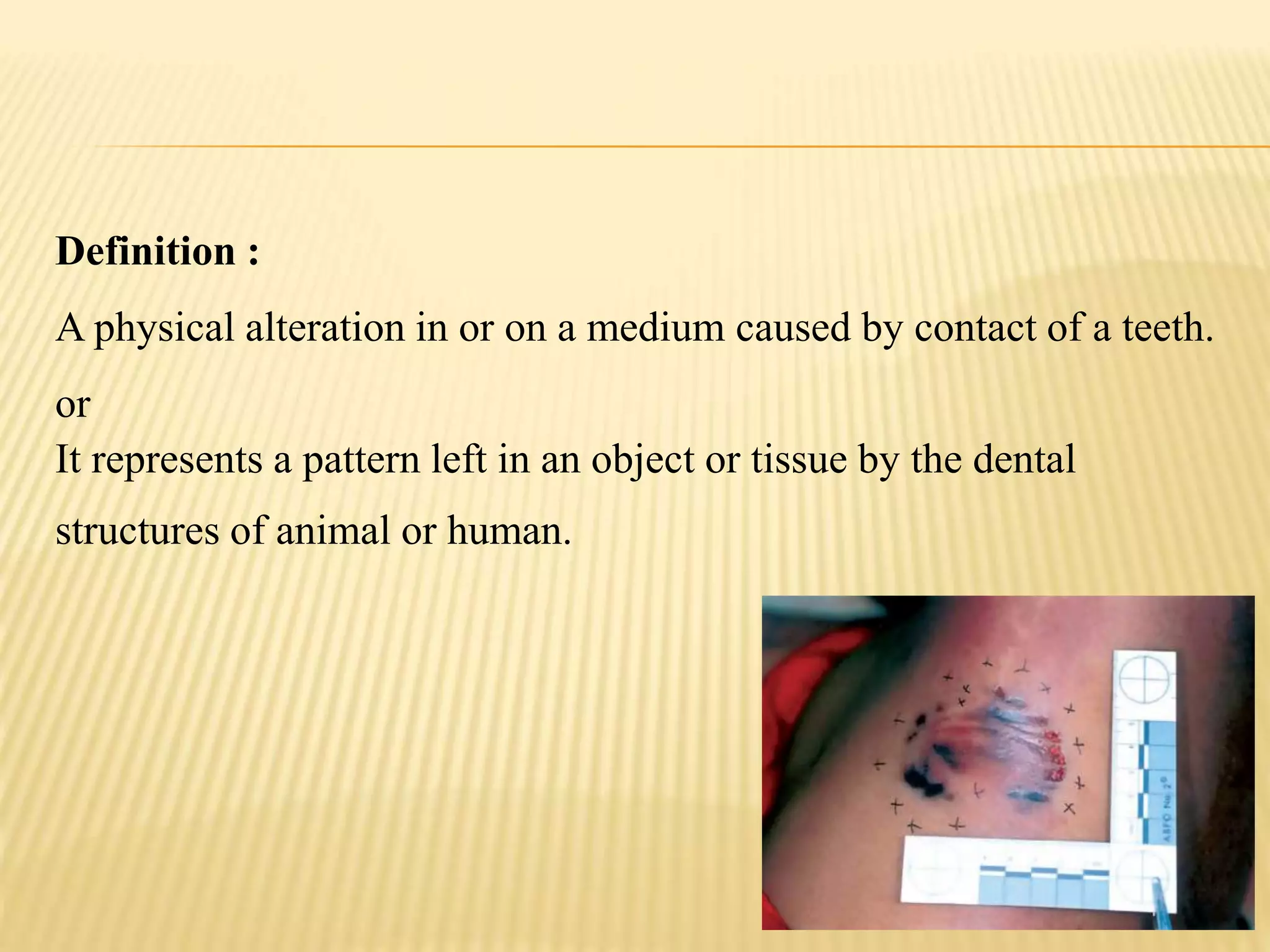
BITE MARK
Definition :
A physical alteration in or on a medium caused by contact of a teeth.
or
It represents a pattern left in an object or tissue by the dental
structures of animal or human.
Defined by MacDonald as
“a mark caused by the teeth either alone or in
combination with other mouth parts”
Classifications
Cameron & Sims- based on type of agent & material
exhibiting it.
Agent Materials
Human Skin, Body Tissue
Animal Food Stuff
Other Material
Mac Donald –Etiologic Classification
 Tooth Pressure Marks –produced by direct application of
pressure by teeth.
 Tongue Pressure Marks – when too much of tissue taken
inside mouth, marks of tongue or rugae will appear.
 Tooth Scrape Marks- due to scraping of teeth across bitten
material, usually by anterior teeth.
WEBSTER’S CLASSIFICATION
 Type I. The food item fractures readily with
limited depth of tooth penetration (e.g. hard
chocolate).
 Type II. Fracture of fragment of food item
with considerable penetration of teeth (e.g.
bite marks in apple and other firm fruits).
 Type III. Complete or near complete
penetration of the food item with slide marks
(e.g. cheese, banana)
HISTORY
 Dr. Jonathan Taft in the 1870 Ohio trial of Ansil L.
Robinson
 Doyle vs. State of Texas, 1954 --- First Modern
Case
 Publication of The Guidelines for Bite Mark
Analysis by the American Board of Forensic
Odontology in 1986
HUMAN BITE MARK APPEARANCE
 circular or elliptical mark (caused by the
upper and lower arches) found on the skin
with a central area of ecchymosis.
 different classes of teeth are usually distinct--
Class charateristics
 Individual charateristics--fractures,
rotations, spacing, etc make the bite mark
distinct.
HUMAN BITE MARK APPEARANCE
 Circular or oval patterned injury consisting of two opposing
U-shaped arches separated at their bases by open spaces
that, in life, represent the throat or posterior portion of the
mouth.
 Along the outer edge or periphery of the impressions of the
arches there is usually a series of abrasions, or contusions,
with or without lacerations, that reflects the size, shape and
arrangement of the class characteristics of the incisal or
occlusal surfaces of the dentition that made the mark
 Central ecchymosis (or contusions), when found, can be
caused by pressure of the teeth, with distortion, leakage, or
rupture of the small vessels and capillaries present. It can
also be caused by the application of negative pressure
caused by sucking or negative pressure produced by the
 Linear abrasions, striations, and contusions can
be caused by the movement of the teeth over
the skin, or by imprinting of the inner surfaces of
the teeth against the skin (lingual markings).
These have also been called drag marks.
 Occasionally a double bite pattern is seen,
where two bites were done quickly in the same
location or the skin slips and the teeth quickly
contacts a second time.
 Compression of the skin surface due to tooth
pressure during a bite initially causes
indentations edema over the bite
subcutaneous bleeding(contusions or
bruises). This is the most common
presentation of bite marks
IDENTIFICATION OF A HUMAN BITE MARK
Sweet has suggested that a human bite mark may be
identifiedby the following characteristics
 Gross Characteristics
A circular or elliptical mark found on the skin with a central area of ecchymosis
 Class Characteristics
Marks produced by different classes of teeth are usually distinct, allowing one to
differentiate the type of tooth within a bite mark
 Individual Characteristics
Features such as fractures, rotations, spacing etc
 The circular/elliptical mark is caused by the
upper and lower arches while the central area of
ecchymosis is apparently due to sucking action
 Incisors produce rectangular marks; canines are
triangular or rectangular depending on the
amount of attrition; premolars and molars are
spherical or point-shaped
The protocol for bite mark evidence collection that follows has been recommended
by the AMERICAN BOARD OF FORENSIC ODONTOLOGY (ABFO)
 Case Demographics
 Name
 Age
 Sex
 Case Number
 Date of Examination
 Name of the Examiner(s)
 Visual Examination
 Photography
 Saliva Swab
 Impressions.
 Impression of the bite area may be made when
tooth indentations exist. The material of choice is
Vinyl Polysiloxane.
 Visual examination
Orientation and location of the mark
Type of injury
Color, size, and shape
Contour, texture, and elasticity of the bite site
Differences between upper and lower arches and between
individual teeth.
 Photography. Photographs provide a permanent record of bite
marks. Therefore no time should be lost in obtaining pictures
 Color and black-and-white photographs from different angles
may be taken.
 Close-up photographs —
 Made with ABFO No. 2 scale
 The camera should be positioned directly over the injury site with the long axis of
the lens perpendicular to the surface of the bitten skin.
 If the bite is on a curved surface and the upper and lower arch marks are wide
apart, separate photographs of each mark should be taken.
 Brown states that when the victim is alive, photographs can be repeated every
24-hours for 3–4 days to record progressive changes in the appearance of bite
mark.
ABFO NO. 2 SCALE
 L-shaped non-flexible instrument
 Two metric scales
 18% color gray scale
 Three circular symbols
 Rectifying grids
Reference scale permits the bite mark images to be measured
and prepared as life-size (i.e., 1:1) representations of the
pattern injury
 Softwares Used:
 Adobe Photoshop
 Mideo Systems CASEWORKSeis
SALIVA SWAB
 It is reasonable to assume that a bite can not be inflicted without leaving
saliva behind. Saliva deposited on skin may have WBCs and sloughed
epithelial cells which may be a source of DNA, enabling a direct link to
the suspect. Care should be taken to not wash the bite area before
saliva swabbing.
 This rehydrates the dried cells in the bitten area.
 The latter prevents degradation of salivary DNA and bacterial growth
 Clark recommends that clothing must also be swabbed for saliva if the
bite has occurred through it
 A cotton swab moistened with distilled water should be used for
swabbing
 The swab is air-dried at room temperature for about 30-minutes, placed
in labeled paper envelopes and stored under refrigeration
 Impressions
 The material of choice is Vinyl Polysiloxane.
 The impression material may be reinforced with
dental stone, autopolymerizing acrylic or
impression compound to prevent dimensional
change
TISSUE SAMPLE
COLLECTION
 With the permission of the Medical Examiner
or coroner, the epidermis, dermis, and
underlying muscle and adipose tissue can be
removed for transillumination analysis.
 Before excision, an acrylic ring or stent must
be secured within 1 inch of the borders of the
injured tissue sample.
 The ring or stent prevents shrinkage and distortion of the
specimen when it is placed into a 4% formalin solution for
fixation.
 The acrylic material is bound to the skin surface with
cyanoacrylate and sutures.
 These tissues samples can be transilluminated by
backlighting.
 This process permits observation of the pattern injury in
the bruised skin by a manner that is not possible when
the tissue is in situ.
EVIDENCE COLLECTION FROM THE SUSPECT
 Photographs of the suspect’s teeth in occlusion and in
open bite
 Maxillary and mandibular impressions made with rubber
base impression material or irreversible hydrocolloid, and
models poured in dental stone.
 Bite registration on a thin sheet of wax may be made if
impressions are unavailable.
 Saliva swabs, preferably from the buccal vestibule, should
be obtained for comparing with the swab collected from
the bite mark.
OVERLAYS
 Indirect Method
 Tracing chewing surfaces of teeth on a sheet of transparent acetate.
 Placing the incisal edges of the study casts on the glass of an office photocopier and
duplicating on special paper.
 Placing an opaque powder, such as barium sulfate, into wax or acrylic test bites and by
obtaining radiographs of these exemplars.
 Direct Method
 Stone cast exemplar of the dentition of a suspect placed over a 1:1 image of a BMPI for
comparison
 Digitized and computer-generated hollow volume overlays are fabricated, enhanced, and
subsequently compared using Adobe Photoshop or other graphic editing programs
CONCLUSIONS IN BITE MARK ANALYSIS
 Definite Biter
 There is reasonable medical certainty to indicate that the bite mark has been
produced by the suspect’s dentition.
 There is concordance of sufficient distinctive, individual characteristics to confer
uniqueness within the population under consideration.
 There is absence of any unexplainable discrepancies.
 Probable Biter
 Bite mark shows some degree of specificity to the suspect’s teeth
by virtue of a sufficient number of matching points, including
some corresponding individual characteristics.
 There is absence of any unexplainable discrepancies
 Possible Biter
 The bite mark and the suspect’s dentition are consistent: although the suspect’s teeth could
have made the bite mark, there are no characteristic matches to be absolutely certain.
 The similarity is non-specific or there is similarity of class characteristics.
 Matching points are general and/or few and there are no incompatible inconsistencies that would
serve to exclude the bite mark as having been caused by the suspect
 Not the Biter
 The bite mark and the suspect’s dentition are not consistent.
 Features on the bite mark indicate that the suspect’s teeth have definitely not caused them
THANK YOU

Bite mark

  • 1.
  • 2.
    Definition : A physicalalteration in or on a medium caused by contact of a teeth. or It represents a pattern left in an object or tissue by the dental structures of animal or human.
  • 3.
    Defined by MacDonaldas “a mark caused by the teeth either alone or in combination with other mouth parts”
  • 4.
    Classifications Cameron & Sims-based on type of agent & material exhibiting it. Agent Materials Human Skin, Body Tissue Animal Food Stuff Other Material
  • 5.
    Mac Donald –EtiologicClassification  Tooth Pressure Marks –produced by direct application of pressure by teeth.  Tongue Pressure Marks – when too much of tissue taken inside mouth, marks of tongue or rugae will appear.  Tooth Scrape Marks- due to scraping of teeth across bitten material, usually by anterior teeth.
  • 6.
    WEBSTER’S CLASSIFICATION  TypeI. The food item fractures readily with limited depth of tooth penetration (e.g. hard chocolate).  Type II. Fracture of fragment of food item with considerable penetration of teeth (e.g. bite marks in apple and other firm fruits).  Type III. Complete or near complete penetration of the food item with slide marks (e.g. cheese, banana)
  • 7.
    HISTORY  Dr. JonathanTaft in the 1870 Ohio trial of Ansil L. Robinson  Doyle vs. State of Texas, 1954 --- First Modern Case  Publication of The Guidelines for Bite Mark Analysis by the American Board of Forensic Odontology in 1986
  • 8.
    HUMAN BITE MARKAPPEARANCE  circular or elliptical mark (caused by the upper and lower arches) found on the skin with a central area of ecchymosis.  different classes of teeth are usually distinct-- Class charateristics  Individual charateristics--fractures, rotations, spacing, etc make the bite mark distinct.
  • 9.
    HUMAN BITE MARKAPPEARANCE  Circular or oval patterned injury consisting of two opposing U-shaped arches separated at their bases by open spaces that, in life, represent the throat or posterior portion of the mouth.  Along the outer edge or periphery of the impressions of the arches there is usually a series of abrasions, or contusions, with or without lacerations, that reflects the size, shape and arrangement of the class characteristics of the incisal or occlusal surfaces of the dentition that made the mark  Central ecchymosis (or contusions), when found, can be caused by pressure of the teeth, with distortion, leakage, or rupture of the small vessels and capillaries present. It can also be caused by the application of negative pressure caused by sucking or negative pressure produced by the
  • 10.
     Linear abrasions,striations, and contusions can be caused by the movement of the teeth over the skin, or by imprinting of the inner surfaces of the teeth against the skin (lingual markings). These have also been called drag marks.  Occasionally a double bite pattern is seen, where two bites were done quickly in the same location or the skin slips and the teeth quickly contacts a second time.
  • 12.
     Compression ofthe skin surface due to tooth pressure during a bite initially causes indentations edema over the bite subcutaneous bleeding(contusions or bruises). This is the most common presentation of bite marks
  • 13.
    IDENTIFICATION OF AHUMAN BITE MARK Sweet has suggested that a human bite mark may be identifiedby the following characteristics  Gross Characteristics A circular or elliptical mark found on the skin with a central area of ecchymosis  Class Characteristics Marks produced by different classes of teeth are usually distinct, allowing one to differentiate the type of tooth within a bite mark  Individual Characteristics Features such as fractures, rotations, spacing etc
  • 14.
     The circular/ellipticalmark is caused by the upper and lower arches while the central area of ecchymosis is apparently due to sucking action  Incisors produce rectangular marks; canines are triangular or rectangular depending on the amount of attrition; premolars and molars are spherical or point-shaped
  • 15.
    The protocol forbite mark evidence collection that follows has been recommended by the AMERICAN BOARD OF FORENSIC ODONTOLOGY (ABFO)  Case Demographics  Name  Age  Sex  Case Number  Date of Examination  Name of the Examiner(s)
  • 16.
     Visual Examination Photography  Saliva Swab  Impressions.  Impression of the bite area may be made when tooth indentations exist. The material of choice is Vinyl Polysiloxane.
  • 17.
     Visual examination Orientationand location of the mark Type of injury Color, size, and shape Contour, texture, and elasticity of the bite site Differences between upper and lower arches and between individual teeth.  Photography. Photographs provide a permanent record of bite marks. Therefore no time should be lost in obtaining pictures  Color and black-and-white photographs from different angles may be taken.
  • 18.
     Close-up photographs—  Made with ABFO No. 2 scale  The camera should be positioned directly over the injury site with the long axis of the lens perpendicular to the surface of the bitten skin.  If the bite is on a curved surface and the upper and lower arch marks are wide apart, separate photographs of each mark should be taken.  Brown states that when the victim is alive, photographs can be repeated every 24-hours for 3–4 days to record progressive changes in the appearance of bite mark.
  • 19.
    ABFO NO. 2SCALE  L-shaped non-flexible instrument  Two metric scales  18% color gray scale  Three circular symbols  Rectifying grids Reference scale permits the bite mark images to be measured and prepared as life-size (i.e., 1:1) representations of the pattern injury
  • 20.
     Softwares Used: Adobe Photoshop  Mideo Systems CASEWORKSeis
  • 21.
    SALIVA SWAB  Itis reasonable to assume that a bite can not be inflicted without leaving saliva behind. Saliva deposited on skin may have WBCs and sloughed epithelial cells which may be a source of DNA, enabling a direct link to the suspect. Care should be taken to not wash the bite area before saliva swabbing.  This rehydrates the dried cells in the bitten area.  The latter prevents degradation of salivary DNA and bacterial growth  Clark recommends that clothing must also be swabbed for saliva if the bite has occurred through it  A cotton swab moistened with distilled water should be used for swabbing  The swab is air-dried at room temperature for about 30-minutes, placed in labeled paper envelopes and stored under refrigeration
  • 22.
     Impressions  Thematerial of choice is Vinyl Polysiloxane.  The impression material may be reinforced with dental stone, autopolymerizing acrylic or impression compound to prevent dimensional change
  • 23.
    TISSUE SAMPLE COLLECTION  Withthe permission of the Medical Examiner or coroner, the epidermis, dermis, and underlying muscle and adipose tissue can be removed for transillumination analysis.  Before excision, an acrylic ring or stent must be secured within 1 inch of the borders of the injured tissue sample.
  • 24.
     The ringor stent prevents shrinkage and distortion of the specimen when it is placed into a 4% formalin solution for fixation.  The acrylic material is bound to the skin surface with cyanoacrylate and sutures.  These tissues samples can be transilluminated by backlighting.  This process permits observation of the pattern injury in the bruised skin by a manner that is not possible when the tissue is in situ.
  • 25.
    EVIDENCE COLLECTION FROMTHE SUSPECT  Photographs of the suspect’s teeth in occlusion and in open bite  Maxillary and mandibular impressions made with rubber base impression material or irreversible hydrocolloid, and models poured in dental stone.  Bite registration on a thin sheet of wax may be made if impressions are unavailable.  Saliva swabs, preferably from the buccal vestibule, should be obtained for comparing with the swab collected from the bite mark.
  • 26.
    OVERLAYS  Indirect Method Tracing chewing surfaces of teeth on a sheet of transparent acetate.  Placing the incisal edges of the study casts on the glass of an office photocopier and duplicating on special paper.  Placing an opaque powder, such as barium sulfate, into wax or acrylic test bites and by obtaining radiographs of these exemplars.  Direct Method  Stone cast exemplar of the dentition of a suspect placed over a 1:1 image of a BMPI for comparison  Digitized and computer-generated hollow volume overlays are fabricated, enhanced, and subsequently compared using Adobe Photoshop or other graphic editing programs
  • 27.
    CONCLUSIONS IN BITEMARK ANALYSIS  Definite Biter  There is reasonable medical certainty to indicate that the bite mark has been produced by the suspect’s dentition.  There is concordance of sufficient distinctive, individual characteristics to confer uniqueness within the population under consideration.  There is absence of any unexplainable discrepancies.  Probable Biter  Bite mark shows some degree of specificity to the suspect’s teeth by virtue of a sufficient number of matching points, including some corresponding individual characteristics.  There is absence of any unexplainable discrepancies
  • 28.
     Possible Biter The bite mark and the suspect’s dentition are consistent: although the suspect’s teeth could have made the bite mark, there are no characteristic matches to be absolutely certain.  The similarity is non-specific or there is similarity of class characteristics.  Matching points are general and/or few and there are no incompatible inconsistencies that would serve to exclude the bite mark as having been caused by the suspect  Not the Biter  The bite mark and the suspect’s dentition are not consistent.  Features on the bite mark indicate that the suspect’s teeth have definitely not caused them
  • 29.