Download as PDF, PPTX
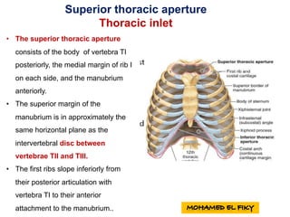
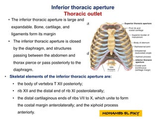
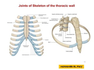
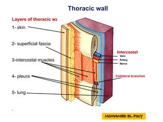
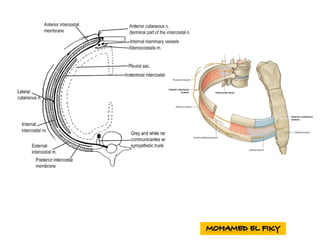
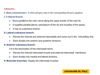
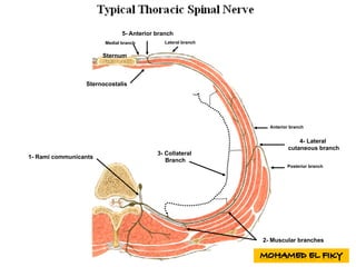
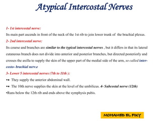
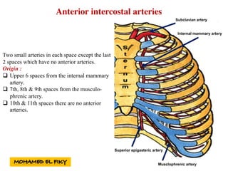
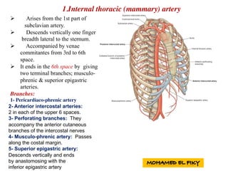
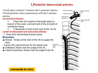

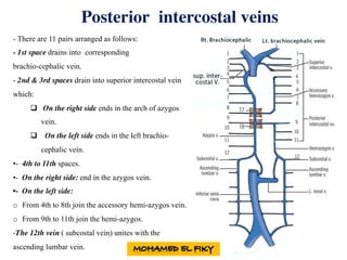


1. The document describes the anatomy of the thoracic wall including bones, joints, muscles, blood vessels, and nerves. 2. It discusses the 12 pairs of ribs, their classification as true, false, or floating ribs, and their articulations with the sternum. 3. The intercostal spaces contain intercostal muscles like the external and internal intercostals that act in respiration, as well as vessels and nerves.


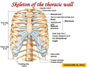
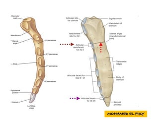
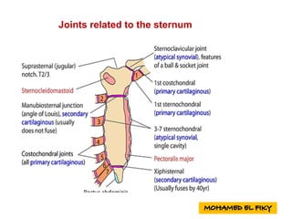
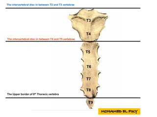
![Anatomy of Male genital organs [auto saved]](https://cdn.slidesharecdn.com/ss_thumbnails/malegenitalorgansauto-saved-200818065025-thumbnail.jpg?width=640&height=640&fit=bounds)


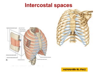
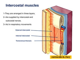
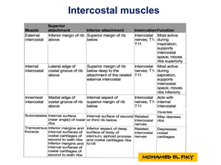
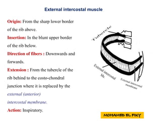
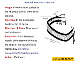
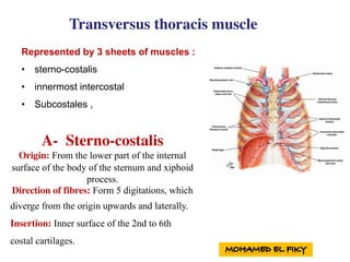
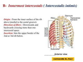
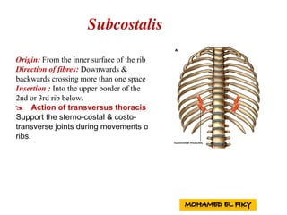
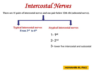
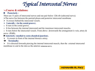
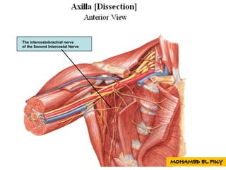
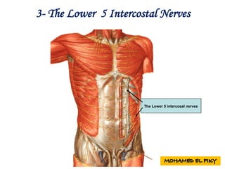
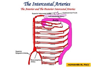
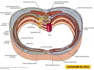
![anatomy of Female genital organs [auto saved]](https://cdn.slidesharecdn.com/ss_thumbnails/femalegenitalorgansauto-saved-200818064612-thumbnail.jpg?width=640&height=640&fit=bounds)


