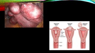
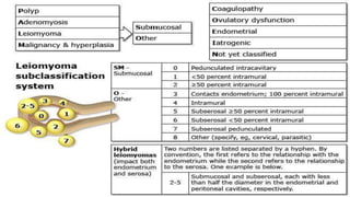
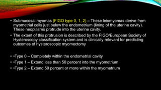
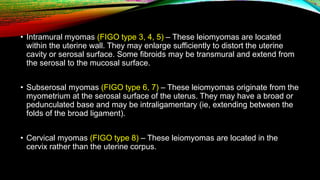
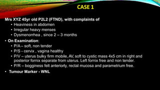
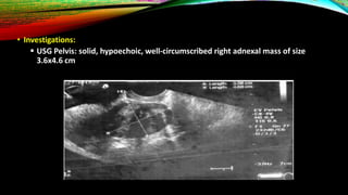
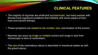
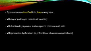
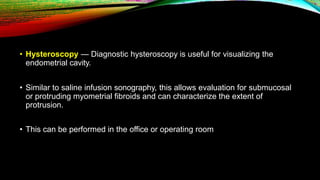
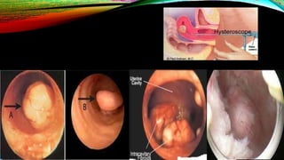
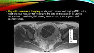
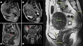
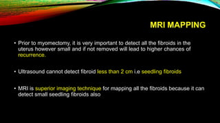
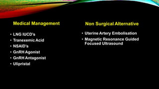
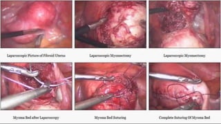
FIBROIDS – CLINICAL
PRESENTATION AND EVALUATION
• Uterine leiomyomas (also referred to as fibroids or myomas) are the most
common pelvic tumor in women
• They are benign monoclonal tumors arising from the smooth muscle cells of
the myometrium.
FIGO CLASSIFICATION
• Submucosal myomas (FIGO type 0, 1, 2) – These leiomyomas derive from
myometrial cells just below the endometrium (lining of the uterine cavity).
These neoplasms protrude into the uterine cavity.
• The extent of this protrusion is described by the FIGO/European Society of
Hysteroscopy classification system and is clinically relevant for predicting
outcomes of hysteroscopic myomectomy
• •Type 0 – Completely within the endometrial cavity
• •Type 1 – Extend less than 50 percent into the myometrium
• •Type 2 – Extend 50 percent or more within the myometrium
• Intramural myomas (FIGO type 3, 4, 5) – These leiomyomas are located
within the uterine wall. They may enlarge sufficiently to distort the uterine
cavity or serosal surface. Some fibroids may be transmural and extend from
the serosal to the mucosal surface.
• Subserosal myomas (FIGO type 6, 7) – These leiomyomas originate from the
myometrium at the serosal surface of the uterus. They may have a broad or
pedunculated base and may be intraligamentary (ie, extending between the
folds of the broad ligament).
• Cervical myomas (FIGO type 8) – These leiomyomas are located in the
cervix rather than the uterine corpus.
BROAD LIGAMENT FIBROID
CERVICAL FIBROID
MULTIPLE FIBROIDS
CASE 1
Mrs XYZ 45yr old P2L2 (FTND), with complaints of
• Heaviness in abdomen
• Irregular heavy menses
• Dysmenorrhea , since 2 – 3 months
• On Examination:
• P/A – soft, non tender
• P/S - cervix , vagina healthy
• P/V – uterus bulky firm mobile, AV, soft to cystic mass 4x5 cm in right and
posterior fornix separate from uterus. Left fornix free and non tender.
• P/R – bogginess felt anteriorly, rectal mucosa and parametrium free.
• Tumour Marker - WNL
• Investigations:
 USG Pelvis: solid, hypoechoic, well-circumscribed right adnexal mass of size
3.6x4.6 cm
• What is your diagnosis?
• BROAD LIGAMENT FIBROID
• How will You differentiate between a True and False Broad Ligament
Fibroid ?
• What will be relation of the Ureter to this fibroid?
True Broad Ligament False Broad Ligament
Originates from the muscle fibres normally
found in the mesometrium (in the round
ligament, ovario-uterine ligament, and the
connective tissue around the uterine and
ovarian vessels)
Arises from the lateral wall of the uterine
corpus or of the cervix, and bulges outward
between the layers of the broad ligament.
Ureter is medial to mass Ureter is lateral to mass
No groove felt between mass and uterus Groove felt between mass and uterus
• In this case
• the right ureter was safeguarded by dissection and enucleation
could be carried out rather easily.
RISK FACTORS
• Race — The incidence rates of fibroids are typically found to be two- to threefold
greater in black women than in white women
• Parity — Parity (having one or more pregnancies extending beyond 20 weeks of
gestation) decreases the chance of fibroid formation
• Early menarche — Early menarche (<10 years old) is associated with an increased
risk of developing fibroids.
• Hormonal contraception — Use of standard or lower dose oral contraceptives (≤35
mcg ethinyl estradiol/day) do not appear to cause fibroids to grow
• Other endocrine factors — Prenatal exposure to diethylstilbestrol is
associated with an increased risk of fibroids
• Smoking – Early studies showed that smoking decreased the risk of having
fibroids, possibly through the inhibition of aromatase
• Others - Diet, Alcohol, Obesity
PATHOGENESIS
• Transformation of normal myocytes into abnormal myocytes, in most instances
through somatic mutations
• Growth of abnormal myocytes into clinically apparent tumors
HISTOLOGY
• Smooth muscle bundles arranged in
whorl patterns
• Abndant amount of Collagen 1 & 3
present
GENETIC PREDISPOSITION
• Uterine leiomyomas are a common phenotype with many underlying
genotypes. Both somatic and inherited mutations account for the majority of
uterine fibroids, with the most common mutations occurring in
the MED12, HMGA1 and HMGA2, FH, collagen type IV, alpha-5 (COL4A5)
and collagen type IV alpha-6 (COL4A6) genes
• Steroid hormone factors that influence leiomyoma development include
upregulation of aromatase, estrogen, and progesterone receptors as well as
a potential role of gonadotropins.
• Stem cells appear to also play a key role in fibroid pathogenesis
CLINICAL FEATURES
• The majority of myomas are small and asymptomatic, but many women with
fibroids have significant problems that interfere with some aspect of their
lives and warrant therapy.
• These symptoms are related to the number, size, and location of the tumors.
• Myomas can occur as single or multiple tumors and range in size from
microscopic to tens of centimeters.
• The size of the myomatous uterus is described in menstrual weeks as with
the gravid uterus.
• Symptoms are classified into three categories :
• ●Heavy or prolonged menstrual bleeding
• ●Bulk-related symptoms, such as pelvic pressure and pain
• ●Reproductive dysfunction (ie, infertility or obstetric complications)
HEAVY OR PROLONGED MENSTRUAL
BLEEDING
• Heavy or prolonged menses is the typical bleeding pattern with leiomyomas
and the most common fibroid symptom
• Submucosal myomas that protrude into the uterine cavity (eg, types 0 and
1)are most frequently related to significant heavy menstrual bleeding
BULK-RELATED SYMPTOMS
• Pelvic pressure or pain — In general, pelvic discomfort is common in women
with fibroids but less common than AUB. If discomfort is present, it is likely to
be chronic, intermittent, dull pressure or pain
• Bowel symptoms – Fibroids that place pressure on the rectum can result in
constipation
• Urinary symptoms – A heterogeneous group of urinary symptoms including
• frequency,
• difficulty emptying the bladder, or, rarely,
• complete urinary obstruction may all occur in up to 60 percent of women with
fibroids .
• Bladder symptoms sometimes arise when an anterior fibroid presses directly
on the bladder or a posterior fibroid pushes the entire uterus forward
INFERTILITY OR OBSTETRIC
COMPLICATIONS
• Leiomyomas that distort the uterine cavity
(submucosal or intramural with an intracavitary
component) result in difficulty conceiving a
pregnancy and an increased risk of miscarriage
• In addition, leiomyomas have been associated with
adverse pregnancy outcomes (eg, placental
abruption, fetal growth restriction, malpresentation,
and preterm labor and birth)
OTHERS
• Painful menses — Painful menses is reported by many women with fibroids
• Painful intercourse — It is controversial whether women with fibroids are more likely
to experience painful intercourse than women without fibroids
• Fibroid degeneration or torsion
DIAGNOSIS
• History Taking –
• It is important to assess the severity of heavy or prolonged menstrual
bleeding.
• A menstrual history is taken and the volume and duration of bleeding elicited
• For women with pelvic pain or pressure, the location, severity, and
characteristics of the pain should be assessed
• Patients should be asked about other potential pain or bulk-related
symptoms, including dyspareunia, urinary retention, or constipation.
• The patient should be asked about infertility, recurrent miscarriage, or
obstetric complications that may be related to fibroids.
• Physical Examination
• A thorough pelvic examination is performed.
• On bimanual pelvic examination, the size, contour, and mobility should be
noted.
• An enlarged, mobile uterus with an irregular contour is consistent with a
leiomyomatous uterus.
• Size, shape, surface, consistency, mobility, fluctuation
PELVIC ULTRASOUND
• Pelvic ultrasound is the first-line study used to evaluate for uterine fibroids.
• Transvaginal ultrasound has high sensitivity (95 to 100 percent) for detecting
myomas in uteri less than 10 gestational weeks' size.
• Precise localization of fibroids is limited in larger uteri or when there are
many tumors.
• Fibroids are seen on ultrasound usually as hypoechoic, well-circumscribed
round masses, frequently with shadowing
• Calcification in a fibroid generally implies that it has degenerated. These
calcifications can be seen on plain film as "popcorn" calcifications in the
pelvis.
• On ultrasound, the calcifications may appear as clumps or rim-like
calcifications within a mass
EVALUATE THE UTERINE CAVITY
• Saline infusion sonography — Saline infusion sonography
(sonohysterography) is an imaging study in which pelvic ultrasound is
performed while saline is infused into the uterine cavity.
• Use of this technique allows identification of submucosal lesions (some of
which may not be seen on routine ultrasonography) and intramural myomas
that protrude into the cavity and characterizes the extent of protrusion into
the endometrial cavity
• Hysteroscopy — Diagnostic hysteroscopy is useful for visualizing the
endometrial cavity.
• Similar to saline infusion sonography, this allows evaluation for submucosal
or protruding myometrial fibroids and can characterize the extent of
protrusion.
• This can be performed in the office or operating room
• Magnetic resonance imaging — Magnetic resonance imaging (MRI) is the
most effective modality for visualizing the size and location of all uterine
myomas and can distinguish among leiomyomas, adenomyosis, and
adenomyomas.
MRI MAPPING
• Prior to myomectomy, it is very important to detect all the fibroids in the
uterus however small and if not removed will lead to higher chances of
recurrence.
• Ultrasound cannot detect fibroid less than 2 cm i.e seedling fibroids
• MRI is superior imaging technique for mapping all the fibroids because it can
detect small seedling fibroids also
DIFFERENTIAL DIAGNOSIS
• Adenomyosis
• Full Bladder
• Ovarian Cyst
• Edometrial Hyperplasia
• Endometrial Polyp
• Paraovarian lesions
• Pelvic Tumors
• Pregnancy
• Hematometra
• Fecolith
MANAGEMENT
• Medical Management
• Non Surgical Alternatives
• Surgical Management
Medical Management
• LNG IUCD’s
• Tranexemic Acid
• NSAID’s
• GnRH Agonist
• GnRH Antagonist
• Ulipristal
Non Surgical Alternative
• Uterine Artery Embolisation
• Magnetic Resonance Guided
Focused Ultrasound
SURGICAL MANAGEMENT
• Laparoscopic Myomectomy
• Laparoscopic Hysterectomy
• Hysteroscopic Myomectomy
2 LAP MYOMECTOMY +
1 HYSTEROSCOPIC
MYOMECTOMY
• Video
SUMMARY
• Uterine leiomyomas are the most common pelvic tumor in women.
• Leiomyomas are benign monoclonal tumors arising from the smooth muscle
cells of the myometrium
• Symptoms attributable to uterine myomas can generally be classified into
three distinct categories: abnormal uterine bleeding (AUB), pelvic pressure
and pain, and reproductive dysfunction
• Transvaginal ultrasound is the most widely used imaging modality for
evaluating fibroids due to its availability and cost-effectiveness.
• Laparoscopic rather than abdominal myomectomy for women with
leiomyomas for whom laparoscopic removal is technically feasible (by size,
number, and location) and who have access to a surgeon with advanced
laparoscopic skills
fibroids-191226053031.pptx

fibroids-191226053031.pptx

  • 1.
  • 3.
    • Uterine leiomyomas(also referred to as fibroids or myomas) are the most common pelvic tumor in women • They are benign monoclonal tumors arising from the smooth muscle cells of the myometrium.
  • 5.
  • 6.
    • Submucosal myomas(FIGO type 0, 1, 2) – These leiomyomas derive from myometrial cells just below the endometrium (lining of the uterine cavity). These neoplasms protrude into the uterine cavity. • The extent of this protrusion is described by the FIGO/European Society of Hysteroscopy classification system and is clinically relevant for predicting outcomes of hysteroscopic myomectomy • •Type 0 – Completely within the endometrial cavity • •Type 1 – Extend less than 50 percent into the myometrium • •Type 2 – Extend 50 percent or more within the myometrium
  • 7.
    • Intramural myomas(FIGO type 3, 4, 5) – These leiomyomas are located within the uterine wall. They may enlarge sufficiently to distort the uterine cavity or serosal surface. Some fibroids may be transmural and extend from the serosal to the mucosal surface. • Subserosal myomas (FIGO type 6, 7) – These leiomyomas originate from the myometrium at the serosal surface of the uterus. They may have a broad or pedunculated base and may be intraligamentary (ie, extending between the folds of the broad ligament). • Cervical myomas (FIGO type 8) – These leiomyomas are located in the cervix rather than the uterine corpus.
  • 8.
  • 9.
  • 10.
  • 11.
    CASE 1 Mrs XYZ45yr old P2L2 (FTND), with complaints of • Heaviness in abdomen • Irregular heavy menses • Dysmenorrhea , since 2 – 3 months • On Examination: • P/A – soft, non tender • P/S - cervix , vagina healthy • P/V – uterus bulky firm mobile, AV, soft to cystic mass 4x5 cm in right and posterior fornix separate from uterus. Left fornix free and non tender. • P/R – bogginess felt anteriorly, rectal mucosa and parametrium free. • Tumour Marker - WNL
  • 12.
    • Investigations:  USGPelvis: solid, hypoechoic, well-circumscribed right adnexal mass of size 3.6x4.6 cm
  • 13.
    • What isyour diagnosis?
  • 14.
  • 15.
    • How willYou differentiate between a True and False Broad Ligament Fibroid ? • What will be relation of the Ureter to this fibroid?
  • 16.
    True Broad LigamentFalse Broad Ligament Originates from the muscle fibres normally found in the mesometrium (in the round ligament, ovario-uterine ligament, and the connective tissue around the uterine and ovarian vessels) Arises from the lateral wall of the uterine corpus or of the cervix, and bulges outward between the layers of the broad ligament. Ureter is medial to mass Ureter is lateral to mass No groove felt between mass and uterus Groove felt between mass and uterus
  • 17.
    • In thiscase • the right ureter was safeguarded by dissection and enucleation could be carried out rather easily.
  • 18.
    RISK FACTORS • Race— The incidence rates of fibroids are typically found to be two- to threefold greater in black women than in white women • Parity — Parity (having one or more pregnancies extending beyond 20 weeks of gestation) decreases the chance of fibroid formation • Early menarche — Early menarche (<10 years old) is associated with an increased risk of developing fibroids. • Hormonal contraception — Use of standard or lower dose oral contraceptives (≤35 mcg ethinyl estradiol/day) do not appear to cause fibroids to grow
  • 19.
    • Other endocrinefactors — Prenatal exposure to diethylstilbestrol is associated with an increased risk of fibroids • Smoking – Early studies showed that smoking decreased the risk of having fibroids, possibly through the inhibition of aromatase • Others - Diet, Alcohol, Obesity
  • 20.
    PATHOGENESIS • Transformation ofnormal myocytes into abnormal myocytes, in most instances through somatic mutations • Growth of abnormal myocytes into clinically apparent tumors
  • 21.
    HISTOLOGY • Smooth musclebundles arranged in whorl patterns • Abndant amount of Collagen 1 & 3 present
  • 22.
    GENETIC PREDISPOSITION • Uterineleiomyomas are a common phenotype with many underlying genotypes. Both somatic and inherited mutations account for the majority of uterine fibroids, with the most common mutations occurring in the MED12, HMGA1 and HMGA2, FH, collagen type IV, alpha-5 (COL4A5) and collagen type IV alpha-6 (COL4A6) genes
  • 23.
    • Steroid hormonefactors that influence leiomyoma development include upregulation of aromatase, estrogen, and progesterone receptors as well as a potential role of gonadotropins. • Stem cells appear to also play a key role in fibroid pathogenesis
  • 24.
    CLINICAL FEATURES • Themajority of myomas are small and asymptomatic, but many women with fibroids have significant problems that interfere with some aspect of their lives and warrant therapy. • These symptoms are related to the number, size, and location of the tumors. • Myomas can occur as single or multiple tumors and range in size from microscopic to tens of centimeters. • The size of the myomatous uterus is described in menstrual weeks as with the gravid uterus.
  • 25.
    • Symptoms areclassified into three categories : • ●Heavy or prolonged menstrual bleeding • ●Bulk-related symptoms, such as pelvic pressure and pain • ●Reproductive dysfunction (ie, infertility or obstetric complications)
  • 26.
    HEAVY OR PROLONGEDMENSTRUAL BLEEDING • Heavy or prolonged menses is the typical bleeding pattern with leiomyomas and the most common fibroid symptom • Submucosal myomas that protrude into the uterine cavity (eg, types 0 and 1)are most frequently related to significant heavy menstrual bleeding
  • 27.
    BULK-RELATED SYMPTOMS • Pelvicpressure or pain — In general, pelvic discomfort is common in women with fibroids but less common than AUB. If discomfort is present, it is likely to be chronic, intermittent, dull pressure or pain • Bowel symptoms – Fibroids that place pressure on the rectum can result in constipation
  • 28.
    • Urinary symptoms– A heterogeneous group of urinary symptoms including • frequency, • difficulty emptying the bladder, or, rarely, • complete urinary obstruction may all occur in up to 60 percent of women with fibroids . • Bladder symptoms sometimes arise when an anterior fibroid presses directly on the bladder or a posterior fibroid pushes the entire uterus forward
  • 29.
    INFERTILITY OR OBSTETRIC COMPLICATIONS •Leiomyomas that distort the uterine cavity (submucosal or intramural with an intracavitary component) result in difficulty conceiving a pregnancy and an increased risk of miscarriage • In addition, leiomyomas have been associated with adverse pregnancy outcomes (eg, placental abruption, fetal growth restriction, malpresentation, and preterm labor and birth)
  • 30.
    OTHERS • Painful menses— Painful menses is reported by many women with fibroids • Painful intercourse — It is controversial whether women with fibroids are more likely to experience painful intercourse than women without fibroids • Fibroid degeneration or torsion
  • 31.
    DIAGNOSIS • History Taking– • It is important to assess the severity of heavy or prolonged menstrual bleeding. • A menstrual history is taken and the volume and duration of bleeding elicited • For women with pelvic pain or pressure, the location, severity, and characteristics of the pain should be assessed
  • 32.
    • Patients shouldbe asked about other potential pain or bulk-related symptoms, including dyspareunia, urinary retention, or constipation. • The patient should be asked about infertility, recurrent miscarriage, or obstetric complications that may be related to fibroids.
  • 33.
    • Physical Examination •A thorough pelvic examination is performed. • On bimanual pelvic examination, the size, contour, and mobility should be noted. • An enlarged, mobile uterus with an irregular contour is consistent with a leiomyomatous uterus. • Size, shape, surface, consistency, mobility, fluctuation
  • 34.
    PELVIC ULTRASOUND • Pelvicultrasound is the first-line study used to evaluate for uterine fibroids. • Transvaginal ultrasound has high sensitivity (95 to 100 percent) for detecting myomas in uteri less than 10 gestational weeks' size. • Precise localization of fibroids is limited in larger uteri or when there are many tumors.
  • 35.
    • Fibroids areseen on ultrasound usually as hypoechoic, well-circumscribed round masses, frequently with shadowing
  • 36.
    • Calcification ina fibroid generally implies that it has degenerated. These calcifications can be seen on plain film as "popcorn" calcifications in the pelvis. • On ultrasound, the calcifications may appear as clumps or rim-like calcifications within a mass
  • 37.
    EVALUATE THE UTERINECAVITY • Saline infusion sonography — Saline infusion sonography (sonohysterography) is an imaging study in which pelvic ultrasound is performed while saline is infused into the uterine cavity. • Use of this technique allows identification of submucosal lesions (some of which may not be seen on routine ultrasonography) and intramural myomas that protrude into the cavity and characterizes the extent of protrusion into the endometrial cavity
  • 38.
    • Hysteroscopy —Diagnostic hysteroscopy is useful for visualizing the endometrial cavity. • Similar to saline infusion sonography, this allows evaluation for submucosal or protruding myometrial fibroids and can characterize the extent of protrusion. • This can be performed in the office or operating room
  • 40.
    • Magnetic resonanceimaging — Magnetic resonance imaging (MRI) is the most effective modality for visualizing the size and location of all uterine myomas and can distinguish among leiomyomas, adenomyosis, and adenomyomas.
  • 42.
    MRI MAPPING • Priorto myomectomy, it is very important to detect all the fibroids in the uterus however small and if not removed will lead to higher chances of recurrence. • Ultrasound cannot detect fibroid less than 2 cm i.e seedling fibroids • MRI is superior imaging technique for mapping all the fibroids because it can detect small seedling fibroids also
  • 43.
    DIFFERENTIAL DIAGNOSIS • Adenomyosis •Full Bladder • Ovarian Cyst • Edometrial Hyperplasia • Endometrial Polyp • Paraovarian lesions • Pelvic Tumors • Pregnancy • Hematometra • Fecolith
  • 44.
    MANAGEMENT • Medical Management •Non Surgical Alternatives • Surgical Management
  • 45.
    Medical Management • LNGIUCD’s • Tranexemic Acid • NSAID’s • GnRH Agonist • GnRH Antagonist • Ulipristal Non Surgical Alternative • Uterine Artery Embolisation • Magnetic Resonance Guided Focused Ultrasound
  • 46.
    SURGICAL MANAGEMENT • LaparoscopicMyomectomy • Laparoscopic Hysterectomy • Hysteroscopic Myomectomy
  • 48.
    2 LAP MYOMECTOMY+ 1 HYSTEROSCOPIC MYOMECTOMY • Video
  • 49.
    SUMMARY • Uterine leiomyomasare the most common pelvic tumor in women. • Leiomyomas are benign monoclonal tumors arising from the smooth muscle cells of the myometrium • Symptoms attributable to uterine myomas can generally be classified into three distinct categories: abnormal uterine bleeding (AUB), pelvic pressure and pain, and reproductive dysfunction • Transvaginal ultrasound is the most widely used imaging modality for evaluating fibroids due to its availability and cost-effectiveness. • Laparoscopic rather than abdominal myomectomy for women with leiomyomas for whom laparoscopic removal is technically feasible (by size, number, and location) and who have access to a surgeon with advanced laparoscopic skills