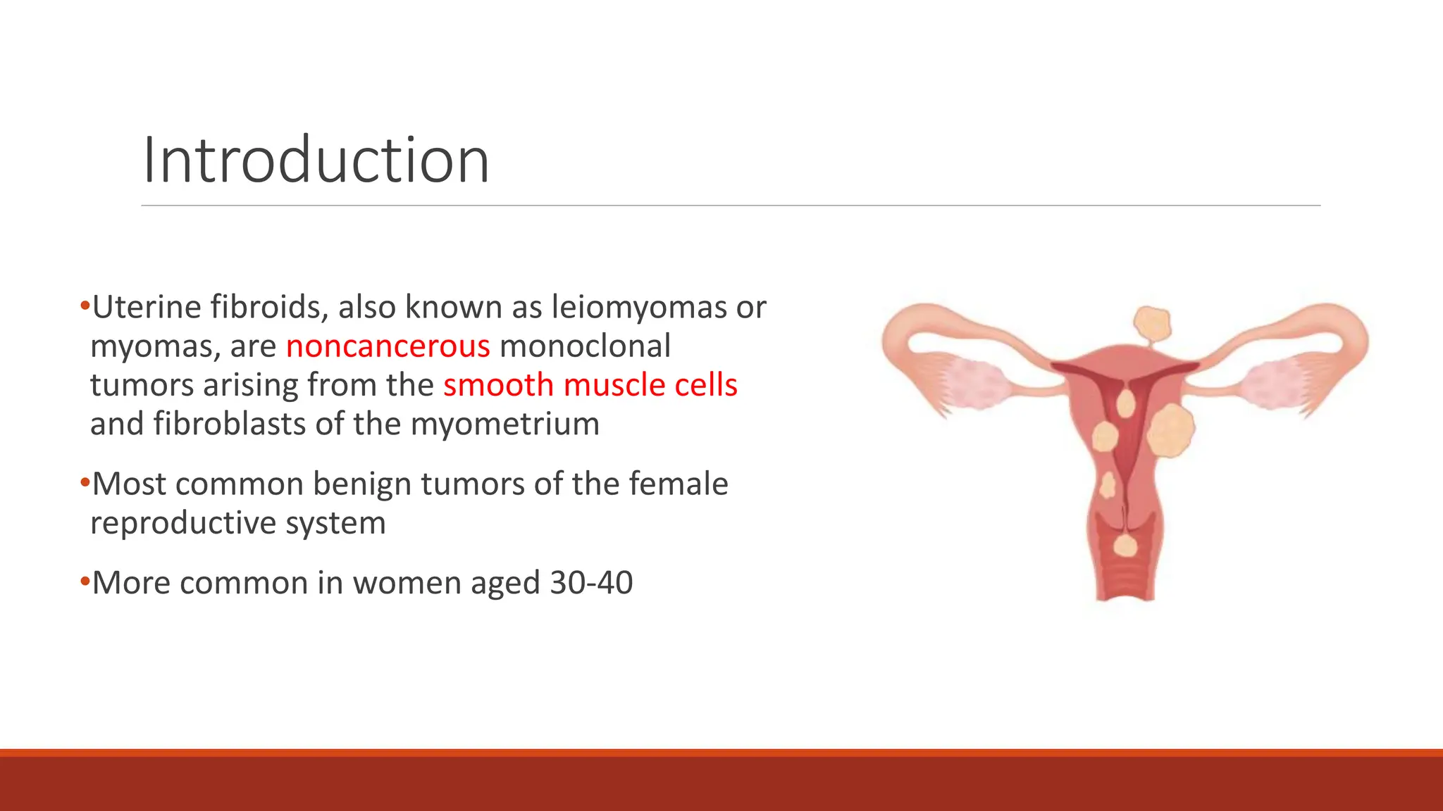
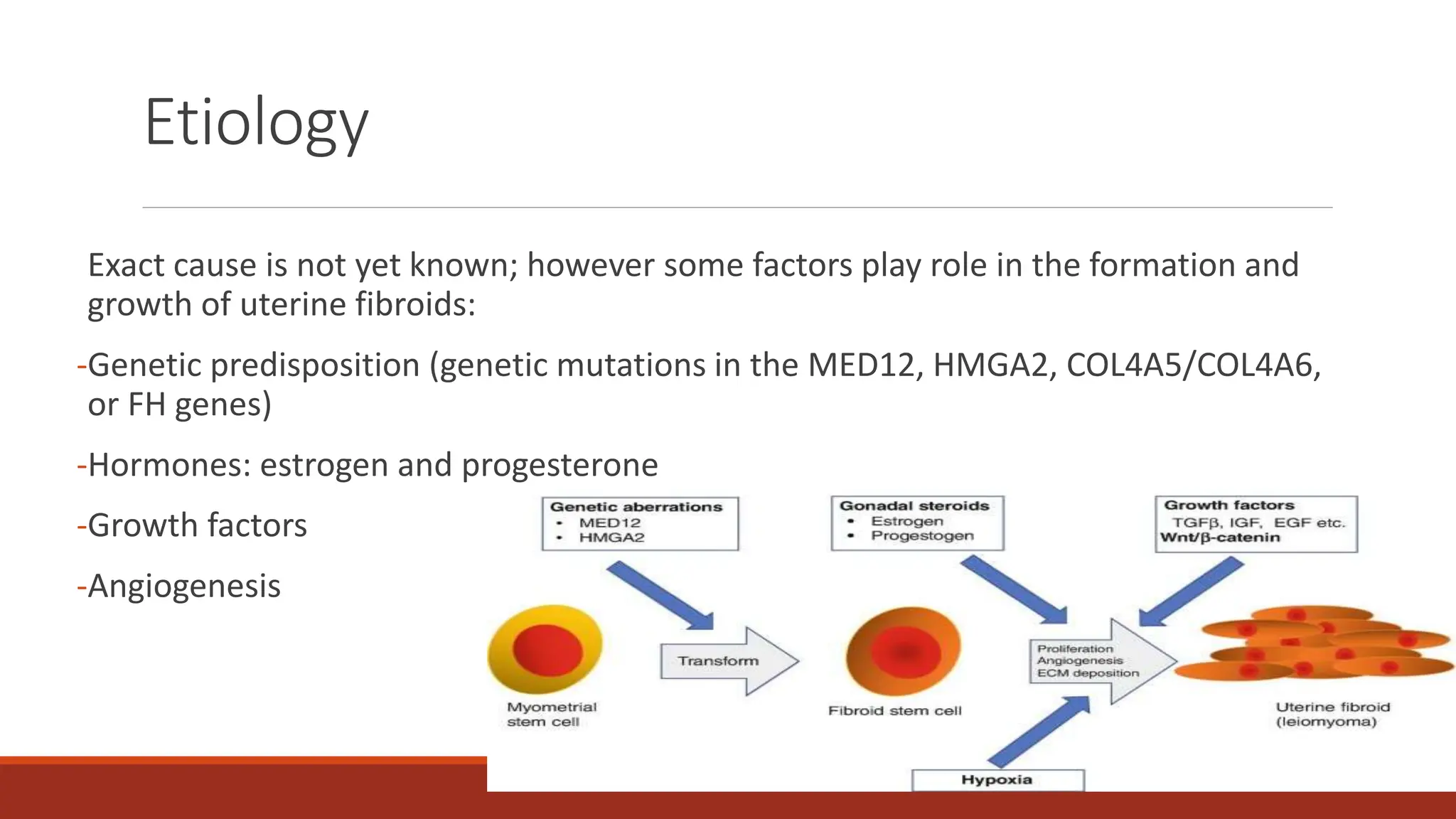
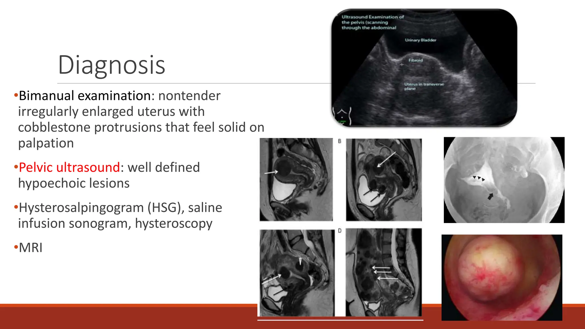
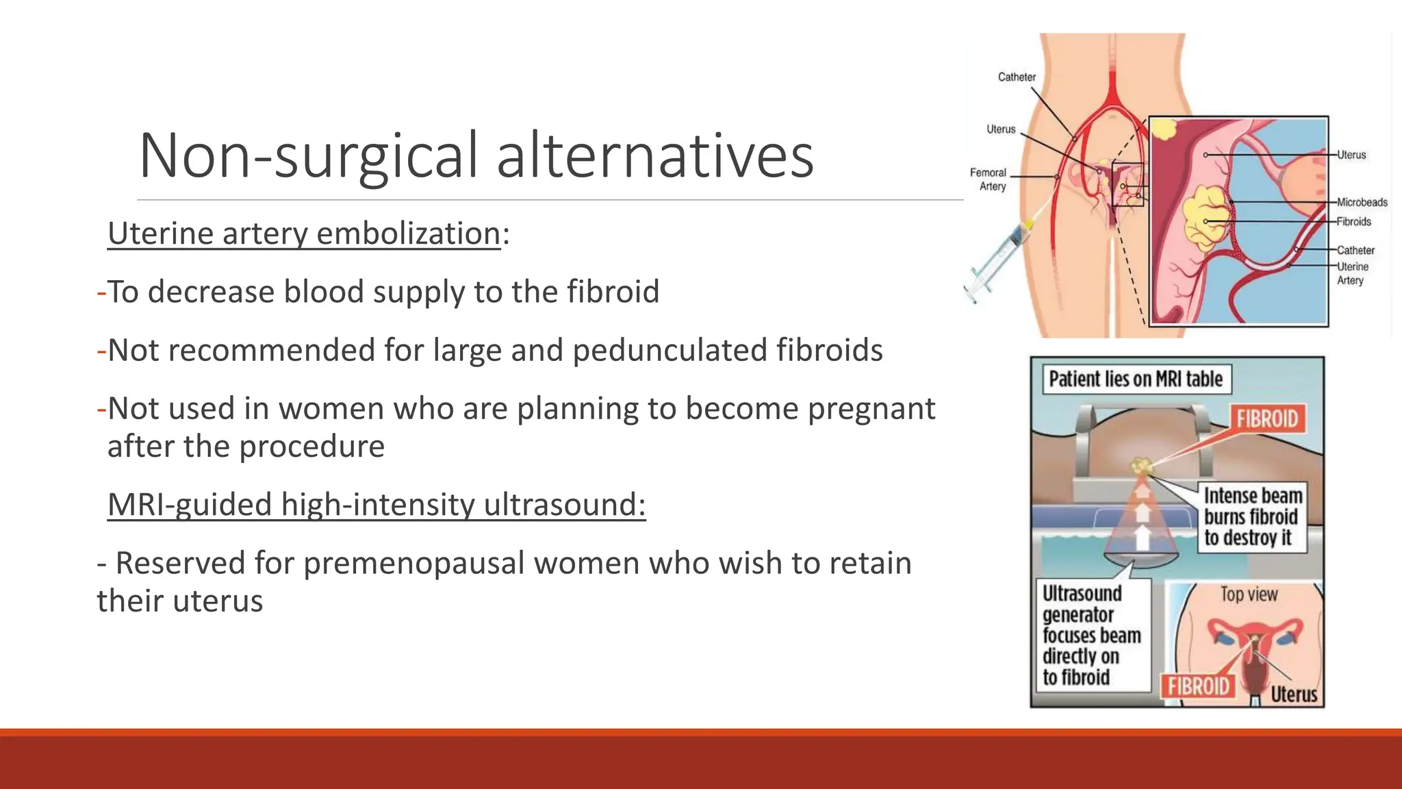
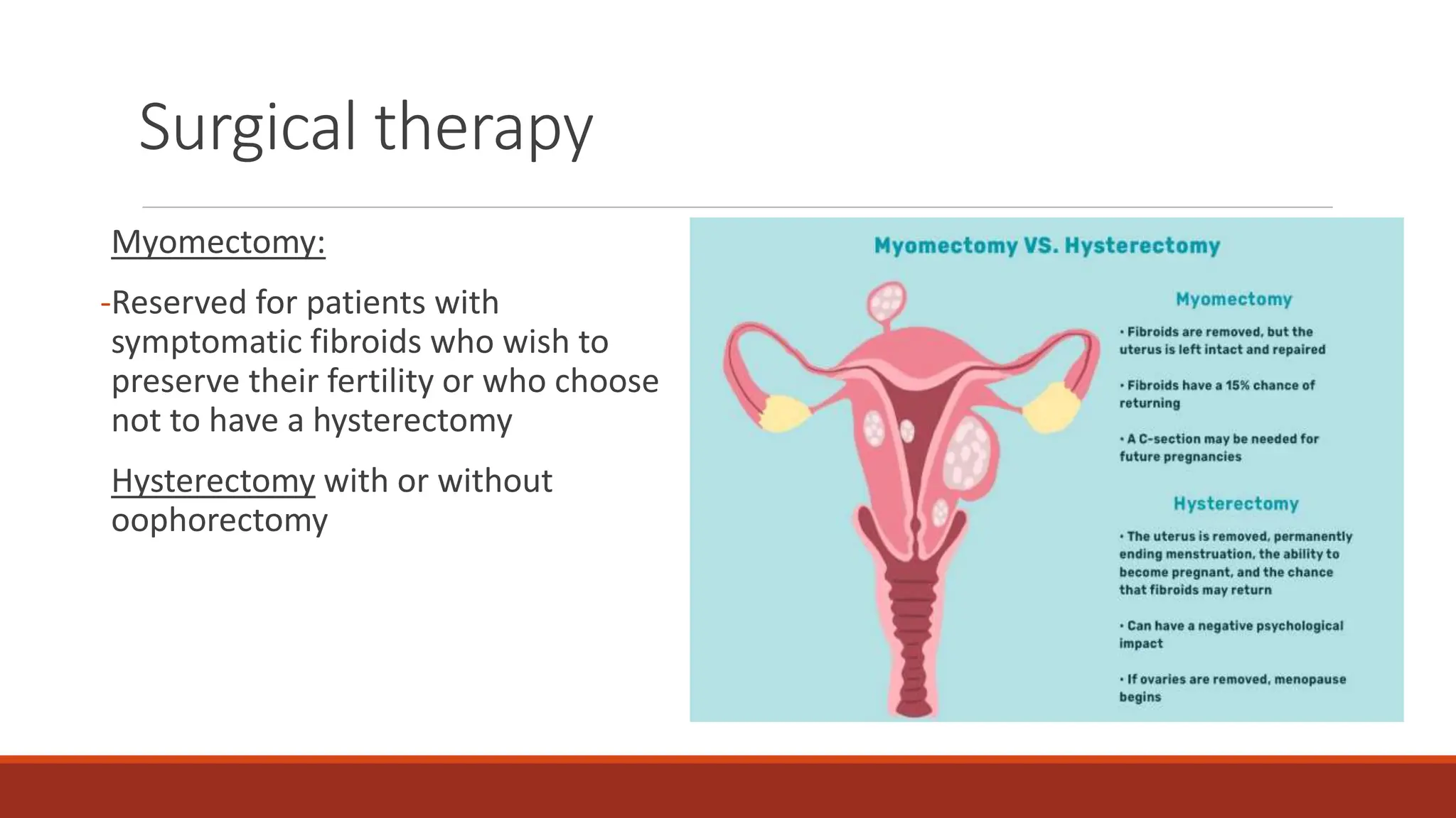
Introduction
•Uterine fibroids, also known as leiomyomas or
myomas, are noncancerous monoclonal
tumors arising from the smooth muscle cells
and fibroblasts of the myometrium
•Most common benign tumors of the female
reproductive system
•More common in women aged 30-40
Types of uterine fibroids
•3 types
•Fibroids contain a large quantity of
extracellular matrix (fibronectin, collagen,
proteoglycan) and are surrounded by a
pseudocapsule of compressed areolar tissue
and smooth muscle cells
•Variable size
Etiology
Exact cause is not yet known; however some factors play role in the formation and
growth of uterine fibroids:
-Genetic predisposition (genetic mutations in the MED12, HMGA2, COL4A5/COL4A6,
or FH genes)
-Hormones: estrogen and progesterone
-Growth factors
-Angiogenesis
Risk factors
Increased risk:
African American
Nulliparity
Early menarche
Perimenopause
Family history
Hypertension
Diabetes
obesity
Alcohol
Decreased risk:
Multiparity
Oral contraception use
Smoking
Exercise
Diet rich in green vegetables
Clinical presentation
Asymptomatic (in 50-60% of cases)
Abnormal uterine bleeding (menorrhagia, metrorrhagia, menometrorrhagia, postcoital spotting)
Iron deficiency anemia: weakness, fatigue, dizziness
Pelvic and lower back pain
Dysmenorrhea, dyspareunia
Pressure-related symptoms: frequency and retention of urine, constipation, hydronephrosis,
venous stasis
Abdominal distention
Infertility
Fibroids in pregnancy
Fibroids increase the risk of:
- Miscarriage
-Preterm labor and delivery
-Fetal malpresentation
-Intrauterine growth restriction
-Dysfunctional labor
- Antepartum and postpartum hemorrhage
- Cesarean delivery
Diagnosis
•Bimanual examination: nontender
irregularly enlarged uterus with
cobblestone protrusions that feel solid on
palpation
•Pelvic ultrasound: well defined
hypoechoic lesions
•Hysterosalpingogram (HSG), saline
infusion sonogram, hysteroscopy
•MRI
Differential diagnosis
Adenomyosis
Endometriosis
Endometrial polyp
Pregnancy; ectopic pregnancy
Leiomyosarcoma
Endometrial carcinoma
Management
•For asymptomatic patients: no treatment required but follow up every 6 months
•Medical therapies: hormonal and nonhormonal options
•Non-surgical alternatives
•Surgical interventions indicated in case of:
-Abnormal uterine bleeding causing anemia
-Severe pelvic pain or secondary amenorrhea
-Uterine size >12 week
-Urinary frequency, retention or hydronephrosis
-Growth after menopause
-Recurrent miscarriage or infertility
-Rapid increase in size
Medical therapy
Nonhormonal drugs: NSAIDs (for dysmenorrhea) and tranexamic acid (to treat heavy
prolonged bleeding)
Hormonal drugs:
-Oral contraceptive pills
-Progestins (medroxyprogesterone acetate, Mirena)
-Mifepristone
-Androgenic steroids (danazol and gestrinone)
-GnRH agonists (nafarelin acetate, leuprolide acetate depot, goserelin acetate): shrink
fibroids and decrease bleeding by decreasing estrogen levels
Non-surgical alternatives
Uterine artery embolization:
-To decrease blood supply to the fibroid
-Not recommended for large and pedunculated fibroids
-Not used in women who are planning to become pregnant
after the procedure
MRI-guided high-intensity ultrasound:
- Reserved for premenopausal women who wish to retain
their uterus
Surgical therapy
Myomectomy:
-Reserved for patients with
symptomatic fibroids who wish to
preserve their fertility or who choose
not to have a hysterectomy
Hysterectomy with or without
oophorectomy
uterine fibroids. bleeding. myoma pptxjj

uterine fibroids. bleeding. myoma pptxjj

  • 1.
    Introduction •Uterine fibroids, alsoknown as leiomyomas or myomas, are noncancerous monoclonal tumors arising from the smooth muscle cells and fibroblasts of the myometrium •Most common benign tumors of the female reproductive system •More common in women aged 30-40
  • 2.
    Types of uterinefibroids •3 types •Fibroids contain a large quantity of extracellular matrix (fibronectin, collagen, proteoglycan) and are surrounded by a pseudocapsule of compressed areolar tissue and smooth muscle cells •Variable size
  • 3.
    Etiology Exact cause isnot yet known; however some factors play role in the formation and growth of uterine fibroids: -Genetic predisposition (genetic mutations in the MED12, HMGA2, COL4A5/COL4A6, or FH genes) -Hormones: estrogen and progesterone -Growth factors -Angiogenesis
  • 4.
    Risk factors Increased risk: AfricanAmerican Nulliparity Early menarche Perimenopause Family history Hypertension Diabetes obesity Alcohol Decreased risk: Multiparity Oral contraception use Smoking Exercise Diet rich in green vegetables
  • 5.
    Clinical presentation Asymptomatic (in50-60% of cases) Abnormal uterine bleeding (menorrhagia, metrorrhagia, menometrorrhagia, postcoital spotting) Iron deficiency anemia: weakness, fatigue, dizziness Pelvic and lower back pain Dysmenorrhea, dyspareunia Pressure-related symptoms: frequency and retention of urine, constipation, hydronephrosis, venous stasis Abdominal distention Infertility
  • 6.
    Fibroids in pregnancy Fibroidsincrease the risk of: - Miscarriage -Preterm labor and delivery -Fetal malpresentation -Intrauterine growth restriction -Dysfunctional labor - Antepartum and postpartum hemorrhage - Cesarean delivery
  • 7.
    Diagnosis •Bimanual examination: nontender irregularlyenlarged uterus with cobblestone protrusions that feel solid on palpation •Pelvic ultrasound: well defined hypoechoic lesions •Hysterosalpingogram (HSG), saline infusion sonogram, hysteroscopy •MRI
  • 8.
    Differential diagnosis Adenomyosis Endometriosis Endometrial polyp Pregnancy;ectopic pregnancy Leiomyosarcoma Endometrial carcinoma
  • 9.
    Management •For asymptomatic patients:no treatment required but follow up every 6 months •Medical therapies: hormonal and nonhormonal options •Non-surgical alternatives •Surgical interventions indicated in case of: -Abnormal uterine bleeding causing anemia -Severe pelvic pain or secondary amenorrhea -Uterine size >12 week -Urinary frequency, retention or hydronephrosis -Growth after menopause -Recurrent miscarriage or infertility -Rapid increase in size
  • 10.
    Medical therapy Nonhormonal drugs:NSAIDs (for dysmenorrhea) and tranexamic acid (to treat heavy prolonged bleeding) Hormonal drugs: -Oral contraceptive pills -Progestins (medroxyprogesterone acetate, Mirena) -Mifepristone -Androgenic steroids (danazol and gestrinone) -GnRH agonists (nafarelin acetate, leuprolide acetate depot, goserelin acetate): shrink fibroids and decrease bleeding by decreasing estrogen levels
  • 11.
    Non-surgical alternatives Uterine arteryembolization: -To decrease blood supply to the fibroid -Not recommended for large and pedunculated fibroids -Not used in women who are planning to become pregnant after the procedure MRI-guided high-intensity ultrasound: - Reserved for premenopausal women who wish to retain their uterus
  • 12.
    Surgical therapy Myomectomy: -Reserved forpatients with symptomatic fibroids who wish to preserve their fertility or who choose not to have a hysterectomy Hysterectomy with or without oophorectomy