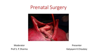
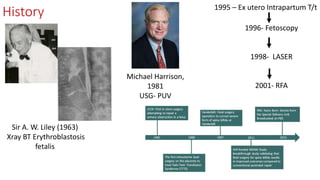
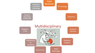
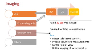
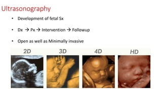
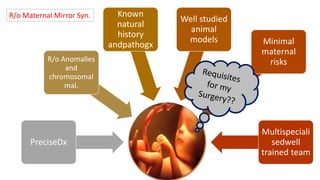
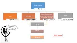
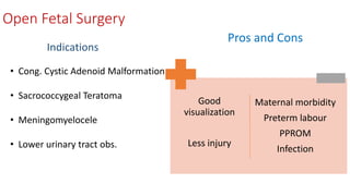
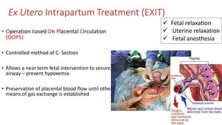
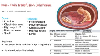
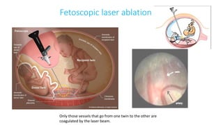
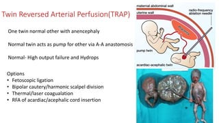
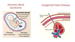
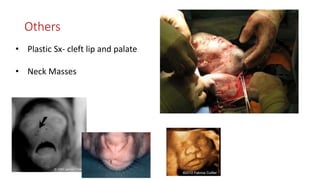
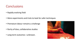
Prenatal Surgery
Moderator
Prof S. P. Sharma
Presenter
Katyayani K Choubey
What is Prenatal Surgery?
• Fetal reconstructive surgery/ antenatal surgery/ fetal surgery/ Maternal-
fetal surgery
• Branch of maternal fetus medicine
• Techniques- to treat birth defects in fetus while in womb
• To stop the progression of congenital diseases
• To protect fetus- demise and disorders by early intervention
The need
3 -11% babies - complex birth defect.
Advances in post-natal therapies  birth defects
cause of morbidity and mortality
Few ds - devastating developmental consequences
benefit from fetal t/t.
option other than expectant mx and termination of
pregnancy.
History
Sir A. W. Liley (1963)
Xray BT Erythroblastosis
fetalis
1995 – Ex utero Intrapartum T/t
Michael Harrison,
1981
USG- PUV
1996- Fetoscopy
1998- LASER
2001- RFA
Multidisciplinary
Fetal
Medicine
Perinatology
Obstetrics
Pediatric
Genetics
Pediatric
Surgery
Fetal
Pathology
Biochemistry
PediatricCy
to-
Genetics
Pediatric
CTVS
Pediatric
Neurology
andNeuroSx
Pediatrics
and Fetal
Cardiology
Imaging
USG
Echocardiography
Ultrafast MRI
Others
2D 3D 4D/HD
Rapid 20 sec MRI is used
No need for fetal immboilisation
Pros
• Better soft-tissue contrast
• Precise volumetric measurements
• Larger field of view
• Better imaging of intracranial str
Ultrasonography
• Development of fetal Sx
• Dx  Px  Intervention  Followup
• Open as well as Minimally invasive
Echocardiography
3 D echo of a heart viewed from
apex
 Evaluate cardiac and extra cardiac ds
Hemodynamic instability
(Hydrops)
 Natural history, pathogx of dses
Criteria
forin
uterosx
PreciseDx
R/o Anomalies
and
chromosomal
mal.
Known
natural
history
andpathogx
Well studied
animal
models Minimal
maternal
risks
Multispeciali
sedwell
trained team
R/o Maternal Mirror Syn.
Fetal Surgery
Types
Open
Conventional
FETENDO
Laparoscopic
Fetal Image-guided
Surgery For Intervention
Or Therapy (FIGS-IT)
Image Guided
RFA
LASER
EXIT
Intra partum
Others
20-28 weeks
Open Fetal Surgery
Mother -GA
Incision- lower abdomen
Pre/Intraop USG done
Placenta visualized
Uterus opened using staplers
Infusion with warmed RL
Seals BV, fixes mb
Prevent contractions,
Maintains volume Fetal Analgesia
Fetal ECG, Pulse oximeter,
ABG
At the end of Sx- Patient still Pregnant Cons
Open Fetal Surgery
Indications
Pros and Cons
Good
visualization
Less injury
Maternal morbidity
Preterm labour
PPROM
Infection
• Cong. Cystic Adenoid Malformation
• Sacrococcygeal Teratoma
• Meningomyelocele
• Lower urinary tract obs.
Minimally Invasive Fetal Endosurgery (FETENDO)
• See and intervene on fetus - very small
endoscopes and USGs- on separate screens
• Percutaneous or Mini- lap
• Length-18 cm scopes diameter -1.2 to 3.5 mm
• visualization angle 0-39 degrees
• GA
FETENDO
Indications
Pros and Cons
Less Invasive
Less Morbidity
Preterm
Skill
Cost
Risk
• Cong. Diaphrag. Hernia
• Twin twin transf. syndr.- Laser/RFA
• Postr. Urethral valve
• Cong. High Airway Obst. Synd.
Fetal Image-guided Surgery For
Intervention Or Therapy (FIGS-IT)
• Manipulating without incision/ endoscopic
view inside the uterus.
real-time cross-sectional USG
guide instruments.
• RA/ LA/ Sedation
FIGS-IT
Indications
Pros and Cons
Least Invasive
Less Morbidity
Preterm
Day care
Skill
Risk
Not for majorprobs
Catheter-shunts
bladder, abdomen, or
chest
RFA/Laser-anomalous
twins,
fetal cardiac defects.
Ex Utero Intrapartum Treatment (EXIT)
• Operation based On Placental Circulation
(OOPS)
• Controlled method of C- Section
• Allows a near term fetal intervention to secure
airway – prevent hypoxemia
• Preservation of placental blood flow until other
means of gas exchange is established
 Fetal relaxation
 Uterine relaxation
 Fetal anesthesia
EXIT
To Airway
Intrinsic
CHAOS
Extrinsic
Teratoma,sev.Micr
ognathia
Iatrogenic
Tracheal clip or
balloon
To Resection
Thoracic, Pulmonary
masses
To Extracorporeal
Mb Oxygenation
Cong. Heart Ds and
CDH
To Separation
Conjoint Twins
Laser
• Fibre in 3 mm sheath
• Near infrared Laser/ Anode/Nd:YAG
• Fired from 1cm distance 1-2cm segment
(visual cessation)
• Selective Fetoscopic Laser Photocoagulation (SFLP)
RFA
Principle - high-frequency alternating
currents l/t temp changes
local coagulation and tissue desiccation.
• 40W  temp 3 prongs 100°C  2-3
minutes.
Applications ??
Congenital conditions treated in utero
Disorder Natural History Benefits Recommended t/t
CDH Lung Hypoplasia Normal anatomy Reverse Open/FETO
LUTO VUR  HNOligohy Lung
Hypoplasia
Removal of uropathy  avoids
irreversible changes
Vesicostomy/VAS/
Valve ablation
CCAM Lung Hypoplasia Hydrops Resection Open/Fetoscopic/ Aspirate/
Laser ablation
SCT Vascular Insufficiency 
Hydrops
Resection Open/laser/fetos.
Placenta Circulation
anomalies
Blood Steal Destroy anastomosis Laser/RFA
CHAOS Lung inflation cardiac
insufficiency  Hydrops
Normal airway Fetscopic Tracheostomy
/Valveplasty/ EXIT
MMC Spinal cord lesion, hydroceph,
ACM, myeloplegia
Amniotic fluid destructive effect
on exposed nerves
Open or Fetoscopic Lesion
coverage
ABS Amputation/ cord constriction Amniotic band dissection
Congenital Diaphragmatic Hernia
 Embryonic phase  abdominal organs
herniate  interfere with lung growth
 developmental arrest- airways &
vasculature.
At birth  pulmonary
hypoplasia, hypertension, immaturity
Lung head
circumference
ratio (LHR) < 1
2 techniques
Open FETO
Fetoscopic endoluminal tracheal occlusion
• Liver up - Kinking of umblical veins
Abandoned
“Plug the lung until it grows”
34 weeks/ EXIT
TOTAL (Tracheal Occlusion To Accelerate Lung Growth) Trial
FETO vs Expectant
Congenital Cystic Adenomatoid Malformation
(CCAM)
• Overgrowth of terminal bronchioles
Macrocystic –>5mm, Fluid
Microcystic – < 5 mm, Solid, Bulky
Large size
Mediastinal shift
Pulmonary hypoplasia
Polyhydroamnios
Nonimmune hydrops
CVR = (Length X
Height X Width X
0.52 )/ HC
CCAM volume
ratio(CVR) > 1.6/
Dominant cyst –
hydrops
Lobectomy/
Segmetectomy
Thoracoamniotic
shunt (TAS)
Two 12-mg im doses
are given 24 hours
apart
Fetal Sx
Lower urinary tract obstructions (LUTO)
• Urethral atresia (UA)
• Posterior Urethral valve (PUV)
• Triad syndrome
• An enlarged fetal bladder + normal amniotic
fluid incomplete obstruction.
USG
• APRPD
• Oligohydramnios
• Loss of CMD
• Renal cortical cysts
Fetal Urine
• Calcium > 8mg/L
• Na+>100meq/L
• Cl- >90meq/L
• β- 2 μglobulin >4 mg/L,
• Urine osmolality >200 mosm/L
Prognosis
Open Sx
The goal – To obtain
sufficient amniotic
fluid - lung growth
and increase chance
of fetal adaptation
Vesicostomy- abdandoned Vesicoamniotic shunt
Fetoscopic Laser Fulgration
In utero Treatment of LUTO
Harrison’s Catheter
CHAOS
Complete glottic web and interarytenoid fusion
• Laryngeal atresia
• Subglottic stenosis
• Laryngeal/ tracheal -rings or
agenesis
Trapping of pulmonary secretions 
hyperinflation of the lungs  increased
intrathoracic pressure  compromised venous
return cardiac output hydrops
EXIT
Sacrococcygeal Teratoma
High output HF
Tumor at sacrum- coccyx
USG: cystic or solid or mixed
• Cystic  asymptomatic
• Solid  lot of blood flow  double work done
by heart high output failure  hydrops
Dx
No High output HF
<32 wks >32 wks
In utero Tx Sx Emergency C- sec
FU with USG
Term C- sec
Treatment Options
Open Tx Resection
RFA
LASER
Meningomyelocele
Treatment Options
Repair Of Meningomyelocele
Twin- Twin Transfusion Syndrome
MCDA twins - unbalanced flow
Donor
• Low flow
• Oligohydramnios
• High output HF
• Brain ischemia
• Small
Recipient
• Fluid overload
• Polyhydramnios
• Congestive HF
• Hydrops fetalis
• Large
Treatment
• Fetoscopic laser ablation - Stage II or greater
• Amnioreduction- limited role
QUINTERO
Only those vessels that go from one twin to the other are
coagulated by the laser beam.
Fetoscopic laser ablation
Twin Reversed Arterial Perfusion(TRAP)
One twin normal other with anencephaly
Normal twin acts as pump for other via A-A anastomosis
Normal- High output failure and Hydrops
Options
• Fetoscopic ligation
• Bipolar cautery/harmonic scalpel division
• Thermal/laser coagualation
• RFA of acardiac/acephalic cord insertion
Amniotic Band
Syndrome
Congenital Heart Disease
Others
• Plastic Sx- cleft lip and palate
• Neck Masses
Ethics
• Two patients, mother and the fetal
patient.
• Balancing risks vs potential benefit.
• Fetal reduction- concern
Current Scenario
• International Fetal Medicine
and Surgical Society (IFMSS)
• North American Fetal Therapy
Network (NAFTNet)
India
Dr. Mohan Abraham, 21-week –
PUV- Laser Fulgration
What lies in future?
Stem cell transplant
• Genetic diseases
• Metabolic disorders
• Hemoglobinopathies,
• Meningomyelocele
• Alpha thalassemia
Future Fix- tissue engineering
Gene Therapy
Conclusions
• Rapidly evolving field
• More experiments and trials to look for safer techniques
• Premature labour remains a challenge
• Rarity of dses, collaborative studies
• Long-term outcomes –unknown.
First fetal surgery survivor finally
meets his doctor / 24 years ago,
UCSF surgeon saved his life in
mom's womb
Dr. Michael Harrison (R) listens to Michael Skinner's lungs during an exam
May 10, 1981, at UCSF Medical Center. Harrison’s catheter
• Magnetic mini mover
• Magnap
• Magnamosis
Thank You

Fetal or Prenatal Surgery

  • 1.
    Prenatal Surgery Moderator Prof S.P. Sharma Presenter Katyayani K Choubey
  • 2.
    What is PrenatalSurgery? • Fetal reconstructive surgery/ antenatal surgery/ fetal surgery/ Maternal- fetal surgery • Branch of maternal fetus medicine • Techniques- to treat birth defects in fetus while in womb • To stop the progression of congenital diseases • To protect fetus- demise and disorders by early intervention
  • 3.
    The need 3 -11%babies - complex birth defect. Advances in post-natal therapies  birth defects cause of morbidity and mortality Few ds - devastating developmental consequences benefit from fetal t/t. option other than expectant mx and termination of pregnancy.
  • 4.
    History Sir A. W.Liley (1963) Xray BT Erythroblastosis fetalis 1995 – Ex utero Intrapartum T/t Michael Harrison, 1981 USG- PUV 1996- Fetoscopy 1998- LASER 2001- RFA
  • 5.
  • 6.
    Imaging USG Echocardiography Ultrafast MRI Others 2D 3D4D/HD Rapid 20 sec MRI is used No need for fetal immboilisation Pros • Better soft-tissue contrast • Precise volumetric measurements • Larger field of view • Better imaging of intracranial str
  • 7.
    Ultrasonography • Development offetal Sx • Dx  Px  Intervention  Followup • Open as well as Minimally invasive
  • 8.
    Echocardiography 3 D echoof a heart viewed from apex  Evaluate cardiac and extra cardiac ds Hemodynamic instability (Hydrops)  Natural history, pathogx of dses
  • 9.
  • 10.
    Fetal Surgery Types Open Conventional FETENDO Laparoscopic Fetal Image-guided SurgeryFor Intervention Or Therapy (FIGS-IT) Image Guided RFA LASER EXIT Intra partum Others 20-28 weeks
  • 11.
    Open Fetal Surgery Mother-GA Incision- lower abdomen Pre/Intraop USG done Placenta visualized Uterus opened using staplers Infusion with warmed RL Seals BV, fixes mb Prevent contractions, Maintains volume Fetal Analgesia Fetal ECG, Pulse oximeter, ABG At the end of Sx- Patient still Pregnant Cons
  • 12.
    Open Fetal Surgery Indications Prosand Cons Good visualization Less injury Maternal morbidity Preterm labour PPROM Infection • Cong. Cystic Adenoid Malformation • Sacrococcygeal Teratoma • Meningomyelocele • Lower urinary tract obs.
  • 13.
    Minimally Invasive FetalEndosurgery (FETENDO) • See and intervene on fetus - very small endoscopes and USGs- on separate screens • Percutaneous or Mini- lap • Length-18 cm scopes diameter -1.2 to 3.5 mm • visualization angle 0-39 degrees • GA
  • 14.
    FETENDO Indications Pros and Cons LessInvasive Less Morbidity Preterm Skill Cost Risk • Cong. Diaphrag. Hernia • Twin twin transf. syndr.- Laser/RFA • Postr. Urethral valve • Cong. High Airway Obst. Synd.
  • 15.
    Fetal Image-guided SurgeryFor Intervention Or Therapy (FIGS-IT) • Manipulating without incision/ endoscopic view inside the uterus. real-time cross-sectional USG guide instruments. • RA/ LA/ Sedation
  • 16.
    FIGS-IT Indications Pros and Cons LeastInvasive Less Morbidity Preterm Day care Skill Risk Not for majorprobs Catheter-shunts bladder, abdomen, or chest RFA/Laser-anomalous twins, fetal cardiac defects.
  • 17.
    Ex Utero IntrapartumTreatment (EXIT) • Operation based On Placental Circulation (OOPS) • Controlled method of C- Section • Allows a near term fetal intervention to secure airway – prevent hypoxemia • Preservation of placental blood flow until other means of gas exchange is established  Fetal relaxation  Uterine relaxation  Fetal anesthesia
  • 18.
    EXIT To Airway Intrinsic CHAOS Extrinsic Teratoma,sev.Micr ognathia Iatrogenic Tracheal clipor balloon To Resection Thoracic, Pulmonary masses To Extracorporeal Mb Oxygenation Cong. Heart Ds and CDH To Separation Conjoint Twins
  • 19.
    Laser • Fibre in3 mm sheath • Near infrared Laser/ Anode/Nd:YAG • Fired from 1cm distance 1-2cm segment (visual cessation) • Selective Fetoscopic Laser Photocoagulation (SFLP)
  • 20.
    RFA Principle - high-frequencyalternating currents l/t temp changes local coagulation and tissue desiccation. • 40W  temp 3 prongs 100°C  2-3 minutes.
  • 21.
  • 22.
    Congenital conditions treatedin utero Disorder Natural History Benefits Recommended t/t CDH Lung Hypoplasia Normal anatomy Reverse Open/FETO LUTO VUR  HNOligohy Lung Hypoplasia Removal of uropathy  avoids irreversible changes Vesicostomy/VAS/ Valve ablation CCAM Lung Hypoplasia Hydrops Resection Open/Fetoscopic/ Aspirate/ Laser ablation SCT Vascular Insufficiency  Hydrops Resection Open/laser/fetos. Placenta Circulation anomalies Blood Steal Destroy anastomosis Laser/RFA CHAOS Lung inflation cardiac insufficiency  Hydrops Normal airway Fetscopic Tracheostomy /Valveplasty/ EXIT MMC Spinal cord lesion, hydroceph, ACM, myeloplegia Amniotic fluid destructive effect on exposed nerves Open or Fetoscopic Lesion coverage ABS Amputation/ cord constriction Amniotic band dissection
  • 23.
    Congenital Diaphragmatic Hernia Embryonic phase  abdominal organs herniate  interfere with lung growth  developmental arrest- airways & vasculature. At birth  pulmonary hypoplasia, hypertension, immaturity Lung head circumference ratio (LHR) < 1
  • 24.
    2 techniques Open FETO Fetoscopicendoluminal tracheal occlusion • Liver up - Kinking of umblical veins Abandoned “Plug the lung until it grows” 34 weeks/ EXIT TOTAL (Tracheal Occlusion To Accelerate Lung Growth) Trial FETO vs Expectant
  • 25.
    Congenital Cystic AdenomatoidMalformation (CCAM) • Overgrowth of terminal bronchioles Macrocystic –>5mm, Fluid Microcystic – < 5 mm, Solid, Bulky Large size Mediastinal shift Pulmonary hypoplasia Polyhydroamnios Nonimmune hydrops CVR = (Length X Height X Width X 0.52 )/ HC CCAM volume ratio(CVR) > 1.6/ Dominant cyst – hydrops
  • 26.
    Lobectomy/ Segmetectomy Thoracoamniotic shunt (TAS) Two 12-mgim doses are given 24 hours apart Fetal Sx
  • 27.
    Lower urinary tractobstructions (LUTO) • Urethral atresia (UA) • Posterior Urethral valve (PUV) • Triad syndrome • An enlarged fetal bladder + normal amniotic fluid incomplete obstruction. USG • APRPD • Oligohydramnios • Loss of CMD • Renal cortical cysts Fetal Urine • Calcium > 8mg/L • Na+>100meq/L • Cl- >90meq/L • β- 2 μglobulin >4 mg/L, • Urine osmolality >200 mosm/L Prognosis
  • 28.
    Open Sx The goal– To obtain sufficient amniotic fluid - lung growth and increase chance of fetal adaptation Vesicostomy- abdandoned Vesicoamniotic shunt Fetoscopic Laser Fulgration In utero Treatment of LUTO Harrison’s Catheter
  • 29.
    CHAOS Complete glottic weband interarytenoid fusion • Laryngeal atresia • Subglottic stenosis • Laryngeal/ tracheal -rings or agenesis Trapping of pulmonary secretions  hyperinflation of the lungs  increased intrathoracic pressure  compromised venous return cardiac output hydrops
  • 30.
  • 31.
    Sacrococcygeal Teratoma High outputHF Tumor at sacrum- coccyx USG: cystic or solid or mixed • Cystic  asymptomatic • Solid  lot of blood flow  double work done by heart high output failure  hydrops Dx No High output HF <32 wks >32 wks In utero Tx Sx Emergency C- sec FU with USG Term C- sec
  • 32.
    Treatment Options Open TxResection RFA LASER
  • 33.
  • 34.
  • 35.
    Twin- Twin TransfusionSyndrome MCDA twins - unbalanced flow Donor • Low flow • Oligohydramnios • High output HF • Brain ischemia • Small Recipient • Fluid overload • Polyhydramnios • Congestive HF • Hydrops fetalis • Large Treatment • Fetoscopic laser ablation - Stage II or greater • Amnioreduction- limited role QUINTERO
  • 36.
    Only those vesselsthat go from one twin to the other are coagulated by the laser beam. Fetoscopic laser ablation
  • 37.
    Twin Reversed ArterialPerfusion(TRAP) One twin normal other with anencephaly Normal twin acts as pump for other via A-A anastomosis Normal- High output failure and Hydrops Options • Fetoscopic ligation • Bipolar cautery/harmonic scalpel division • Thermal/laser coagualation • RFA of acardiac/acephalic cord insertion
  • 38.
  • 39.
    Others • Plastic Sx-cleft lip and palate • Neck Masses
  • 40.
    Ethics • Two patients,mother and the fetal patient. • Balancing risks vs potential benefit. • Fetal reduction- concern
  • 41.
    Current Scenario • InternationalFetal Medicine and Surgical Society (IFMSS) • North American Fetal Therapy Network (NAFTNet)
  • 42.
    India Dr. Mohan Abraham,21-week – PUV- Laser Fulgration
  • 43.
    What lies infuture? Stem cell transplant • Genetic diseases • Metabolic disorders • Hemoglobinopathies, • Meningomyelocele • Alpha thalassemia Future Fix- tissue engineering Gene Therapy
  • 44.
    Conclusions • Rapidly evolvingfield • More experiments and trials to look for safer techniques • Premature labour remains a challenge • Rarity of dses, collaborative studies • Long-term outcomes –unknown.
  • 45.
    First fetal surgerysurvivor finally meets his doctor / 24 years ago, UCSF surgeon saved his life in mom's womb Dr. Michael Harrison (R) listens to Michael Skinner's lungs during an exam May 10, 1981, at UCSF Medical Center. Harrison’s catheter • Magnetic mini mover • Magnap • Magnamosis
  • 47.