This document discusses the history and current state of fetal surgery. It begins with a brief history of fetal surgery from the first documented nonhuman fetal surgery in 1884 to the development of techniques in the 1940s-1960s. Key developments include the first reported human fetal surgery in 1965 and the advent of ultrasound technology in the late 1960s, which enabled less invasive fetal procedures. The document outlines the conditions currently treated with closed (non-hysterotomy) surgical therapies, including fetal transfusion and lower urinary tract obstruction. It emphasizes the importance of a multidisciplinary team approach and counseling for families considering fetal surgery.

![had cystoscopically guided therapy, either posterior
urethral valve ablation by laser or hydroablation or
urethral stent placement. The remainder had vesi-
coamnionic shunts placed after cystoscopy demon-
strated no fixable lesion or the cystoscopically
directed intervention was not successful. Intervention
improved overall perinatal survival compared with no
intervention (odds ratio [OR] 3.82, 95% confidence
interval [CI] 2.14–6.82), but once terminations and
fetal demises were removed from the data set, the only
group that demonstrated benefit from intervention
was the subgroup with poor prognosis (OR 9.36,
95% CI 1.41–62.05). There was no significant
improvement in the rate of survival with normal post-
natal renal function in the group with good prognosis
(OR 2.98, 95% CI 0.45–19.62). No comparison of the
group with good prognosis and the subgroup with
poor prognosis could be made, because no fetuses in
the group with poor prognosis survived with normal
renal function. The authors concluded that prenatal
intervention for lower urinary tract obstruction is
associated with increased perinatal survival (particu-
larly in the subgroup with poor prognosis), but that
intervention is associated with increased incidence of
impaired postnatal renal function.
Despite the relative ease (compared with fetal
cystoscopy) of shunt placement, vesico-amnionic shunt
placement is associated with significant fetal morbidity.
Initial reports documented 4% mortality and 44%
complication rates.36
A more recent series illustrated
the associated morbidities: two of nine catheters had
to be re-inserted; only six of nine fetuses survived
(there was one termination and two deaths attributable
to pulmonary hypoplasia) and three of the six survivors
had some degree of renal impairment (two had end-
stage renal disease and one had mild impairment).37
Fetal cystoscopy, the other modality used to
address lower urinary tract obstruction, offers
improved sensitivity in detecting posterior urethral
valves compared with ultrasonography (87–100%
detection using cystoscopy compared with 45% using
ultrasonography).38,39
It also offers the theoretical
advantage of preserving normal fetal bladder cycling,
which is essential for normal postnatal bladder func-
tion. The most comprehensive analysis of the effect of
fetal cystoscopy on postnatal outcomes is the meta-
analysis of four eligible studies including 63 patients
that was performed by Morris et al38
in 2011.
Although cystoscopy was associated with an increase
in perinatal survival compared with no treatment (OR
20.51, 95% CI 3.87–108.69), it offered no improve-
ment in perinatal survival over vesico-amnionic shunt
(OR 1.49, 95% CI 0.13–16.97).
Although most authorities agree that fetal inter-
vention should be based on an understanding of the
natural history of the disorder being treated, there
are little data in this regard for lower urinary tract
obstruction. One of the only studies to address long-
term outcome relative to age at diagnosis is the study
by Ylinen et al,40
in which a cohort of 46 Finnish
neonates, 23 of whom had antenatal diagnoses
and 23 had postnatal diagnoses, were followed for
a mean of 12.5 years. These investigators found
no significant difference between the cohort with
antenatal diagnoses and the cohort with postnatal
diagnoses with respect to poor renal function
(complete renal failure or end-stage renal disease,
mean glomerular filtration rate, age of advancing to
end-stage renal disease, initial or highest creatinine,
presence of vesicoureteral reflux, or incidence of
renal dysplasia). The authors acknowledged that it
is still not clear whether the real damage associated
with lower urinary tract obstruction is caused by the
disturbed urodynamics resulting from antenatal
obstruction or whether the renal dysplasia develops
as a result of the same insult that caused the obstruc-
tion, and thus co-exists with but is not caused by the
lower urinary tract obstruction. Until the etiology
of lower urinary tract obstruction–associated renal
dysplasia is clarified, it will not be possible to accu-
rately identify appropriate candidates for antenatal
intervention.
The almost inescapable conclusion arising from
these data is that we have not as yet demonstrated the
efficacy of intrauterine intervention for lower urinary
tract obstruction. The literature is problematic
because it consists of small case series with widely
differing selection criteria for intervention. Although
meta-analyses overcome some of these limitations,
a prospective randomized trial is needed. The
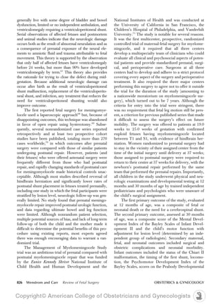
percutaneous shunting in low urinary tract obstruc-
tion (PLUTO) trial41
proposed to randomize 150 fe-
tuses with lower urinary tract obstruction to either
vesico-amnionic shunt or conservative noninterven-
tional care. It began in 2009 but closed because of
slow enrollment after enlisting only 31 patients.
Although the trial did not reach recruitment goals,
it found that shunted fetuses had better survival than
nonshunted fetuses (relative risk [RR] 3.30, 95% CI
1.02–9.62; P5.03). The study reached no conclu-
sions regarding the benefit (or lack thereof) of vesi-
coamnionic shunting on long-term renal function.
The much-needed high-quality data that we hoped
to see from the PLUTO trial, data that we could
use to inform our choice of interventions, remain
elusive.
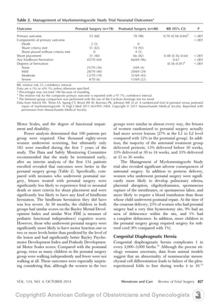
VOL. 124, NO. 4, OCTOBER 2014 Wenstrom and Carr Review of Fetal Surgery 821](https://image.slidesharecdn.com/cirugia-fetal-150826044414-lva1-app6892/85/Cirugia-fetal-5-320.jpg)
![are left to compare case series and collections of
case series, most of which evaluated hydropic and
nonhyropic fetuses separately. The 2007 report of
Rustico56
included data from 203 published cases of
antenatally treated isolated fetal pleural effusion,
which indicated a survival rate of 77% to 82% in non-
hydropic fetuses and 50% to 62% in hydropic fetuses
treated with thoracentesis or thoracoamniotic shunt-
ing. The survival rate was 60% in the pleurodesis
group, with no difference between hydropic and non-
hydropic fetuses, but the numbers were quite small.
Duerloo et al62
reviewed 108 hydropic fetuses with
pleural effusion culled from the published literature,
treated with thoracentesis, thoracoamniotic shunting,
or pleurodesis. They found very similar survival rates
of 60% to 80% regardless of intervention. Pellegrini
et al63
summarized their large single-center experience
and reported an 85% survival rate in shunted nonhy-
dropic fetuses and a 47% survival rate in shunted hy-
dropic fetuses, for an overall survival rate of 52%. In
2012, Yang et al64
published the largest series of pleu-
rodesis for fetal pleural effusion. Of 49 fetuses with
bilateral pleural effusions attributed to chylothorax,
4 had spontaneous resolution (8.2%) and 14 (31.1%)
did not survive to birth (10 [22.2%] had an intrauter-
ine fetal death and 4 [8.9%] terminated after unsatis-
factory results after pleurodesis). After successful
pleurodesis procedures, the rate of long-term survival
was 14.8% (4/27) for hydropic fetuses and was 66.7%
(12/18) for nonhydropic fetuses.
Our understanding of the natural history of fetal
pleural effusions remains imperfect. Natural history
observational studies suggest that untreated fetal pleural
effusions are associated with 63% to 73% survival in
nonhydropic cases and 35% to 50% in hydropic cases.56
However, in most published series, in utero interven-
tion appears to be associated with a survival rate of 60%
to 85% for isolated nonhydropic fetal pleural effusions
and 50% to 60% for hydropic fetuses. As noted pre-
viously, in most series it is unclear if survival with or
without antenatal treatment was achieved by elective
preterm delivery, so the effect of antenatal treatment
on the prolongation of pregnancy is unknown. At pres-
ent, the available data suggest that intervening in cases
of isolated nonhydropic pleural effusion offers, at most,
a small increase in survival.
CONDITIONS TREATED WITH OPEN
SURGICAL THERAPIES
Technique
“Open” fetal surgery refers to the fact that a hysterot-
omy is performed to gain access to the fetus, and
creating the hysterotomy might be considered the
most challenging part of the surgery. The uterine inci-
sion must be placed well away from the placental
edge, which is located intraoperatively using ultraso-
nography, but must also allow ready access to the
fetus. Once the optimal site is chosen, two full-
thickness stay sutures are placed through the uterus
and into the amniotic cavity at one edge of the
planned incision site, fixing the membranes to the
uterine wall, and the uterine cavity is entered with
a trocar. A uterine stapling device with absorbable
staples is then inserted through the opening created
by the trocar, engaged, and fired along the planned
incision line; the staples fix the membranes to the
uterine wall so that they can be incorporated into
the closure, thus preventing membrane separation.
The edge of the incision corresponding to the trocar
site, which is not covered by staples, is then rendered
hemostatic with a running lock stitch of absorbable
suture. One serious complication that can occur dur-
ing this part of the procedure is bleeding between the
membranes and the uterus, leading to a subchorionic
hematoma, which could potentially dissect the mem-
branes away from the uterine wall. Recognizing this
problem early allows sutures to be placed to tamponade
the bleeding vessels. Ideally, the fetus is positioned
directly beneath the incision site with only minimal
manipulation, and a catheter for the infusion of warm
saline is placed into the uterus to maintain amniotic
fluid volume and prevent umbilical cord compression
and fetal cooling. The fetal heart rate is monitored ultra-
sonographically throughout the procedure, with fetal
resuscitation in the form of position change, increased
amnioinfusion, or maternal measures provided as
needed.
OPEN SURGICAL THERAPIES
Myelominingocele
Neural tube defects, including anencephaly, encepha-
locele, and myelominingocele, are the most common
congenital structural defects worldwide. Before folic
acid supplementation, neural tube defects affected 1–2
per 1,000 pregnancies The fortification of cereal and
grain products in the United States (begun in 1996,
mandatory by January 1998) has been associated with
a 31% decrease in the incidence of neural tube de-
fects.65
Myelominingocele is the result of incomplete
closure of the neural tube, resulting in defective ver-
tebrae that permit the neural placode, meninges, or
both to herniate out of the spinal canal, allowing the
open dura mater to fuse laterally to the dermis and the
open pia pater to fuse to the epidermis.66
The spinal cord
is damaged at the site of and distal to the defect and, as
a consequence, survivors with meningomyelocele
VOL. 124, NO. 4, OCTOBER 2014 Wenstrom and Carr Review of Fetal Surgery 825](https://image.slidesharecdn.com/cirugia-fetal-150826044414-lva1-app6892/85/Cirugia-fetal-9-320.jpg)