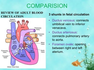
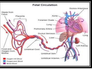
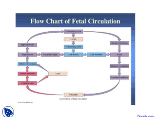
The fetal circulation allows the developing fetus to exchange materials with its mother through structures like the umbilical cord and placenta. There are three major shunts in the fetal circulatory system - the ductus venosus, ductus arteriosus, and foramen ovale - that direct highly-oxygenated blood from the placenta away from the lungs and toward vital organs like the brain and heart. At birth, the loss of placental blood flow and beginning of breathing cause pressure changes and closure of the shunts, redirecting blood flow to the lungs for oxygenation and establishing the postnatal circulatory pattern.
The female reproductive system contains two main parts: the uterus, which hosts the developing fetus, produces vaginal and uterine secretions, and passes the anatomically male sperm through to the fallopian tubes; and the ovaries, which produce the anatomically female egg cells.
TAPVC defines the anomaly in which the pulmonary veins have no connection with the left atrium. Rather, the pulmonary veins connect directly to one of the systemic veins (TAPVC) or drain in to right atrium.
A PFO or ASD is present essentially in those who survive after birth
When pulmonary veins drain anomalously into the right atrium either because of complete absence of the interatrial septum or malattachment of the septum primum , then it is known as total anomalous pulmonary venous drainage.
When some or all of the pulmonary veins drain anomalously in to RA or its tributaries without being abnormally connected, the terms partially anomalous pulmonary venous drainage (PAPVD) or totally anomalous pulmonary venous drainage (TAPVD) with normal pulmonary venous connections are used.
Describe the normal fetal circulation and mention the changes that occur in it is placental stage and after birth. Fetal circulation is composed of placenta, umbilical cord, heart and systemic blood vessels.
A major difference between the fetal circulation and postnatal circulation is that the lungs are not used during the fetal stage resulting in the presence of shunts to move oxygenated blood and nutrients from the placenta to the fetal tissue.
At birth, the start of breathing and the severance of the umbilical cord prompt various changes that quickly transform fetal circulation into postnatal circulation.
When the embryo develops into the fetus, it creates a functional cardiovascular system that cooperates with the mother's system.
During birth, there are functional physiological changes that transform the shared system into an individual one for the fetus.
In the fetus main filtration site for plasma nutrients and wastes in the placenta, which is outside of the body cavity.
In adults, the circulation occurs entirely inside the body.
The blood that flow to through the fetus is actually more complicated than after the baby is born (normal heart).
This is because the mother (the placenta) is doing the work that the baby's lungs will do after birth.
The placenta accepts the blood without oxygen from the fetus through blood vessels that leave the fetus through the umbilical cord (Umbilical arteries , there are two of them).
When blood goes through the placenta it pick up oxygwn.
The oxygen rich blood then returns to the fetus via the third vessels in the umbilical cord (Umbilical vein).
The oxygen rich blood that enters the fetus passes through the fetal liver and enters the right side of the heart.
The oxygen rich blood goes through one of the two extra connections in the fetal heart that will close after the baby is born.
The hole between the top two heart chmbers (right and left atrium) is called "Patent Foramen Ovale (PFO).
This hole allows the oxygen rich blood to go form the right atrium to left atrium and then to the left ventricle and out the aorta.
As a result the blood with the most oxygen gets to the brain.
Blood coming back from the fetus's body also enters the right atrium, but the fetus is able to send this oxygen poor blood from the right atrium to the right ventricle (the chamber that normally pumps blood to the lungs).
most of the blood that leaves the right ventricle in the fetus bypass the lungs through the second of the extra fetal connections known as the ductus arteriosus.
The ductus arteriosus sends the oxygen poor blood to the organs in the lower half of the fetal body. This also allows for the oxygen poor blood to leave the fetus through the umbilical arteries and get back to the placenta to pick up oxygen.
Since the patent foramen ovale and ductus arteriosus are normal findings in the fetus, it is impossible to predict whether or not these connections will close normally after birth in a normal fetal heart.
Fetal Circulation by Barkha Devi,Lecturer,Sikkim Manipal College of NursingBarkha Devi
This PowerPoint will provide you a short a sweet lecture about fetal circulation. Please give me your feed back .
-Discuss anatomy and physiology of fetal circulation
-Compare and contrast fetal circulation to infant circulation
-Define specialized structures of fetal circulation
Blood from the placenta is carried to the fetus by the umbilical vein. In humans, less than a third of this enters the fetal ductus venosus and is carried to the inferior vena cava, while the rest enters the liver proper from the inferior border of the liver. The branch of the umbilical vein that supplies the right lobe of the liver first joins with the portal vein. The blood then moves to the right atrium of the heart. In the fetus, there is an opening between the right and left atrium (the foramen ovale), and most of the blood flows through this hole directly into the left atrium from the right atrium, thus bypassing pulmonary circulation. The continuation of this blood flow is into the left ventricle, and from there it is pumped through the aorta into the body. Some of the blood moves from the aorta through the internal iliac arteries to the umbilical arteries, and re-enters the placenta, where carbon dioxide and other waste products from the fetus are taken up and enter the maternal circulation.
This is a presentation I had made for giving a seminar on Fetal Circulation in the first year of my MBBS course in Maharashtra.
Please share it with your juniors and colleagues.Thank You
Presentation By Tashif Jilani
This ppt explaining the basics of psychiatric nursing including its scope, team members, role and qualities of a nurse to all nursing and allied health students
Welcome to TechSoup New Member Orientation and Q&A (May 2024).pdfTechSoup
In this webinar you will learn how your organization can access TechSoup's wide variety of product discount and donation programs. From hardware to software, we'll give you a tour of the tools available to help your nonprofit with productivity, collaboration, financial management, donor tracking, security, and more.
2024.06.01 Introducing a competency framework for languag learning materials ...Sandy Millin
http://sandymillin.wordpress.com/iateflwebinar2024
Published classroom materials form the basis of syllabuses, drive teacher professional development, and have a potentially huge influence on learners, teachers and education systems. All teachers also create their own materials, whether a few sentences on a blackboard, a highly-structured fully-realised online course, or anything in between. Despite this, the knowledge and skills needed to create effective language learning materials are rarely part of teacher training, and are mostly learnt by trial and error.
Knowledge and skills frameworks, generally called competency frameworks, for ELT teachers, trainers and managers have existed for a few years now. However, until I created one for my MA dissertation, there wasn’t one drawing together what we need to know and do to be able to effectively produce language learning materials.
This webinar will introduce you to my framework, highlighting the key competencies I identified from my research. It will also show how anybody involved in language teaching (any language, not just English!), teacher training, managing schools or developing language learning materials can benefit from using the framework.
The Roman Empire A Historical Colossus.pdfkaushalkr1407
The Roman Empire, a vast and enduring power, stands as one of history's most remarkable civilizations, leaving an indelible imprint on the world. It emerged from the Roman Republic, transitioning into an imperial powerhouse under the leadership of Augustus Caesar in 27 BCE. This transformation marked the beginning of an era defined by unprecedented territorial expansion, architectural marvels, and profound cultural influence.
The empire's roots lie in the city of Rome, founded, according to legend, by Romulus in 753 BCE. Over centuries, Rome evolved from a small settlement to a formidable republic, characterized by a complex political system with elected officials and checks on power. However, internal strife, class conflicts, and military ambitions paved the way for the end of the Republic. Julius Caesar’s dictatorship and subsequent assassination in 44 BCE created a power vacuum, leading to a civil war. Octavian, later Augustus, emerged victorious, heralding the Roman Empire’s birth.
Under Augustus, the empire experienced the Pax Romana, a 200-year period of relative peace and stability. Augustus reformed the military, established efficient administrative systems, and initiated grand construction projects. The empire's borders expanded, encompassing territories from Britain to Egypt and from Spain to the Euphrates. Roman legions, renowned for their discipline and engineering prowess, secured and maintained these vast territories, building roads, fortifications, and cities that facilitated control and integration.
The Roman Empire’s society was hierarchical, with a rigid class system. At the top were the patricians, wealthy elites who held significant political power. Below them were the plebeians, free citizens with limited political influence, and the vast numbers of slaves who formed the backbone of the economy. The family unit was central, governed by the paterfamilias, the male head who held absolute authority.
Culturally, the Romans were eclectic, absorbing and adapting elements from the civilizations they encountered, particularly the Greeks. Roman art, literature, and philosophy reflected this synthesis, creating a rich cultural tapestry. Latin, the Roman language, became the lingua franca of the Western world, influencing numerous modern languages.
Roman architecture and engineering achievements were monumental. They perfected the arch, vault, and dome, constructing enduring structures like the Colosseum, Pantheon, and aqueducts. These engineering marvels not only showcased Roman ingenuity but also served practical purposes, from public entertainment to water supply.
Read| The latest issue of The Challenger is here! We are thrilled to announce that our school paper has qualified for the NATIONAL SCHOOLS PRESS CONFERENCE (NSPC) 2024. Thank you for your unwavering support and trust. Dive into the stories that made us stand out!
The Art Pastor's Guide to Sabbath | Steve ThomasonSteve Thomason
What is the purpose of the Sabbath Law in the Torah. It is interesting to compare how the context of the law shifts from Exodus to Deuteronomy. Who gets to rest, and why?
Model Attribute Check Company Auto PropertyCeline George
In Odoo, the multi-company feature allows you to manage multiple companies within a single Odoo database instance. Each company can have its own configurations while still sharing common resources such as products, customers, and suppliers.
Ethnobotany and Ethnopharmacology:
Ethnobotany in herbal drug evaluation,
Impact of Ethnobotany in traditional medicine,
New development in herbals,
Bio-prospecting tools for drug discovery,
Role of Ethnopharmacology in drug evaluation,
Reverse Pharmacology.
2. MEANING
• The circulatory system of fetus, called the fetal
circulation, exists only in the fetus & contains
special structure that allows the developing fetus
to exchange material with its mother.
• Umbilical cord:
• 2 umbilical arteries(carries non-o2 blood from
fetus to placenta).
• 1 umbilical vein arteries(carries o2 blood from
placenta to fetus).
3. COMPARISION
REVIEW OF ADULT BLOOD
CIRCULATION
3 shunts in fetal circulation
• Ductus venosus: connects
umbilical vein to inferior
venacava.
• Ductus arteriosus:
connects pulmonary artery
to aorta.
• Foramen ovale: opening
between right and left
aterium.
4.
5.
6.
7. FETAL CIRCULATION
• Before birth, blood from the placenta, about
80% saturated with oxygen, returns to the
fetus by way of the umbilical vein.
• On approaching the liver, most of this blood
flows through the ductus venosus directly
into the inferior vena cava, bypassing the
liver.
• After a short course in the inferior vena
cava, where placental blood mixes with
deoxygenated blood returning from the
lower limbs, it enters the right atrium.
8. FETAL CIRCULATION
• Here it is guided toward the oval foramen by
the valve of the inferior vena cava, and most
of the blood passes directly into the left
atrium.
• From the left atrium, where it mixes with a
small amount of desaturated blood
returning from the lungs, blood enters the
left ventricle and ascending aorta.
• Since the coronary and carotid arteries are
the first branches of the ascending aorta,
the heart musculature and the brain are
supplied with well-oxygenated blood.
9. FETAL CIRCULATION
• A small amount from the IVC is prevented
from entering the left atrium and remains in
the right atrium.
• It mixes with desaturated blood returning
from the head and arms by way of the
superior vena cava.
• Desaturated blood from the superior vena
cava flows by way of the right ventricle into
the pulmonary trunk.
10. FETAL CIRCULATION
• During fetal life, resistance in the pulmonary
vessels is high, such that most of this blood
passes directly through the ductus
arteriosus into the descending aorta, where
it mixes with blood from the proximal aorta.
• After coursing through the descending
aorta, blood flows toward the placenta by
way of the two umbilical arteries.
• The oxygen saturation in the umbilical
arteries is approximately 52-58%.
11. FETAL CIRCULATION
• Theoretically, mixing may occur in the following
places :
1)In the liver by mixture with a small amount of
blood returning from the portal system.
2)In the inferior vena cava which carries
deoxygenated blood returning from the lower
extremities, pelvis, and kidneys.
3)In the right atrium by mixture with blood returning
from the head and limbs.
4)In the left atrium by mixture with blood returning
from the lungs.
5)At the entrance of the ductus arteriosus into the
descending aorta.
12. CIRCULATORY CHANGES AT BIRTH
• Changes in the vascular system at birth are
caused by cessation of placental blood flow and
the beginning of respiration.
• Since the ductus arteriosus closes by muscular
contraction of its wall, the amount of blood
flowing through the lung vessels increases
rapidly.
• This, in turn, raises pressure in the left atrium.
• Simultaneously, pressure in the right atrium
decreases as a result of interruption of placental
blood flow.
• The septum primum is then apposed to the
septum secundum and functionally the oval
13. Foramen ovale Closes shortly after birth,
fuses completely in first year.
Ductus arteriousus Closes soon after birth,
becomes ligamentum
arteriousum in about 3
months.
Ductus venosus Ligamentum venosum
Umbilical arteries Medial umbilical ligaments
Umbilical vein Ligamentum teres
14.
15. Loss of placenta also leads to:
1. First breath
2. Lungs expand and fluid
is expelled
3. Decreased pulmonary
resistance
4. Increased pressure in
left atrium
5. Closure of foramen
ovale
• Increased systemic
resistance
• Pressure in right atrium
decreased
• Change from right to left
shunting to left to right
blood flow
• Increased O2 levels in
pulmonary circulation
• Closure of the ductus
arteriosus
15