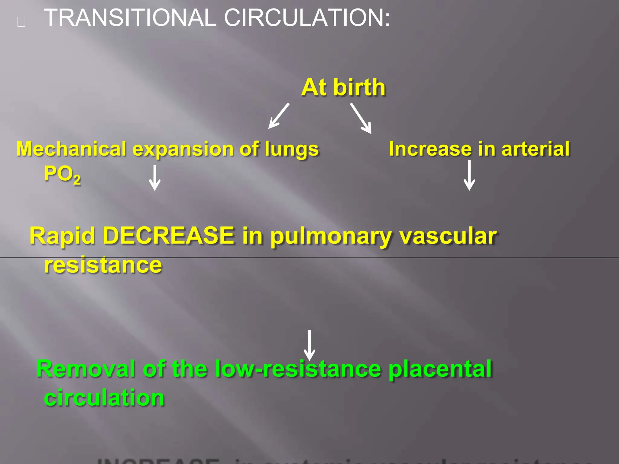
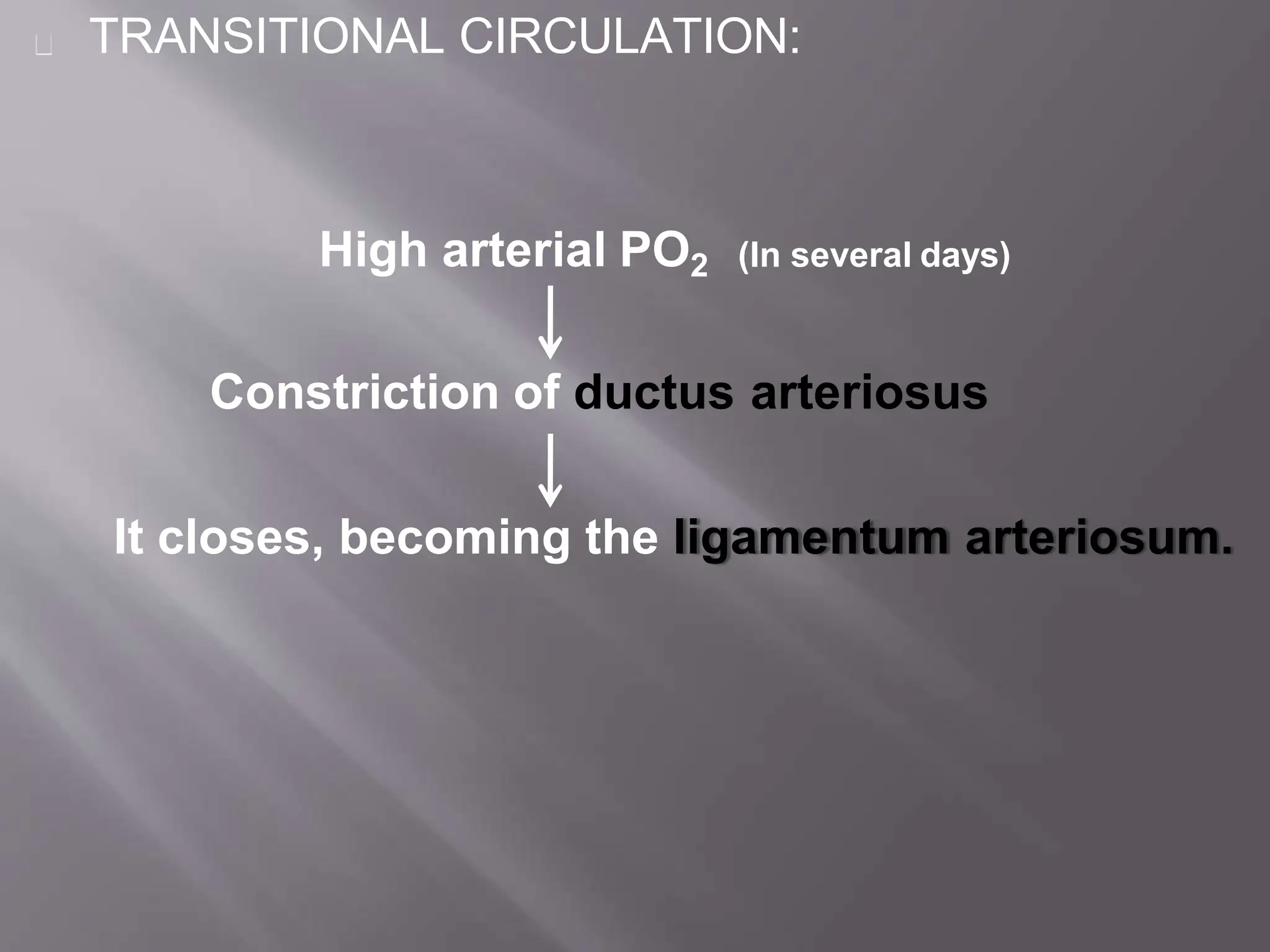
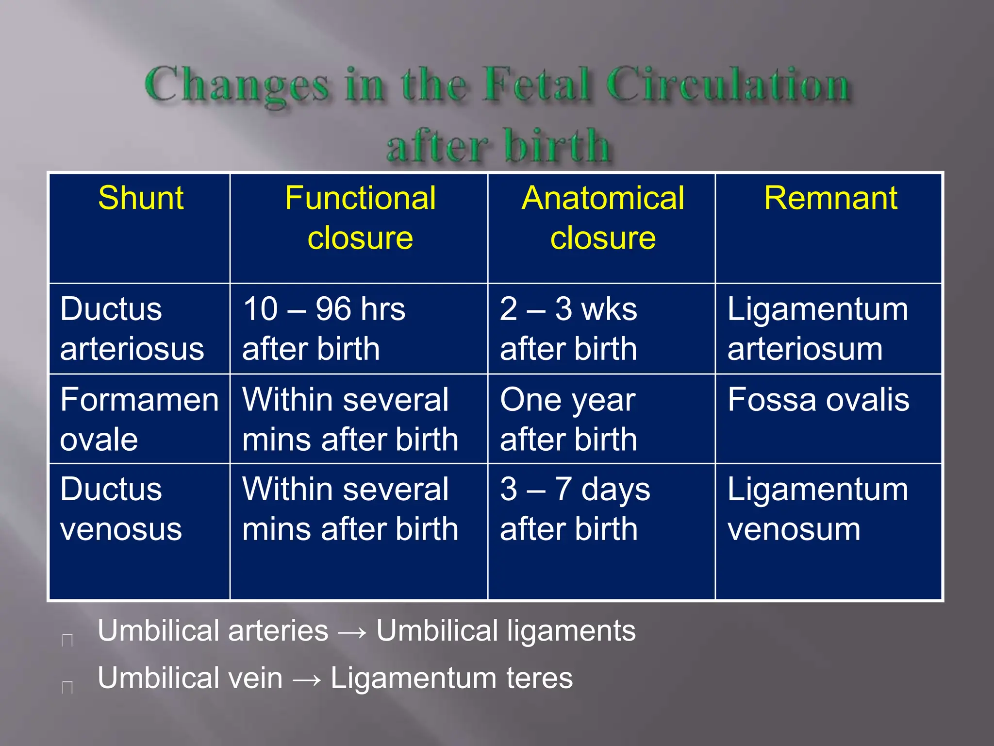
Call Girls Electronic City Just Call 7001305949 Top Class Call Girl Service A...
CMC Fetal Circulation.pptx Dr Biswajit Mukherjee
1.
2. The fetal circulation is the circulatory system of
a human fetus, often encompassing the entire
fetoplacental circulation which includes
the umbilical cord and the blood vessels within
the placenta that carry fetal blood.
3.
4. -Begins to develop toward the end of the
third week.
-Heart starts to beat at the beginning of the
fourth week.
-The critical period of heart development is
from 20 day to 50 day after fertilization.
-Many critical events occur during cardiac
development, and any deviation from this
normal pattern can cause congenital heart
defects, if development of heart
doesn't occur properly.
5.
6. Foetal circulation consequently differs from the
adult one predominantly due to the presence
of 3 major vascular shunts:
Ductus venosus: between the umbilical vein
and IVC
Foramen ovale: between the right and left
atrium
Ductus arteriosus: between the pulmonary
artery and descending aorta
8. :
1.Placenta:
Has the lowest vascular resistance in the fetus.
Receives the largest amount of combined (Rt + Lt)
Ventricular Output (55%)
9. 1. Superior Vena Cava:
Drains the upper part of the body,including the brain (15% of
combined ventricular output).
Most of SVC blood goes to the Right Ventricle.
10. 3. Inferior Vena Cava:
Drains lower part of body and
placenta (70% of combined
ventricular output)
Part of IVC blood with high O2
goes into LA via Foramen Ovale.
Remaining IVC blood enter RV
and Pulmonary artery.
Since blood is oxygenated
in the placenta, Oxygen
saturation in IVC
(PO2 = 26-28%) is higher
than that in SVC (12-14%).
11. COURSE OF FETAL CIRCULATION:
Most of SVC blood (less oxygenated blood) goes into RV.
Most of IVC blood (high O2 concentration) is directed by the Crista
Dividens to the LA through Foramen ovale.
Rest of IVC blood enters RV & pulmonary artery.
Less oxygenated blood in Pulmonary artery flows through Ductus
Arteriosus to descending aorta and then to placenta for oxygenation.
12. COURSE OF FETAL CIRCULATION:
The Result is:
Brain and coronary circulation receive blood with higher
concentration (PO2 = 28 mm Hg) than the lower part of the
body (PO2 = 24 mm Hg)
13. FETAL CIRCULATION: The pathway:
Placenta Oxygenated blood Umbilical vein
Hepatic circulation Bypasses liver & joins IVC
via ductus venosus
Partially mixes with poorly oxygenated
IVC
blood derived from lower part of fetal
body
14. FETAL CIRCULATION:
Combined lower body blood plus umbilical venous blood
flow (PO2 of ≈26–28 mm Hg) passes through IVC to the
Right atrium and is preferentially directed across the
foramen ovale to the left atrium.
The blood then flows into the left ventricle and is ejected
into the ascending aorta.
Fetal SVC blood, which is considerably less oxygenated
(PO2 of 12–14 mm Hg), enters the Right atrium and
preferentially traverses the tricuspid valve, rather than
the foramen ovale, and flows primarily to the right
ventricle.
15. FETAL CIRCULATION:
From the right ventricle Pulmonary artery.
Because the pulmonary arterial circulation is
vasoconstricted, only about 10% of right ventricular
outflow enters the lungs.
The rest 90% blood (which has a PO2 of ≈18–22 mm Hg)
bypasses the lungs and flows through the ductus
arteriosus into the descending aorta to perfuse the lower
part of the fetal body.
It then returns to the placenta via the two
umbilical arteries.
16. Thus, upper part of fetal body (including coronary & cerebral arteries
and those to upper extremities) is perfused exclusively from the Left
ventricle with blood that has a slightly higher PO2 , than the blood
perfusing the lower part of the fetal body, which is derived mostly
from the Right ventricle.
Only a small volume of blood from the ascending aorta (10% of fetal
cardiac output) flows across the aortic isthmus to the descending
aorta.
17. Thus, upper part of fetal body (including coronary & cerebral arteries
and those to upper extremities) is perfused exclusively from the Left
ventricle with blood that has a slightly higher PO2 , than the blood
perfusing the lower part of the fetal body, which is derived mostly
from the Right ventricle.
Only a small volume of blood from the ascending aorta (10% of fetal
cardiac output) flows across the aortic isthmus to the descending
aorta.
18. SVC upper
LA LV Aorta Ductus arteriosus
Foramen ovale RV
body
IVC
50% through
ductus venosus
50% to
Portal circulation
Umbilical Vein
Oxy.blood
PLACENTA
21. FETAL CIRCULATION:
The total fetal cardiac output—the combined output of
both the left and right ventricles—is ≈ 350 mL/kg/min.
Descending aortic blood flow :
-65% returns to placenta;
-Remaining 35% perfuses the fetal organs &
tissues.
Right ventricular output is about 1.3 times the left
ventricular flow.
Thus, during fetal life the right ventricle
-is pumping against systemic blood pressure
-is performing greater volume of work than LV.
22.
23. The change from fetal to postnatal circulation
happens very quickly.
Changes are initiated by baby’s first breath.
24. TRANSITIONAL CIRCULATION:
At birth
Increase in arterial
Mechanical expansion of lungs
PO2
Rapid DECREASE in pulmonary vascular
resistance
Removal of the low-resistance placental
circulation
25. TRANSITIONAL CIRCULATION:
Right ventricle output now flows entirely into the
pulmonary circulation.
Pulmonary vascular resistance becomes lower
than systemic vascular resistance,
Shunt through ductus arteriosus reverses &
becomes left to right.
27. TRANSITIONAL CIRCULATION:
Increased volume of pulmonary blood flow
returning to left atrium
Increases left atrial volume and pressure
Closure of foramen ovale (functionally)
(Although the foramen may remain probe patent)
Becomes Fossa Ovalis
28. Removal of the placenta from the circulation
Also results in closure of the ductus venosus.
The left ventricle is now coupled to the high-resistance
systemic circulation its wall thickness and mass
begin to increase.
In contrast, the right ventricle is now coupled to the low-
resistance pulmonary circulation its wall thickness
and mass decrease slightly.
29. Foetal circulation: The left ventricle in the fetus pumped
blood only to the upper part of the body and brain
After birth, LV must deliver the entire systemic cardiac
output (≈450 mL/kg/min). (almost 200% increase in
output)
This marked increase in left ventricular performance is
achieved through a combination of hormonal and
metabolic signals, including an INCREASE IN :
-The level of circulating catecholamines and
-The myocardial receptors (β-adrenergic)
(through which catecholamines have their effect)
30. Umbilical arteries → Umbilical ligaments
Umbilical vein → Ligamentum teres
Shunt Functional
closure
Anatomical
closure
Remnant
Ductus
arteriosus
10 – 96 hrs
after birth
2 – 3 wks
after birth
Ligamentum
arteriosum
Formamen
ovale
Within several
mins after birth
One year
after birth
Fossa ovalis
Ductus
venosus
Within several
mins after birth
3 – 7 days
after birth
Ligamentum
venosum