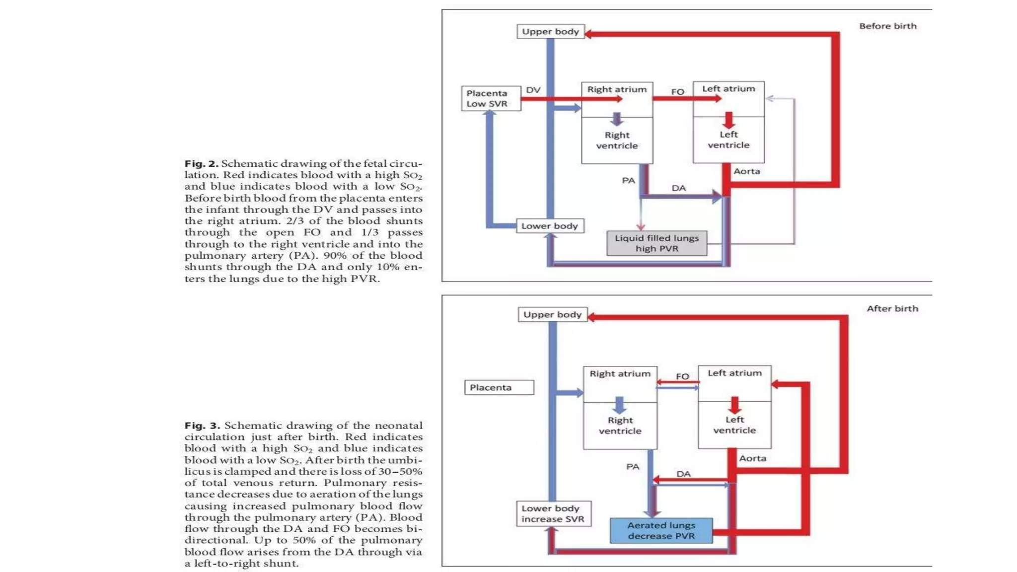
FETAL CIRCULATION AND CHANGES
AT BIRTH
Dr Jeevan Joseph Jose
FETAL CIRCULATION-
INTRODUCTION
• Fetal circulation differs from adult circulation
• Fetus- organ for gas and nutrient exchange and removing waste products and carbon dioxide .–Placenta
• Adult-Lungs
INTRODUCTION
. Fetal circulation- bypasses the lung Ductus arteriosus
The liver is also bypassed -Ductus Venosus
Blood can travel from the right atrium to the left atrium via the Foramen ovale.
Normal fetal heart rate is between 110 and 160 peats per minute.
Compared to adults, fetuses have decreased ventricular filling and reduced contractility.
ISSUES OF CONCERN
Preterm have 50% more ventricular cardiac muscle mass than those born at term.
Preterm babies are at increased risk of ischemic heart disease and heart failure
Shunts fail to close after birth; the baby can be born with congenital heart defects - signs and symptoms;
The yolk sac initiates erythropoiesis until the liver can take over at five weeks gestation, the bone marrow
finally contributes at six months gestation.
Fetal hemoglobin also possesses a higher affinity for oxygen when compared to maternal hemoglobin.
Fetal tissue has adapted ways to unload the oxygen from the higher affinity hemoglobin in the fetal tissue by
creating an acidic environment.
By the age of 4 to 6 months, the baby will have an adult hemoglobin level and no fetal hemoglobin. After birth,
FEATUS O2 SATURATION
O2 saturation at featus IVC 70-80 %
RA ~55%
LA~ 65%
Systemic circulation 60%
Deoxygenated blood saturation ~40%
CIRCULATION AT MATERNO-
PLACENTAL LEVEL
• Placenta is anchored to the wall of mothers uterus
• Chorion act as a barrier between the maternal and fetal circulation
FETAL CIRCULATION
● Oxygenated blood passes from placenta to fetus via UMBILICAL VEIN
● Umbilical vein –passes superiorly beneath anterior abdominal wall –distributes blood to liver –via DUCTUS
VENOSUS to IVC
● Most blood passes via the ductus venosus
● Small percentage of blood go to liver to mix with blood from portal circulation
● Ductus venous blood enters the IVC –its flow is controlled by a sphincteric mechanisam
● In IVC for a short distance , the placental oxygenated blood mixes blood from lower limbs
● Then it enters the right atrium
● Right Atrium most blood is directed to left atrium via FORAMEN OVALE
● Small amount of blood remains in right atrium and mixes with desaturated blood coming from head and arms by
SVC
● This is then directed to the right ventricle
● The blood in left atrium mixes with a small amount of desaturated blood in the lungs and goes to Left ventricle
then to Ascending Aorta
● Coronary and Carotid arteries are branches of ascending aorta ,heart and brain receive well oxygenated blood.
● Desaturated blood from SVC in the Right ventricle flows into the pulmonaryb trunk
● In fetal life ,Pulmonary Vascular resistance is High ,Hence this blood passes through the ductus arteriosus
into the descending aorta ,where it mixes with some oxygenated blood from the Ascending aorta
● This blood supplies the lower body and returns to placenta via the umbilical arteries
• Oxygen Saturation in Umbilical arteries is about 40-50%
• In fetal life only 10 % of Cardiac output enters Pulmonary arteries remainder is directed to the aorta
● About 60% of aortic blood flow enters the umbilical arteries and is redirected to the placenta
CHANGES AT BIRTH
● When the umbilical cord is clamped
● Umbilical vessels are occluded
● Massive reduction in venous return to the right heart
• Foramen Ovale Closes as pressure in the Right Atrium Falls
• The increased systemic resistance also raises the pressure in the left atrium to be higher than the right
atrium, and this causes the foramen ovale to close
• Functional closure of foramen ovale and ductus arteriosus takes place at birth anatomical closure by
fibrosis occurs days later
• Ductus Arteriosum – Ligamentum Arteriosum
• Ductus arteriosus closes.With first breath ,lungs get aerated reduced pulmonary artery pressures and pulmonary artery vasodialatation
• Hence right ventricular output increases and the ductus arteriosus closes
After birth ,The smooth muscle in the ductus arteriosus responds to the oxygen by increasing calcium channel activity, causing constriction and
ultimately shunt closure.
• Prostaglandins from the placenta keep the shunts in the fetus open. During birth, the shunts usually close due to the loss of prostaglandins from
placental separation and increased oxygen due to respiration
● Umbilical arteries close at birth
● Proximal part remains open as superior vesical arteries
● Distal part form thte lateral umbilical ligaments
● Umbilical veins- Ligamentum Teres
● DUCTUS VENOSUS – LIGAMENTUM VENOSUM
PATHOPHYSIOLOGY
Fetal circulation, the right side of the heart has higher pressures than the left side of the heart.
This pressure difference allows the shunts to remain open.
In postnatal circulation, when the baby takes its first breath, pulmonary resistance decreases, and blood flow
through the placenta ceases. Blood commences flowing through the lungs, and the pressure on the left side
becomes higher than on the right.
PATHOPHYSIOLOGY
Congenital heart defects arise when shunts fail to close after birth.
These defects can be cyanotic or acyanotic.
Cyanotic heart defects are typically from right-to-left shunts in blood after birth.
Acyanotic heart defects are typically left-to-right shunts in blood after birth.
THANKS YOU Thank you 😄

FETAL CIRCULATION AND CHANGES AT BIRTH.pptx

  • 1.
    FETAL CIRCULATION ANDCHANGES AT BIRTH Dr Jeevan Joseph Jose
  • 3.
    FETAL CIRCULATION- INTRODUCTION • Fetalcirculation differs from adult circulation • Fetus- organ for gas and nutrient exchange and removing waste products and carbon dioxide .–Placenta • Adult-Lungs
  • 4.
    INTRODUCTION . Fetal circulation-bypasses the lung Ductus arteriosus The liver is also bypassed -Ductus Venosus Blood can travel from the right atrium to the left atrium via the Foramen ovale. Normal fetal heart rate is between 110 and 160 peats per minute. Compared to adults, fetuses have decreased ventricular filling and reduced contractility.
  • 5.
    ISSUES OF CONCERN Pretermhave 50% more ventricular cardiac muscle mass than those born at term. Preterm babies are at increased risk of ischemic heart disease and heart failure Shunts fail to close after birth; the baby can be born with congenital heart defects - signs and symptoms;
  • 6.
    The yolk sacinitiates erythropoiesis until the liver can take over at five weeks gestation, the bone marrow finally contributes at six months gestation. Fetal hemoglobin also possesses a higher affinity for oxygen when compared to maternal hemoglobin. Fetal tissue has adapted ways to unload the oxygen from the higher affinity hemoglobin in the fetal tissue by creating an acidic environment. By the age of 4 to 6 months, the baby will have an adult hemoglobin level and no fetal hemoglobin. After birth,
  • 7.
    FEATUS O2 SATURATION O2saturation at featus IVC 70-80 % RA ~55% LA~ 65% Systemic circulation 60% Deoxygenated blood saturation ~40%
  • 8.
    CIRCULATION AT MATERNO- PLACENTALLEVEL • Placenta is anchored to the wall of mothers uterus • Chorion act as a barrier between the maternal and fetal circulation
  • 9.
    FETAL CIRCULATION ● Oxygenatedblood passes from placenta to fetus via UMBILICAL VEIN ● Umbilical vein –passes superiorly beneath anterior abdominal wall –distributes blood to liver –via DUCTUS VENOSUS to IVC
  • 10.
    ● Most bloodpasses via the ductus venosus ● Small percentage of blood go to liver to mix with blood from portal circulation
  • 11.
    ● Ductus venousblood enters the IVC –its flow is controlled by a sphincteric mechanisam ● In IVC for a short distance , the placental oxygenated blood mixes blood from lower limbs
  • 12.
    ● Then itenters the right atrium ● Right Atrium most blood is directed to left atrium via FORAMEN OVALE ● Small amount of blood remains in right atrium and mixes with desaturated blood coming from head and arms by SVC ● This is then directed to the right ventricle
  • 13.
    ● The bloodin left atrium mixes with a small amount of desaturated blood in the lungs and goes to Left ventricle then to Ascending Aorta ● Coronary and Carotid arteries are branches of ascending aorta ,heart and brain receive well oxygenated blood.
  • 14.
    ● Desaturated bloodfrom SVC in the Right ventricle flows into the pulmonaryb trunk ● In fetal life ,Pulmonary Vascular resistance is High ,Hence this blood passes through the ductus arteriosus into the descending aorta ,where it mixes with some oxygenated blood from the Ascending aorta
  • 15.
    ● This bloodsupplies the lower body and returns to placenta via the umbilical arteries
  • 16.
    • Oxygen Saturationin Umbilical arteries is about 40-50% • In fetal life only 10 % of Cardiac output enters Pulmonary arteries remainder is directed to the aorta
  • 17.
    ● About 60%of aortic blood flow enters the umbilical arteries and is redirected to the placenta
  • 18.
    CHANGES AT BIRTH ●When the umbilical cord is clamped ● Umbilical vessels are occluded ● Massive reduction in venous return to the right heart
  • 20.
    • Foramen OvaleCloses as pressure in the Right Atrium Falls • The increased systemic resistance also raises the pressure in the left atrium to be higher than the right atrium, and this causes the foramen ovale to close • Functional closure of foramen ovale and ductus arteriosus takes place at birth anatomical closure by fibrosis occurs days later
  • 21.
    • Ductus Arteriosum– Ligamentum Arteriosum • Ductus arteriosus closes.With first breath ,lungs get aerated reduced pulmonary artery pressures and pulmonary artery vasodialatation • Hence right ventricular output increases and the ductus arteriosus closes After birth ,The smooth muscle in the ductus arteriosus responds to the oxygen by increasing calcium channel activity, causing constriction and ultimately shunt closure. • Prostaglandins from the placenta keep the shunts in the fetus open. During birth, the shunts usually close due to the loss of prostaglandins from placental separation and increased oxygen due to respiration
  • 23.
    ● Umbilical arteriesclose at birth ● Proximal part remains open as superior vesical arteries ● Distal part form thte lateral umbilical ligaments
  • 24.
    ● Umbilical veins-Ligamentum Teres
  • 25.
    ● DUCTUS VENOSUS– LIGAMENTUM VENOSUM
  • 26.
    PATHOPHYSIOLOGY Fetal circulation, theright side of the heart has higher pressures than the left side of the heart. This pressure difference allows the shunts to remain open. In postnatal circulation, when the baby takes its first breath, pulmonary resistance decreases, and blood flow through the placenta ceases. Blood commences flowing through the lungs, and the pressure on the left side becomes higher than on the right.
  • 27.
    PATHOPHYSIOLOGY Congenital heart defectsarise when shunts fail to close after birth. These defects can be cyanotic or acyanotic. Cyanotic heart defects are typically from right-to-left shunts in blood after birth. Acyanotic heart defects are typically left-to-right shunts in blood after birth.
  • 28.