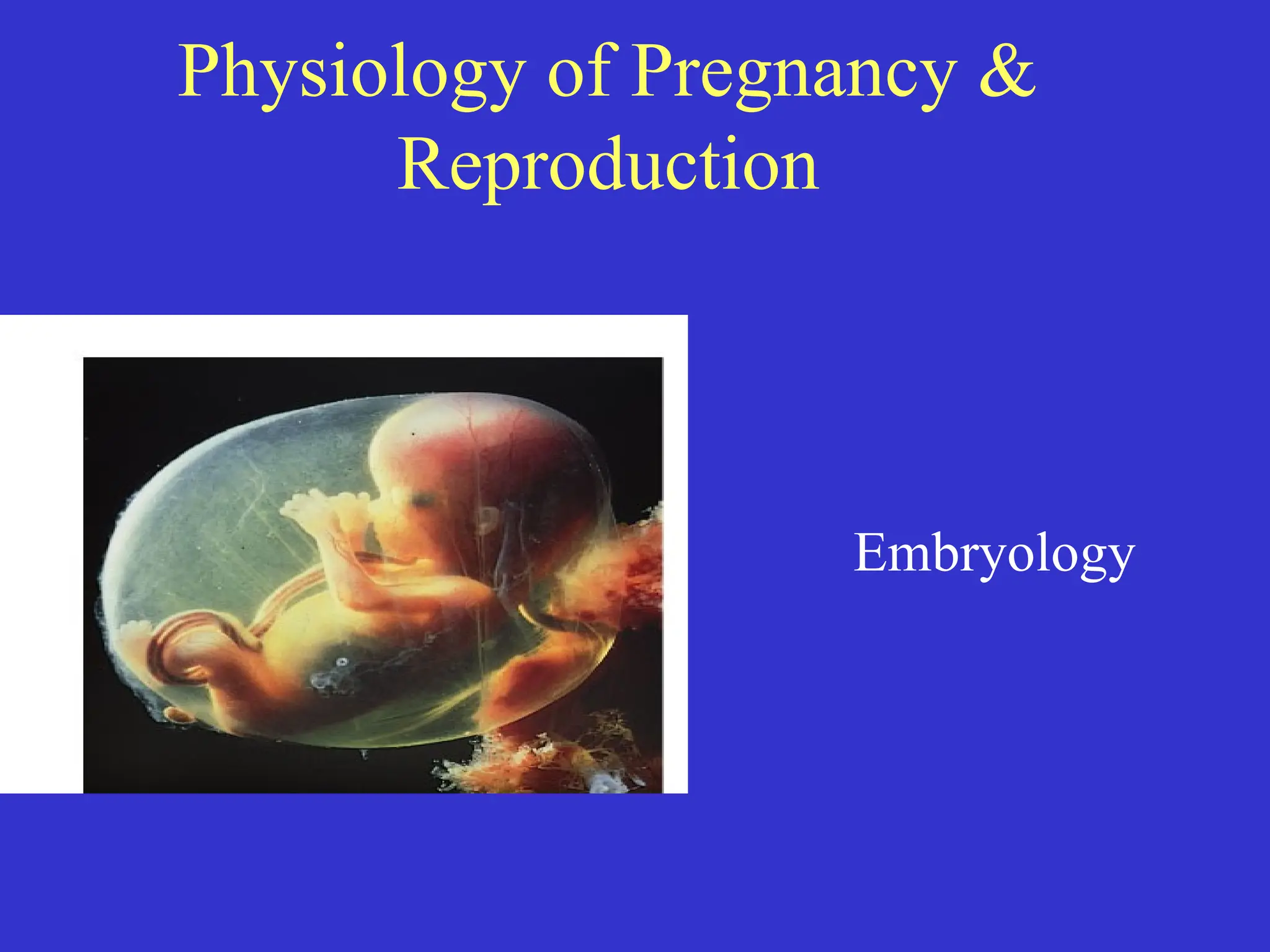
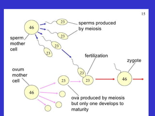
Embryology
• Definition: thestudy of the origin and
development of an organism
• Prenatal period: before birth
– 38 weeks from conception to birth (average) “fetal”
age
3.
Traditional (artificial) division:
•“Embryonic” period: first 8 weeks
– All major organs formed
• “Fetal” period: remaining 30 weeks
– Organs grow larger and become more complex
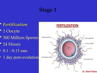
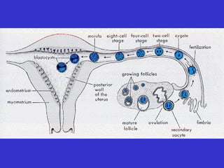
Fertilization
• SPERM +EGG(OOCYTE) = ZYGOTE
• The fertilization process takes about 24
hours.
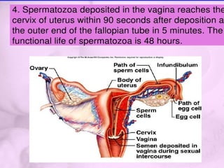
• Sperm life = 48 hours
– It takes about ten hours to navigate the female
productive track, moving up the vaginal canal,
through the cervix, and into the fallopian tube
where fertilization begins.
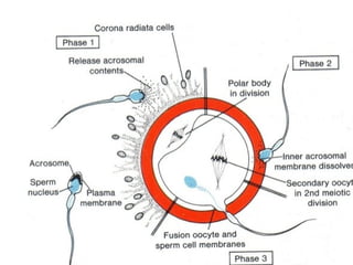
Fertilization
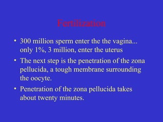
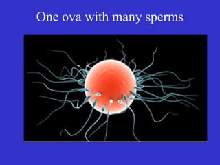
• 300 millionsperm enter the the vagina...
only 1%, 3 million, enter the uterus
• The next step is the penetration of the zona
pellucida, a tough membrane surrounding
the oocyte.
• Penetration of the zona pellucida takes
about twenty minutes.
Fertilization
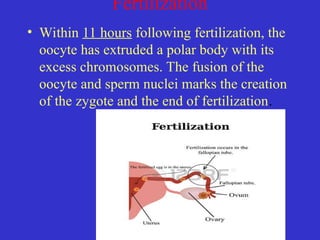
• Within 11hours following fertilization, the
oocyte has extruded a polar body with its
excess chromosomes. The fusion of the
oocyte and sperm nuclei marks the creation
of the zygote and the end of fertilization.
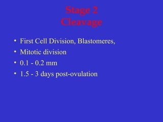
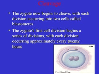
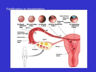
Cleavage
• The zygotenow begins to cleave, with each
division occurring into two cells called
blastomeres
• The zygote's first cell division begins a
series of divisions, with each division
occurring approximately every twenty
hours
19.
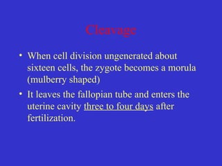
Cleavage
• When celldivision ungenerated about
sixteen cells, the zygote becomes a morula
(mulberry shaped)
• It leaves the fallopian tube and enters the
uterine cavity three to four days after
fertilization.
20.
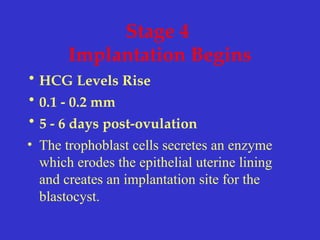
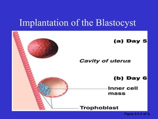
Stage 4
Implantation Begins
•HCG Levels Rise
• 0.1 - 0.2 mm
• 5 - 6 days post-ovulation
• The trophoblast cells secretes an enzyme
which erodes the epithelial uterine lining
and creates an implantation site for the
blastocyst.
21.
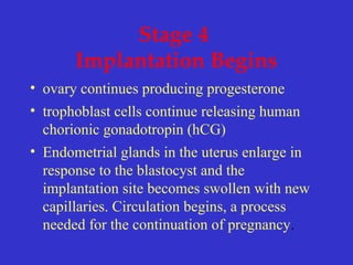
Stage 4
Implantation Begins
•ovary continues producing progesterone
• trophoblast cells continue releasing human
chorionic gonadotropin (hCG)
• Endometrial glands in the uterus enlarge in
response to the blastocyst and the
implantation site becomes swollen with new
capillaries. Circulation begins, a process
needed for the continuation of pregnancy.
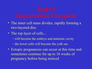
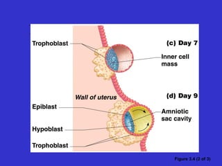
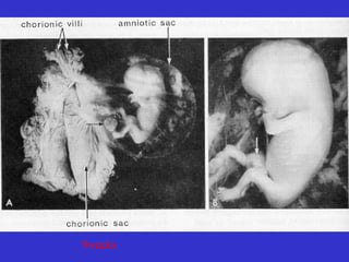
Stage 5
Implantation Complete
•The inner cell mass divides, rapidly forming a
two-layered disc
• The top layer of cells...
– will become the embryo and amniotic cavity
– the lower cells will become the yolk sac.
• Ectopic pregnancies can occur at this time and
sometimes continue for up to 16 weeks of
pregnancy before being noticed
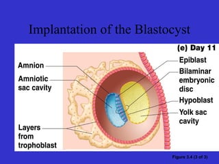
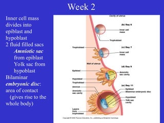
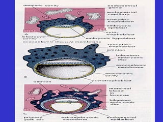
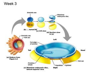
Week 2
Inner cellmass
divides into
epiblast and
hypoblast
2 fluid filled sacs
Amniotic sac
from epiblast
Yolk sac from
hypoblast
Bilaminar
embryonic disc:
area of contact
(gives rise to the
whole body)
29.
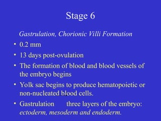
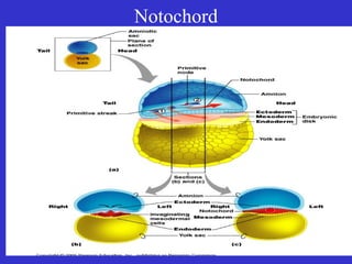
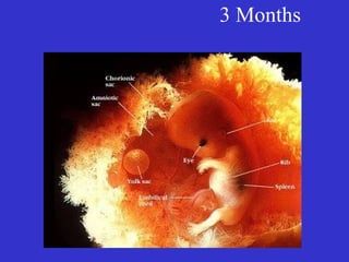
Stage 6
Gastrulation, ChorionicVilli Formation
• 0.2 mm
• 13 days post-ovulation
• The formation of blood and blood vessels of
the embryo begins
• Yolk sac begins to produce hematopoietic or
non-nucleated blood cells.
• Gastrulation three layers of the embryo:
ectoderm, mesoderm and endoderm.
31.
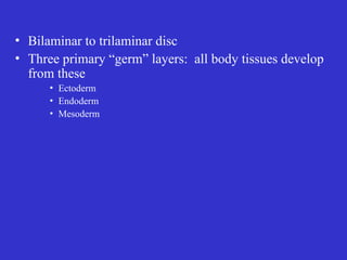
• Bilaminar totrilaminar disc
• Three primary “germ” layers: all body tissues develop
from these
• Ectoderm
• Endoderm
• Mesoderm
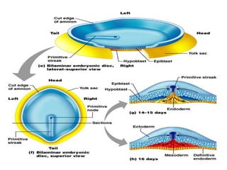
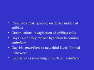
• Primitive streak(groove) on dorsal surface of
epiblast
• Grastrulation: invagination of epiblast cells
• Days 14-15: they replace hypoblast becoming
endoderm
• Day 16: mesoderm (a new third layer) formed
in between
• Epiblast cells remaining on surface: ectoderm
35.
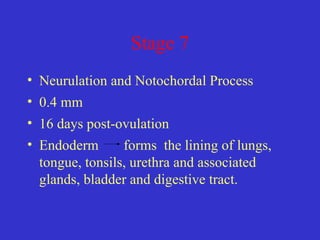
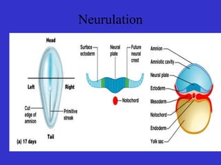
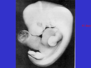
Stage 7
• Neurulationand Notochordal Process
• 0.4 mm
• 16 days post-ovulation
• Endoderm forms the lining of lungs,
tongue, tonsils, urethra and associated
glands, bladder and digestive tract.
36.
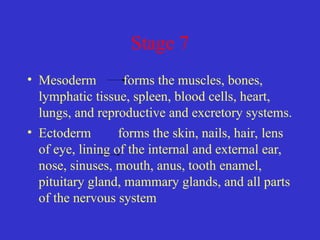
Stage 7
• Mesodermforms the muscles, bones,
lymphatic tissue, spleen, blood cells, heart,
lungs, and reproductive and excretory systems.
• Ectoderm forms the skin, nails, hair, lens
of eye, lining of the internal and external ear,
nose, sinuses, mouth, anus, tooth enamel,
pituitary gland, mammary glands, and all parts
of the nervous system
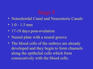
Stage 8
• NotochordalCanal and Neurenteric Canals
• 1.0 - 1.5 mm
• 17-19 days post-ovulation
• Neural plate with a neural groove
• The blood cells of the embryo are already
developed and they begin to form channels
along the epithelial cells which form
consecutively with the blood cells.
Stage 9
• Appearanceof Somites(condensations of
mesoderm, appear on either side of the neural
groove
• 1.5 - 2.5 mm
• 19 - 21 days post-ovulation
• Primitive streak
• Endocardial (muscle) cells begin to fuse
and form into the early embryo's two heart
tubes.
43.
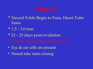
Stage 10
• NeuralFolds Begin to Fuse, Heart Tube
fuses
• 1.5 - 3.0 mm
• 21 - 23 days post-ovulation
• Cardiac muscle contraction begins
• Eye & ear cells are present
• Neural tube starts closing
45.
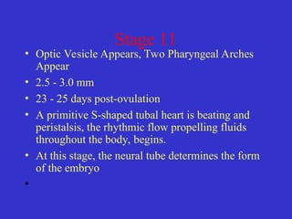
Stage 11
• OpticVesicle Appears, Two Pharyngeal Arches
Appear
• 2.5 - 3.0 mm
• 23 - 25 days post-ovulation
• A primitive S-shaped tubal heart is beating and
peristalsis, the rhythmic flow propelling fluids
throughout the body, begins.
• At this stage, the neural tube determines the form
of the embryo
•
46.
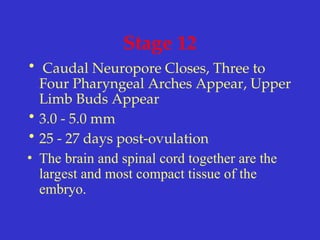
Stage 12
• CaudalNeuropore Closes, Three to
Four Pharyngeal Arches Appear, Upper
Limb Buds Appear
• 3.0 - 5.0 mm
• 25 - 27 days post-ovulation
• The brain and spinal cord together are the
largest and most compact tissue of the
embryo.
47.
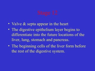
Stage 12
• Valve& septa appear in the heart
• The digestive epithelium layer begins to
differentiate into the future locations of the
liver, lung, stomach and pancreas.
• The beginning cells of the liver form before
the rest of the digestive system.
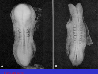
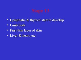
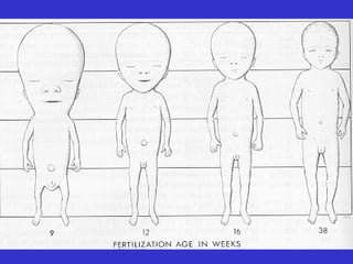
Stage 15
• 6to 8 weeks post fertilization
• Further development of nervous system,
heart.
• Innervation, the distribution of nerves,
begins in the lower limb buds.
57.
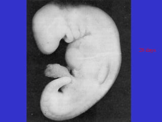
Stage 17
• approximately41 postovulatory days
• A Four Chambered Heart and a Sense of
Smell
• Primitive germ cells arrive at the genital
area and will respond to genetic instructions
to develop into either female or male
genitals.
58.
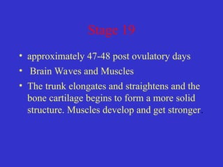
Stage 19
• approximately47-48 post ovulatory days
• Brain Waves and Muscles
• The trunk elongates and straightens and the
bone cartilage begins to form a more solid
structure. Muscles develop and get stronger.
59.
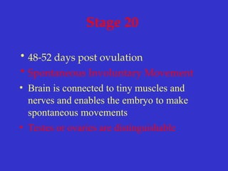
Stage 20
• 48-52days post ovulation
• Spontaneous Involuntary Movement
• Brain is connected to tiny muscles and
nerves and enables the embryo to make
spontaneous movements
• Testes or ovaries are distinguishable
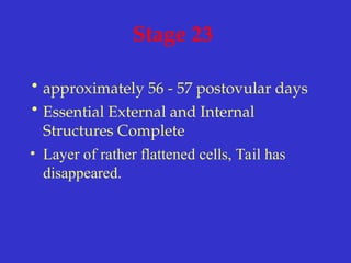
Stage 23
• approximately56 - 57 postovular days
• Essential External and Internal
Structures Complete
• Layer of rather flattened cells, Tail has
disappeared.
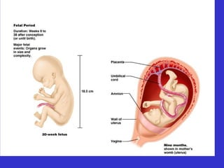
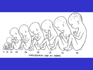
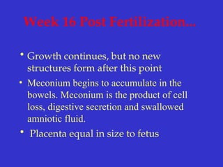
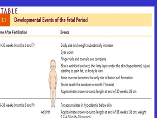
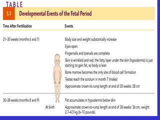
Week 16 PostFertilization...
• Growth continues, but no new
structures form after this point
• Meconium begins to accumulate in the
bowels. Meconium is the product of cell
loss, digestive secretion and swallowed
amniotic fluid.
• Placenta equal in size to fetus
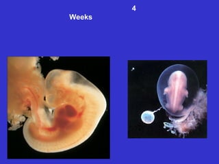
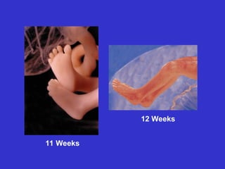
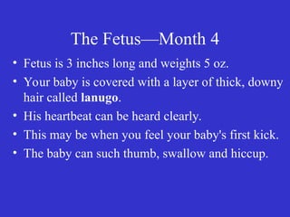
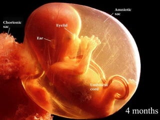
The Fetus—Month 4
•Fetus is 3 inches long and weights 5 oz.
• Your baby is covered with a layer of thick, downy
hair called lanugo.
• His heartbeat can be heard clearly.
• This may be when you feel your baby's first kick.
• The baby can such thumb, swallow and hiccup.
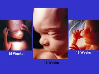
Week 18 PostFertilization...
• A dramatic growth period for the fetus.
• Fetus has phases of sleep and waking and
may prefer a favorite sleep position.
• Ovaries containing primitive egg cells &
uterus present
• Brown fat & vernix
• Placenta is fully formed and grows in
diameter though not in thickness.
80.
Week 20
• Fetussucks thumb
• Extremely rapid brain growth (which lasts
until five years after birth) begins.
• Testes of male fetuses begin descending
from the pelvis into the scrotum.
• Arms and legs move with more force, as
muscles strengthen.
81.
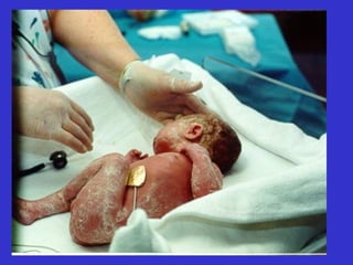
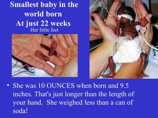
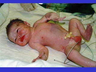
Smallest baby inthe
world born
At just 22 weeks
• She was 10 OUNCES when born and 9.5
inches. That's just longer than the length of
your hand. She weighed less than a can of
soda!
Her little feet
82.
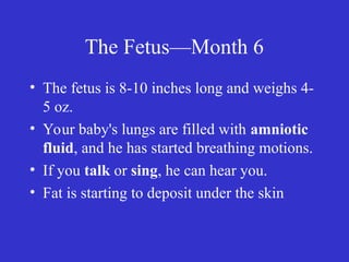
The Fetus—Month 6
•The fetus is 8-10 inches long and weighs 4-
5 oz.
• Your baby's lungs are filled with amniotic
fluid, and he has started breathing motions.
• If you talk or sing, he can hear you.
• Fat is starting to deposit under the skin
Week 26
• Lungsmay be mature enough to breath air!
• Fetal body is two to three percent body fat.
• Eyes are partially open and eyelashes
present.
• Sucking and swallowing improves.
85.
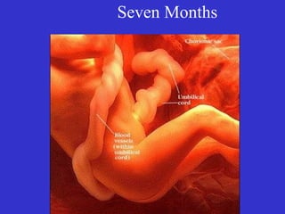
The Fetus—Month 7
•Fetus is 10-12 inches long and weighs about
1-2 pounds.
• Fetus is active and then rests.
• The baby now uses the four senses of
vision, hearing, taste and touch
Week 30
• Bodygrowth slows down
• The iris is colored and the pupil reflexes
responding to light.
88.
Week 32
• Fetusrests on uterus - no longer floating.
• Eyes open during alert times and close
during sleep. Eye color is usually blue,
regardless of the permanent color as
pigmentation is not fully developed
90.
The Fetus—Month 8
•The fetus is 14-16 inches long and weighs
2-3 pounds
• Layers of fat are piling on.
• Fetus has probably turned head-down in
preparation for birth.
• Fetus may react to noises with a jerking
action
Week 34
• Headmay now position (head-down) into
pelvis before labor.
• Gastrointestinal system is very immature
and will stay that way until three or fourth
years after birth.
• Fetus stores about 15% of weight in fat to
keep temperature of body warm
93.
The Fetus—Month 9
•Fetus is about 17-18 inches long and weighs 5-6
pounds
• Skin is smooth because of the fat
• Baby’s movement slows down due to lack of room
• “Lightening” occurs when the baby drops in the
pelvis
• Disease fighting antibodies are taken from the
mother’s blood
94.
40 weeks
• Fullterm
•Fifteen percent of body is fat, eighty
percent of which is underneath the skin, the
other twenty percent around the organs.
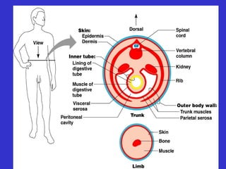
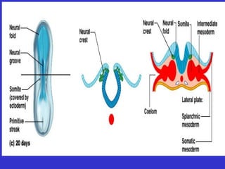
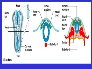
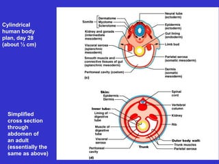
#40 Notochord signals overlying ectoderm
Formation begins of spinal cord and brain (neurulation)
Neural plate to neural groove to neural tube: pinched off into body
#42 Mesoderm begins to differentiate
Lateral to notochord, week 3
Extends cranially and caudally (from head to tail or crown to rump)
Division of mesoderm into three regions
Somites: 40 pairs of body segments (repeating units, like building blocks) by end week 4
Intermediate mesoderm: just lateral to somites
Lateral plate: splits to form coelom (“cavity”)
#44 Closure of neural tube: begins at end of week 3; complete by end of week 4 (folic acid important for this step)
Extends cranially (eventually brain) and caudally (spinal cord)
Neural crest, lateral ectodermal cells, pulled along and form sensory nerve cells and other structures


![CTEV [ clubfoot] DR ARUN LAL ,DR MOHAMED ASHRAF travancore medical college k...](https://cdn.slidesharecdn.com/ss_thumbnails/ctevclubfootdrarunlaldrmohamedashraftravancoremedicalcollegekollamkeralaindia-260208063247-18fc466c-thumbnail.jpg?width=640&height=640&fit=bounds)


