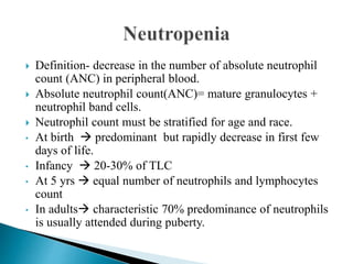
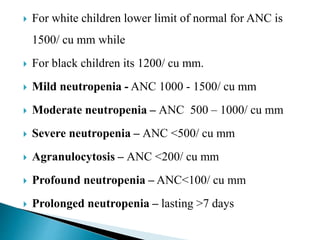
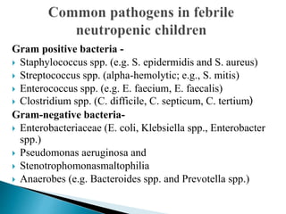
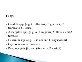
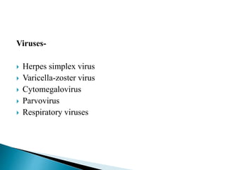
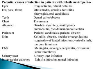
Dr. Dilipkumar Choudhary
Department of Pediatrics
 Definition- decrease in the number of absolute neutrophil
count (ANC) in peripheral blood.
 Absolute neutrophil count(ANC)= mature granulocytes +
neutrophil band cells.
 Neutrophil count must be stratified for age and race.
• At birth  predominant but rapidly decrease in first few
days of life.
• Infancy  20-30% of TLC
• At 5 yrs  equal number of neutrophils and lymphocytes
count
• In adults characteristic 70% predominance of neutrophils
is usually attended during puberty.
 For white children lower limit of normal for ANC is
1500/ cu mm while
 For black children its 1200/ cu mm.
 Mild neutropenia - ANC 1000 - 1500/ cu mm
 Moderate neutropenia – ANC 500 – 1000/ cu mm
 Severe neutropenia – ANC <500/ cu mm
 Agranulocytosis – ANC <200/ cu mm
 Profound neutropenia – ANC<100/ cu mm
 Prolonged neutropenia – lasting >7 days
 The relative lower limit of normal in blacks likely
reflects the prevalence of the Duffy negative (Fy-/-)
blood group, which is selectively enriched in
populations in malaria belt of Africa and is associated
with ANCs 200-600/ cu mm less than those who are
Duffy positive.
 Development of fever in a neutropenic patient.
 Defined as single oral/axillary temperature >38.3˚C
(101˚F) or two consecutive temperature >38˚C (100˚F)
in a 12 hr period for at least 1hr with ANC <500/cu mm
or <1000 cu mm with expected decline to <500/cu mm
during the next 48 hrs.
 Common in children on chemotherapy and the most
common oncologic emergency.
 The risk of infection is clearly increased when ANC drop
below 1000/mm3, with a marked increase when the ANC is
below 500/mm3.
 Neutropenic patient can have serious life threatening
infections even in the absence of fever and may not present
with any localizing symptoms and signs of infection such as
exudate, fluctuance and regional lymphadenopathy,
sometimes only fever remain the only consistent early sign.
 Approximately 48 to 60% patients who become febrile
have an established or occult infection.
 Common sites of infection are the alimentary tract (i.e.,
mouth, pharynx, esophagus, large and small bowel and
rectum), perirectal abscess, sinuses, throat, ear (otitis
media), lungs and skin( cellulitis, furunculosis).
 Catheter and IV sites are potential source of infection.
Gram positive bacteria -
 Staphylococcus spp. (e.g. S. epidermidis and S. aureus)
 Streptococcus spp. (alpha-hemolytic; e.g., S. mitis)
 Enterococcus spp. (e.g. E. faecium, E. faecalis)
 Clostridium spp. (C. difficile, C. septicum, C. tertium)
Gram-negative bacteria-
 Enterobacteriaceae (E. coli, Klebsiella spp., Enterobacter
spp.)
 Pseudomonas aeruginosa and
 Stenotrophomonasmaltophilia
 Anaerobes (e.g. Bacteroides spp. and Prevotella spp.)
Fungi-
 Candida spp. (e.g. C. albicans, C. glabrata, C.
tropicalis, C. krusei)
 Aspergillus spp. (e.g. A. fumigatus, A. flavus, and A.
terreus)
 Fusarium spp. (e.g. F. solani and F. oxysporum)
 Cryptococcus neoformans
 Pneumocystis jiroveci (formerly, P. carinii)
Viruses-
 Herpes simplex virus
 Varicella-zoster virus
 Cytomegalovirus
 Parvovirus
 Respiratory viruses
Potential causes of infection in patients with febrile neutropenia-
Eyes Conjunctivitis, orbital cellulitis
Ear, nose, throat Otitis media, sinusitis, tonsillitis
pharyngitis, oral candidiasis
Teeth Dental caries/abscess
Chest Pneumonia
Abdomen Diarrhea, dysentery, neutropenic-
enterocolitis, pseudomembranous colitis
Perineum Perianal candidiasis, perianal abscess
Skin Cellulitis, abscess, nodular or target lesions
suggestive of fungal infections, varicella rash,
purpura fulminans
CNS Meningitis, meningoencephalitis, cavernous
sinus thrombosis
Urinary tract Urinary tract infection
Intravascular catheters Exit site infection, tunnel infection
History
 Fever -onset, duration and severity
 Associated localizing symptoms: Ear, nose, throat,
respiratory, gastrointestinal tract, musculoskeletal and
urinary system.
 Phase of chemotherapy (intensive vs. non intensive)
 Duration since the last chemotherapy
 Recent hospitalization and antibiotics received, if any
Examination
 Vitals- in every patient of suspected febrile
neutropenia, the patient may appear well despite being
in a state of hemodynamic compromise.
 Detailed physical examination focusing on possible
sites of infection must be undertaken. Sites that are
commonly overlooked include oral cavity, ear, sinuses,
skin, nails, perianal area, intravascular catheter
insertion site and the site of bone marrow aspiration.
Investigations
First line investigations- to be performed in every case
(1).Complete blood count, including differential leukocyte count and
ANC
(2). Serum electrolytes, urea and creatinine
(3). Blood culture: Obtain as early as possible and always before the
administration of antibiotics. Two sets of blood cultures from separate
venipuncture sites should ideally be drawn. In the presence of a
central venous catheter, a blood culture should be obtained from each
lumen of the catheter and another from peripheral vein.
(4). Chest radiograph: mandatory for all, initial x-ray may be non
informative but must be taken as a baseline for comparison with later
films.
(5). Cultures from any other site, as clinically relevant. This includes
stool, urine, cerebrospinal fluid, skin, respiratory secretions or pus.
Second line investigations- Dictated by the clinical course:
(1).CT scan of chest/paranasal sinuses may be indicated in
patients with suspected fungal infection.
(2).Bronchoalveolar lavage: If pneumonia is non-resolving/
non-responding.
(3).Skin biopsy, from skin nodules, if any.
 Despite advances in diagnostic methods, infection is
documented only in 30-40% patients.
Risk stratification is crucial in determining the
appropriate-
 Choice of antimicrobials,
 Route of administration (intravenous vs. oral),
 Setting (inpatient vs. outpatient) and
 Duration.
 The patients can be classified as low or high risk
Low risk High risk
Neutropenia expected to resolve in
about 7 days
Indicators:
• Afebrile for 24hrs
• Clinically well, hemodynamically
stable
• Sterile blood culture
• Control of local infection
• Evidence of bone marrow recovery
with rising polymorphs count/
platelets/ ANC
• Non-intensive phase of
chemotherapy e.g., maintenance
phase of chemotherapy
• Malignancy in remission
• ANC≥100/mm3 and likely to rise
within the next 7 d.
Neutropenia >7days
Indicators
• Fever persisting
• Culture positive
• No evidence of bone marrow
recovery
• Recent intensive chemotherapy
• Profound neutropenia (ANC<100
cells/mm3), anticipated to extend
for >7 d.
• Evidence of hypotension,
respiratory distress or hypoxemia.
• Mucositis interfering with oral
intake or resulting in diarrhea
 Presence of fever >39˚C, associated hypotension, ANC<100
cu mm, duration of neutropenia >7days  frequently
associated with bacteremia.
 Santolaya, et al found serum CRP more than 90 mg/L,
hypotension, relapsed leukemia, platelet count less than
50,000/mm3 and recent chemotherapy to be useful predictors
of serious bacterial infection.
 Klaassen et al found an absolute monocyte count less than 100
cell/mm3, co-morbidity and abnormal chest radiograph to
correlate with high risk for significant bacterial infection.
 For adults- the Multinational Association of Supportive Care
in Cancer (MASCC) score is used for risk stratification.
Patient with score with ≥21 are at lower risk of complications.
General considerations-
 Pro- active steps must be taken to reduce incidence of
hospital acquired sepsis.
 Barrier nursing practise- frequent hand washing, the
use of alcohol based hand rub in between patients and
wearing gloves must be ensured .
 Use of IV fluids, central lines, foley’s catheter etc. must
be restricted, if possible.
 Administration of IV fluids for minor reasons should be
avoided.
 Nasogastric feeding should be encouraged in patients with
anorexia or mucositis.
 Rectal enemas, suppositories, and rectal examinations are
contraindicated in neutropenic patients
 High-risk’ patients are to be hospitalized and administered
broad-spectrum intravenous antibiotics
 Empirical treatment should begin as soon as possible, even
before the results of cultures are available.
 Important considerations during initial empirical
therapy-
• Risk stratification
• Any focal signs and symptoms, site of infection (e.g.,
lung)
• Antimicrobial susceptibilities of local pathogens
• Recent hospitalization, recent antibiotic exposure or
prophylactic drugs
• Past history of infection (especially fungal or resistant
bacterial such as ESBL or MRSA infection)
• Most common potentially infecting organism based on
type of immune defect
• Co-morbid conditions.
 Care-takers are advised not to administer paracetamol
at home as it may mask fever and can lead to delay in
seeking medical care.
 Administer the first dose of antibiotics without any
delay. Delay in initiating antimicrobials significantly
increases the morbidity and mortality.
 Anti-pseudomonal agents-
penicillin (piperacillin-tazobactum), antipseudomonal
cephalosporin (cefoperazone-sulbactum) or
carbepenems (meropenem or imipenem-cilastatin) or
cefepime is recommended as first line by Infectious
Disease Society of America
BUT
no significant differences in treatment failure, including
antibiotic modification, infection-related mortality, or
adverse events were observed while comparing anti-
pseudomonas penicillin± aminoglycoside regimen with
carbapenem monotherapy in a recent meta analysis.
 Empirically  combination of antipseudomonal
antibiotic (ceftazidime, cefaperazone/ salbactam) + an
aminoglycoside is used as first line choice.
 Switch to second line drugs: Vancomycin and
Carbepenems (meropenem or imipenem-cilastatin)
after 48–72 h, if fever is unrelenting and there is no
improvement in the clinical condition.
 Colistin- reserved as a third line drug.
 If the cultures yield a specific pathogen, the regimen
should be modified accordingly
 For low risk FN  oral amoxicillin- clavulinate with
ofloxacin OR cetrioxone with amikacin
 Indications for need of vancomycin in initial regimen if
patient has -
• Hypotension or evidence of septic shock
• Obvious catheter related infection
• H/O colonization with MRSA
• High risk for viridans streptococci (severe mucositis/ AML /
prior use of quinolone prophylaxis).
 If indwelling line insitu or no response in 48 hrs 
antistaphylococcal antibiotic should be added.
 If fever persist for 4-5 days  antifungal (eg. Amphotericin
B) should be added.
 Discontinuation should always be kept in mind to minimize
development of bacterial resistance
Add antistaphylococcal drug and if febrile at 4-5 days add antifungal
Febrile neutropenia
Initial evaluation
Physical examination
investigations
Start treatment
Ceftazidime + amynolycoside
iv
Monitor
-Fever
-ANC
Febrile afetr48hrs Afebrile
Afebrile
ANC>500/cu mm ANC<500/cu mm
Low risk High risk
Stop
therapy
when
afebrile
for
>48hrs
Switch to
oral
antibiotic
Low risk High risk
Give oral
antibiotic
for 7 days
Continue
antibiotic
for 10-14
days
 If fever takes long to respond , the cultures are positive
or ANC is <100/cu mm on admission antibiotics are
given for 10-14 days or till ANC increases >500/cu
mm.
 P. jiroveci can cause pneumonia regardless of
neutrophil count, prophylaxis with trimethoprim-
sulfamethoxazole against PCP is an effective
preventive strategy and should be provided to all
children undergoing active treatment for malignancy.
Hematopoietic growth factor-
 G-CSF (Filgrastim) @5mcg/kg/day in addition to
antibiotics is useful in children with complicated febrile
neutropenia (pneumonia, hypotension, invasive fungal
infection or multi organ dysfunction), it results in-
• more rapid neutrophil recovery
• relatively fewer days of antibiotic use
• Shortens length of hospital stay
• Reduces mortality and morbidity
But G-CSF has no role in the management of children
with uncomplicated febrile neutropenia.
Thank you

Febrile neutropenia by dr. dilip

  • 1.
  • 2.
     Definition- decreasein the number of absolute neutrophil count (ANC) in peripheral blood.  Absolute neutrophil count(ANC)= mature granulocytes + neutrophil band cells.  Neutrophil count must be stratified for age and race. • At birth  predominant but rapidly decrease in first few days of life. • Infancy  20-30% of TLC • At 5 yrs  equal number of neutrophils and lymphocytes count • In adults characteristic 70% predominance of neutrophils is usually attended during puberty.
  • 3.
     For whitechildren lower limit of normal for ANC is 1500/ cu mm while  For black children its 1200/ cu mm.  Mild neutropenia - ANC 1000 - 1500/ cu mm  Moderate neutropenia – ANC 500 – 1000/ cu mm  Severe neutropenia – ANC <500/ cu mm  Agranulocytosis – ANC <200/ cu mm  Profound neutropenia – ANC<100/ cu mm  Prolonged neutropenia – lasting >7 days
  • 4.
     The relativelower limit of normal in blacks likely reflects the prevalence of the Duffy negative (Fy-/-) blood group, which is selectively enriched in populations in malaria belt of Africa and is associated with ANCs 200-600/ cu mm less than those who are Duffy positive.
  • 5.
     Development offever in a neutropenic patient.  Defined as single oral/axillary temperature >38.3˚C (101˚F) or two consecutive temperature >38˚C (100˚F) in a 12 hr period for at least 1hr with ANC <500/cu mm or <1000 cu mm with expected decline to <500/cu mm during the next 48 hrs.
  • 6.
     Common inchildren on chemotherapy and the most common oncologic emergency.  The risk of infection is clearly increased when ANC drop below 1000/mm3, with a marked increase when the ANC is below 500/mm3.  Neutropenic patient can have serious life threatening infections even in the absence of fever and may not present with any localizing symptoms and signs of infection such as exudate, fluctuance and regional lymphadenopathy, sometimes only fever remain the only consistent early sign.
  • 7.
     Approximately 48to 60% patients who become febrile have an established or occult infection.  Common sites of infection are the alimentary tract (i.e., mouth, pharynx, esophagus, large and small bowel and rectum), perirectal abscess, sinuses, throat, ear (otitis media), lungs and skin( cellulitis, furunculosis).  Catheter and IV sites are potential source of infection.
  • 8.
    Gram positive bacteria-  Staphylococcus spp. (e.g. S. epidermidis and S. aureus)  Streptococcus spp. (alpha-hemolytic; e.g., S. mitis)  Enterococcus spp. (e.g. E. faecium, E. faecalis)  Clostridium spp. (C. difficile, C. septicum, C. tertium) Gram-negative bacteria-  Enterobacteriaceae (E. coli, Klebsiella spp., Enterobacter spp.)  Pseudomonas aeruginosa and  Stenotrophomonasmaltophilia  Anaerobes (e.g. Bacteroides spp. and Prevotella spp.)
  • 9.
    Fungi-  Candida spp.(e.g. C. albicans, C. glabrata, C. tropicalis, C. krusei)  Aspergillus spp. (e.g. A. fumigatus, A. flavus, and A. terreus)  Fusarium spp. (e.g. F. solani and F. oxysporum)  Cryptococcus neoformans  Pneumocystis jiroveci (formerly, P. carinii)
  • 10.
    Viruses-  Herpes simplexvirus  Varicella-zoster virus  Cytomegalovirus  Parvovirus  Respiratory viruses
  • 11.
    Potential causes ofinfection in patients with febrile neutropenia- Eyes Conjunctivitis, orbital cellulitis Ear, nose, throat Otitis media, sinusitis, tonsillitis pharyngitis, oral candidiasis Teeth Dental caries/abscess Chest Pneumonia Abdomen Diarrhea, dysentery, neutropenic- enterocolitis, pseudomembranous colitis Perineum Perianal candidiasis, perianal abscess Skin Cellulitis, abscess, nodular or target lesions suggestive of fungal infections, varicella rash, purpura fulminans CNS Meningitis, meningoencephalitis, cavernous sinus thrombosis Urinary tract Urinary tract infection Intravascular catheters Exit site infection, tunnel infection
  • 12.
    History  Fever -onset,duration and severity  Associated localizing symptoms: Ear, nose, throat, respiratory, gastrointestinal tract, musculoskeletal and urinary system.  Phase of chemotherapy (intensive vs. non intensive)  Duration since the last chemotherapy  Recent hospitalization and antibiotics received, if any
  • 13.
    Examination  Vitals- inevery patient of suspected febrile neutropenia, the patient may appear well despite being in a state of hemodynamic compromise.  Detailed physical examination focusing on possible sites of infection must be undertaken. Sites that are commonly overlooked include oral cavity, ear, sinuses, skin, nails, perianal area, intravascular catheter insertion site and the site of bone marrow aspiration.
  • 14.
    Investigations First line investigations-to be performed in every case (1).Complete blood count, including differential leukocyte count and ANC (2). Serum electrolytes, urea and creatinine (3). Blood culture: Obtain as early as possible and always before the administration of antibiotics. Two sets of blood cultures from separate venipuncture sites should ideally be drawn. In the presence of a central venous catheter, a blood culture should be obtained from each lumen of the catheter and another from peripheral vein. (4). Chest radiograph: mandatory for all, initial x-ray may be non informative but must be taken as a baseline for comparison with later films. (5). Cultures from any other site, as clinically relevant. This includes stool, urine, cerebrospinal fluid, skin, respiratory secretions or pus.
  • 15.
    Second line investigations-Dictated by the clinical course: (1).CT scan of chest/paranasal sinuses may be indicated in patients with suspected fungal infection. (2).Bronchoalveolar lavage: If pneumonia is non-resolving/ non-responding. (3).Skin biopsy, from skin nodules, if any.  Despite advances in diagnostic methods, infection is documented only in 30-40% patients.
  • 16.
    Risk stratification iscrucial in determining the appropriate-  Choice of antimicrobials,  Route of administration (intravenous vs. oral),  Setting (inpatient vs. outpatient) and  Duration.  The patients can be classified as low or high risk
  • 17.
    Low risk Highrisk Neutropenia expected to resolve in about 7 days Indicators: • Afebrile for 24hrs • Clinically well, hemodynamically stable • Sterile blood culture • Control of local infection • Evidence of bone marrow recovery with rising polymorphs count/ platelets/ ANC • Non-intensive phase of chemotherapy e.g., maintenance phase of chemotherapy • Malignancy in remission • ANC≥100/mm3 and likely to rise within the next 7 d. Neutropenia >7days Indicators • Fever persisting • Culture positive • No evidence of bone marrow recovery • Recent intensive chemotherapy • Profound neutropenia (ANC<100 cells/mm3), anticipated to extend for >7 d. • Evidence of hypotension, respiratory distress or hypoxemia. • Mucositis interfering with oral intake or resulting in diarrhea
  • 18.
     Presence offever >39˚C, associated hypotension, ANC<100 cu mm, duration of neutropenia >7days  frequently associated with bacteremia.  Santolaya, et al found serum CRP more than 90 mg/L, hypotension, relapsed leukemia, platelet count less than 50,000/mm3 and recent chemotherapy to be useful predictors of serious bacterial infection.  Klaassen et al found an absolute monocyte count less than 100 cell/mm3, co-morbidity and abnormal chest radiograph to correlate with high risk for significant bacterial infection.  For adults- the Multinational Association of Supportive Care in Cancer (MASCC) score is used for risk stratification. Patient with score with ≥21 are at lower risk of complications.
  • 19.
    General considerations-  Pro-active steps must be taken to reduce incidence of hospital acquired sepsis.  Barrier nursing practise- frequent hand washing, the use of alcohol based hand rub in between patients and wearing gloves must be ensured .  Use of IV fluids, central lines, foley’s catheter etc. must be restricted, if possible.
  • 20.
     Administration ofIV fluids for minor reasons should be avoided.  Nasogastric feeding should be encouraged in patients with anorexia or mucositis.  Rectal enemas, suppositories, and rectal examinations are contraindicated in neutropenic patients  High-risk’ patients are to be hospitalized and administered broad-spectrum intravenous antibiotics  Empirical treatment should begin as soon as possible, even before the results of cultures are available.
  • 21.
     Important considerationsduring initial empirical therapy- • Risk stratification • Any focal signs and symptoms, site of infection (e.g., lung) • Antimicrobial susceptibilities of local pathogens • Recent hospitalization, recent antibiotic exposure or prophylactic drugs • Past history of infection (especially fungal or resistant bacterial such as ESBL or MRSA infection) • Most common potentially infecting organism based on type of immune defect • Co-morbid conditions.
  • 22.
     Care-takers areadvised not to administer paracetamol at home as it may mask fever and can lead to delay in seeking medical care.  Administer the first dose of antibiotics without any delay. Delay in initiating antimicrobials significantly increases the morbidity and mortality.
  • 23.
     Anti-pseudomonal agents- penicillin(piperacillin-tazobactum), antipseudomonal cephalosporin (cefoperazone-sulbactum) or carbepenems (meropenem or imipenem-cilastatin) or cefepime is recommended as first line by Infectious Disease Society of America BUT no significant differences in treatment failure, including antibiotic modification, infection-related mortality, or adverse events were observed while comparing anti- pseudomonas penicillin± aminoglycoside regimen with carbapenem monotherapy in a recent meta analysis.
  • 24.
     Empirically combination of antipseudomonal antibiotic (ceftazidime, cefaperazone/ salbactam) + an aminoglycoside is used as first line choice.  Switch to second line drugs: Vancomycin and Carbepenems (meropenem or imipenem-cilastatin) after 48–72 h, if fever is unrelenting and there is no improvement in the clinical condition.  Colistin- reserved as a third line drug.  If the cultures yield a specific pathogen, the regimen should be modified accordingly  For low risk FN  oral amoxicillin- clavulinate with ofloxacin OR cetrioxone with amikacin
  • 25.
     Indications forneed of vancomycin in initial regimen if patient has - • Hypotension or evidence of septic shock • Obvious catheter related infection • H/O colonization with MRSA • High risk for viridans streptococci (severe mucositis/ AML / prior use of quinolone prophylaxis).  If indwelling line insitu or no response in 48 hrs  antistaphylococcal antibiotic should be added.  If fever persist for 4-5 days  antifungal (eg. Amphotericin B) should be added.  Discontinuation should always be kept in mind to minimize development of bacterial resistance
  • 26.
    Add antistaphylococcal drugand if febrile at 4-5 days add antifungal Febrile neutropenia Initial evaluation Physical examination investigations Start treatment Ceftazidime + amynolycoside iv Monitor -Fever -ANC Febrile afetr48hrs Afebrile
  • 27.
    Afebrile ANC>500/cu mm ANC<500/cumm Low risk High risk Stop therapy when afebrile for >48hrs Switch to oral antibiotic Low risk High risk Give oral antibiotic for 7 days Continue antibiotic for 10-14 days
  • 28.
     If fevertakes long to respond , the cultures are positive or ANC is <100/cu mm on admission antibiotics are given for 10-14 days or till ANC increases >500/cu mm.  P. jiroveci can cause pneumonia regardless of neutrophil count, prophylaxis with trimethoprim- sulfamethoxazole against PCP is an effective preventive strategy and should be provided to all children undergoing active treatment for malignancy.
  • 29.
    Hematopoietic growth factor- G-CSF (Filgrastim) @5mcg/kg/day in addition to antibiotics is useful in children with complicated febrile neutropenia (pneumonia, hypotension, invasive fungal infection or multi organ dysfunction), it results in- • more rapid neutrophil recovery • relatively fewer days of antibiotic use • Shortens length of hospital stay • Reduces mortality and morbidity But G-CSF has no role in the management of children with uncomplicated febrile neutropenia.
  • 30.