Downloaded 10 times
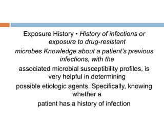


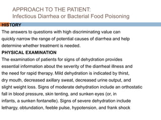
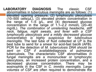
![DIAGNOSIS When bacterial meningitis is suspected,
blood cultures should be immediately obtained and
empirical antimicrobial and adjunctive
dexamethasone therapy initiated without delay
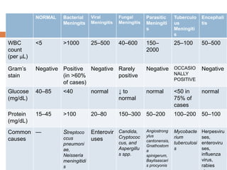
The diagnosis of bacterial meningitis is made by
examination of the CSF, The classic CSF
abnormalities in bacterial meningitis (Table 164-2)
are (1) polymorphonuclear (PMN) leukocytosis
(>100 cells/μL in 90%), (2) decreased glucose
concentration (<2.2 mmol/L [<40 mg/dL] and/ or
CSF/serum glucose ratio of <0.4 in ~60%), (3)
increased protein concentration (>0.45 g/L [>45
mg/dL] in 90%), and (4) increased opening
pressure (>180 mmH2O in 90%). CSF bacterial
cultures are positive in >80% of patients, and CSF
Gram’s stain demonstrates organisms in >60%.](https://image.slidesharecdn.com/infectiousdiseases-200530210143/85/Infectious-diseases-139-320.jpg)
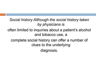
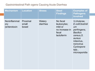
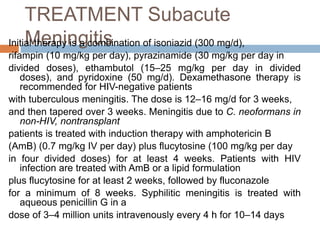
![PROGNOSIS Mortality rate is 3–7% for meningitis caused by
H. influenzae, N. meningitidis, or group B streptococci;
15% for that due to L. monocytogenes; and 20% for S.
pneumoniae. In general, the risk of death from bacterial
meningitis increases with (1) decreased level of
consciousness on admission, (2) onset of seizures within
24 h of admission, (3) signs of increased ICP, (4) young
age (infancy) and age >50, (5) the presence of comorbid
conditions including shock and/or the need for mechanical
ventilation, and (6) delay in the initiation of treatment.
Decreased CSF glucose concentration (<2.2 mmol/L [<40
mg/dL]) and markedly increased CSF protein concentration
(>3 g/L [> 300 mg/dL]) have been predictive of increased
mortality and poorer outcomes in some series. Moderate or
severe sequelae occur in ~25% of survivors, although the
exact incidence varies with the infecting organism.
Common sequelae include decreased intellectual function,
memory impairment, seizures, hearing loss and dizziness,
and gait disturbances.](https://image.slidesharecdn.com/infectiousdiseases-200530210143/85/Infectious-diseases-144-320.jpg)


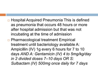
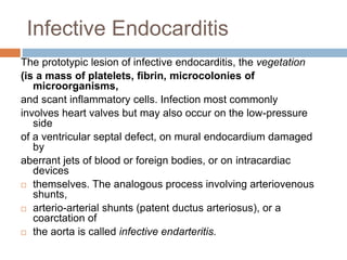
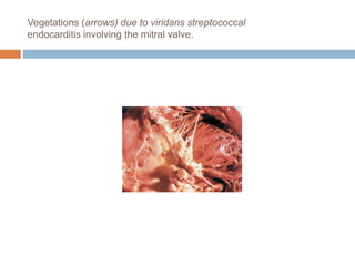
This document provides information on approaching and evaluating patients with potential infectious diseases. It discusses taking an exposure and social history, performing a physical exam focusing on vital signs, lymph nodes, skin, and foreign bodies. Diagnostic testing options are outlined including lab tests, imaging, and pathogen-specific tests. Empirical antibiotic therapy is recommended for common infections like pneumonia based on presentation. Community-acquired pneumonia causes are discussed. Hospital-acquired pneumonia treatment typically involves antibiotics until culture results are available. Infective endocarditis typically involves bacterial vegetation on heart valves.


